 Tools
Tools
 Malignant Eyelid Lesions
Introduction
Malignant eyelid lesions are often initially identified during routine examinations by eye care providers, primary care clinicians, or dermatologists. The most common eyelid malignancies include basal cell carcinoma (BCC), squamous cell carcinoma (SCC), sebaceous carcinoma, melanoma, and Merkel cell carcinoma (MCC). Although less common periocular malignancies exist, any cutaneous malignancy has the potential to involve the eyelid or periocular region and should be considered during evaluation.
A detailed history and a thorough examination are essential when assessing a suspicious eyelid lesion. Clinicians should evaluate lesion duration, growth rate, tenderness, discharge, bleeding, ulceration, lash loss, and recurrence. Risk factors such as excessive ultraviolet exposure, prior skin cancer, immunosuppression, and nonhealing cutaneous lesions should increase suspicion for malignancy. Careful examination under magnification with slit-lamp biomicroscopy or dermoscopy improves lesion characterization and may reduce unnecessary surgical procedures while facilitating earlier diagnosis and treatment.[1][2]
When malignancy is suspected, prompt referral for biopsy and histopathologic evaluation is warranted. Management depends on tumor type, extent, and patient-specific factors and may include surgical excision, radiotherapy, chemotherapy, immunotherapy, cryotherapy, or laser-based treatment modalities.
Etiology
Register For Free And Read The Full Article
 Search engine and full access to all medical articles
- 10 free questions in your specialty
- Free CME/CE Activities
- Free daily question in your email
- Save favorite articles to your dashboard
- Emails offering discounts
Learn more about a Subscription to StatPearls Point-of-Care
Etiology
Clinicians should recognize that prolonged and cumulative UV radiation exposure is the strongest risk factor for the most common eyelid malignancies, including BCC, SCC, and melanoma.[3] Understanding the mechanisms by which UV radiation induces cellular damage and carcinogenesis can improve recognition of additional risk factors and support earlier implementation of preventive counseling and risk-factor modification strategies.
Although a detailed review of UV-induced molecular injury is beyond the scope of this article, a brief overview of the underlying mechanisms is clinically relevant. UV-A and UV-B radiation contribute to carcinogenesis through complementary pathways. UV-A radiation promotes the formation of reactive oxygen species that damage cellular structures and induce oxidative stress. In contrast, UV-B radiation directly damages DNA through thymidine dimer formation, resulting in mutagenic cellular changes.[4][5] DNA damage may impair normal cellular repair and regulatory mechanisms, leading to mutations that inactivate tumor suppressor genes or promote uncontrolled activation of oncogenes, ultimately contributing to malignant transformation.[6] Additional risk factors for eyelid malignancies may increase susceptibility to UV-induced genetic injury, impair immune surveillance of malignant cells, or promote carcinogenesis through other DNA-damaging mechanisms. These risk factors may be modifiable, such as environmental or behavioral exposures, or non-modifiable, including inherited genetic predisposition.[3] The following sections review lesion-specific pathophysiology and risk factors associated with malignant eyelid lesions. Please see StatPearls' companion resource, "Skin Cancer," for further information. Unique eyelid malignancies that are not primarily associated with UV-induced damage are discussed separately within their respective sections.
Actinic Keratosis
Actinic keratosis, also known as solar keratosis, is considered a premalignant skin lesion for both SCC and BCC, although SCC is more common. Actinic keratosis, as with SCC and BCC, typically arises from prolonged exposure to UV radiation. Other variables also influence their development, including male gender, older age, baldness, and a history of melanoma or SCC .[3] UV-induced mutations in the p53 gene have been observed in actinic keratosis. The p53 tumor suppressor gene is critical to cell apoptosis and the prevention of tumorigenesis; a mutation in this gene leads to the uncontrolled growth of malignant cells.[4][5]
Given the relation with UV radiation, lesions typically arise on the sun-exposed regions of the body—that is, the dorsum of the hands, shoulders, and face. These lesions can occur alone or as multiple lesions and tend to have overlying yellow/brown scales. Adjacent skin may show signs of sun damage, including telangiectasias and yellow discoloration. Occasionally, heavily pigmented actinic keratosis may resemble lentigo maligna.[3]
Basal Cell Carcinoma
BCC is the most common skin malignancy worldwide, with approximately 80% of lesions occurring in the head/neck region and 20% on the eyelids.[4] In one study, two-thirds of SCCs and one-third of BCCs were shown to arise from previously clinically diagnosed actinic keratosis in a high-risk population.[5] Risk factors align with those for actinic keratosis and with increased cumulative UV exposure, including age, lack of sun-protective measures, and fair skin type. Additionally, smoking has been implicated as a major risk factor for BCC. Several studies have identified mutations in the PTCH1 gene of the sonic hedgehog pathway and in the p53 tumor suppressor gene in patients with BCC. Interestingly, targeted therapies that inhibit the sonic hedgehog pathway, such as Sonidegib, have shown promising results in BCC treatment.[7] Lastly, several inherited diseases or syndromes predispose individuals to developing BCC; some are listed below.
Systemic conditions that may predispose a patient to BCC include:
- Sturge Weber/Port-wine stain
- Basal cell nevus syndrome
- Gorlin-Goltz syndrome
- Rombo syndrome
- Bazex-Dupré-Christol syndrome
- Xeroderma pigmentosum
For a more in-depth discussion on cutaneous BCC, the authors refer you to the StatPearls article dedicated to this; we will primarily focus on aspects specific to eyelid BCC.
Squamous Cell Carcinoma and Keratoacanthoma
Eyelid SCC is characterized by abnormal proliferation of cutaneous squamous cells and encompasses a spectrum of disorders ranging from precursors, such as actinic keratosis, to SCC. UV radiation plays a critical role in lesion genesis. Other unique causes include arsenic exposure, human papillomavirus infection, trauma, and HIV infection. SCC can also commonly occur after organ transplantation, with a reported incidence of 30% for lung transplants and 26% for heart transplant patients over 5 years.[8][9] This finding is most likely related to post-transplant immunosuppressants, which reduce the immune system's ability to identify and destroy cancerous cells.
In a recent genetic sequencing analysis of SCC lesions, including ocular SCC, many of the familiar genes were implicated, such as p53, RB1, and CDKN2A. Further stratification of these findings supports treating ocular SCC similarly to SCC elsewhere, given the shared genetic abnormalities.[10] The associated systemic diseases listed below are related to alterations at these genetic loci, such as Fanconi anemia and the p53 gene.
Keratoacanthomas can be considered a well-differentiated variant of SCC, whereas actinic keratoses are precancerous squamous lesions. Keratoacanthoma often presents as a dome-shaped lesion with central hyperkeratosis and ulceration. Clinically, it is characterized by rapid growth followed by spontaneous involution. However, no pathognomonic features differentiate SCC from keratoacanthoma.
Systemic conditions that may predispose patients to keratoacanthoma or SCC include:
- Oculocutaneous albinism
- Epidermolysis bullosa
- Fanconi anemia
- Ferguson-Smith syndrome
- Muir-Torre syndrome
- Mibelli-type porokeratosis
- Rothmund-Thomson syndrome
- Keratitis-ichthyosis-deafness syndrome
- Bloom syndrome
- Epidermodysplasia verruciformis
- Xeroderma pigmentosum
Sebaceous Carcinoma
Sebaceous carcinoma of the eyelid typically presents as a yellow, painless mass along the eyelid margin originating from sebaceous glands such as meibomian glands or glands of Zeis.[6] The clinical appearance of sebaceous carcinoma may resemble a chalazion or hordeolum, but the clinical history may differ. Additionally, given the origin of sebaceous carcinoma, these lesions may be located on the eyelid margin, caruncle, or the tarsal conjunctiva, demonstrating the importance of double-eversion of the upper lid during the ophthalmic examination.[7][8] Although the exact pathogenesis of sebaceous carcinoma is unknown, multiple associations have been documented in the literature. Muir-Torre syndrome is an autosomal dominant disorder with cutaneous malignancies such as sebaceous carcinoma and internal systemic malignancies such as colorectal and endometrial carcinoma.[11] Muir-Torre syndrome is more commonly associated with sebaceous carcinoma located outside of the periocular region.[12][13][14] Tumor locations, history of immunosuppression, history of radiotherapy, perineural involvement, and pathologic features should all be considered during risk stratification.[15][16][17][18]
Mutations in the TP53 gene have also been implicated in the development of periocular sebaceous carcinoma. A review of 15 patients with sebaceous carcinoma of the eyelid demonstrated that 66.7% contained point mutations in the p53 gene.[19] In Li-Fraumeni syndrome, a germline mutation in TP53 is associated with the development of sebaceous carcinoma.[20]
The high gene mutation rate of p53 may aid in obtaining an immunohistochemical diagnosis of sebaceous carcinoma. Alterations in the expression of ERBB2, adhesion proteins (ie, E-cadherin), and epithelial-to-mesenchymal transition markers have also been demonstrated.[21]
Melanoma
Eyelid melanoma shares many features with cutaneous melanoma. Risk factors include excessive sun exposure, presence of dysplastic nevi, a personal history of melanoma, older age, and Fitzpatrick skin types I-II.[22][[23]][24] The pathophysiology of melanoma is multifaceted, and knowledge of the various responsible mutations continues to grow. UV signatures are one of the most well-documented features of melanoma. These signatures are defined as C-to-T transitions at dipyrimidine sites.[25] In addition to these UV signatures, other mutations must be present for transformation to melanoma, including activating mutations (eg, BRAF), tertiary mutations, invasive mutations, and immune-system evasion mutations.[26][27]
Approximately 50% of patients with advanced melanoma have BRAF mutations. The RAF isoforms are proteins involved in the activation of downstream pathways; the BRAF mutation leads to an 800-fold increase in kinase activity, leading to increased cellular proliferation.[28] Given the existing body of research on the molecular pathophysiology of melanoma, further discussion is reserved for a dedicated article.
Systemic conditions that may predispose a patient to this include:
- Familial atypical multiple mole melanoma syndrome
- Familial melanoma
- Xeroderma pigmentosum
Merkel Cell Carcinoma
MCC is a poorly understood and multifarious skin lesion. Merkel cells are mechanoreceptors and were historically thought to be the source of MCC. However, MCC is now known to have both epithelial and neuroendocrine origins; for this reason, epidermal stem cells, dermal stem cells, and B cells have also been suggested as alternative cell of origin.[29][30] A notable aspect of the pathophysiology of MCC is that it is among the few cutaneous malignancies associated with a virus, namely, Merkel cell polyomavirus.[31][32] In fact, approximately 80% of MCC samples are associated with Merkel cell polyomavirus. MCC lesions are further classified by viral positivity, as they exhibit distinct characteristics and mutation-related changes. One of the more notable differences is that virus-positive cell lines exhibit a downregulation of MHC-I proteins on the cell surface, inhibiting targeted tumor cell destruction.[33][34][32]
Immunosuppression is a well-documented and significant risk factor for the development of cutaneous MCC; however, its incidence remains unclear. There is a demonstrated link between UV exposure and increased rates of MCC in sun-exposed skin. Additionally, the use of psoralens and associated UV-A light has been associated with an increased incidence of MCC.[35]
Epidemiology
From a global cancer perspective, nonmelanoma skin malignancies are extremely common yet generally associated with low lethality, which often results in their exclusion from large cancer registries. Consequently, robust worldwide estimates of their incidence are limited compared with those of cancers such as lung, breast, and colorectal cancer. Beyond malignant melanoma, large, well-characterized global studies specifically addressing malignant eyelid lesions are even more limited.
However, a recent large retrospective study from the United States using the American Academy of Ophthalmology's Intelligent Research in Sight (IRIS) Registry analyzed nearly 72 million patient records and provided high-quality epidemiologic data on malignant eyelid lesions. The prevalence of any eyelid cancer was 145.1 per 100,000 individuals, with BCC and SCC demonstrating the highest individual prevalence rates at 87.9 and 11.1 per 100,000, respectively. The prevalence of multiple eyelid cancers was 11.2 per 100,000. Demographic factors most strongly associated with malignant eyelid lesions included older age, non-Hispanic White race, male sex, and a history of smoking.[36]
Other large studies evaluating biopsied eyelid lesions, including benign, premalignant, and malignant tumors, reported prevalence rates ranging from 85.7% to 92.5% for benign lesions, 1.1% to 6.0% for premalignant lesions, and 1.5% to 13.1% for malignant lesions. [9][37][38] Among malignant lesions, 48.5% to 60% originated from epidermal cells, whereas 11.8% to 34.6% arose from adnexal cells. Consistent with findings from the IRIS Registry, BCC and SCC were the most frequently identified malignant eyelid tumors. Common benign lesions included melanocytic nevi, squamous cell papilloma, seborrheic keratoses, and epidermal cysts.[9][37][38]
Actinic Keratosis
Understanding premalignant lesions is of great importance to surgeons, as early recognition provides greater opportunity for successful intervention. One of the most prevalent premalignant lesions is actinic keratosis. A 2024 meta-analysis of global actinic keratosis prevalence revealed that nearly 14% of the general population is affected by these lesions, making an estimated 1.1 billion people affected worldwide.[6] However, the prevalence of actinic keratosis may vary widely by location and population risk factors, as observed when comparing the 23% prevalence rate in the South Wales Skin Cancer Study with 60% in patients in Australia.[39][40] As discussed earlier, risk factors for actinic keratosis include male gender, significant cumulative sun exposure, older age, baldness, a personal history of melanoma or SCC, and Fitzpatrick skin types I and II.[39][41] Individuals with actinic keratosis carry a significantly higher risk of developing keratoacanthomas, with studies estimating a greater than 5-fold increased risk.[42]
Basal Cell Carcinoma
In the United States and other Western countries, BCC is the most common malignancy of the eyelid, with a prevalence of 0.088%.[36] Using data from the English Cancer Registries, Saleh et al reported that the incidence of BCC in England was 0.0045%.[43]
As indicated above, regional variations exist, though BCC remains among the top 2 eyelid malignancies in all studied countries. For example, in India, BCC is the second most common eyelid malignancy (24%) after sebaceous carcinoma (53%).[44]
Squamous Cell Carcinoma and Keratoacanthoma
In the United States and other Western countries, SCC is the second most common malignancy of the eyelid after BCC, accounting for <5% of all eyelid malignancies. The American Academy of Ophthalmology's IRIS Registry estimates a prevalence at 0.011%. However, it is still 40 times less common than BCC. SCC typically occurs in patients older than 50, though it can present at any age.
In other countries, SCC remains among the top 3 eyelid malignancies, though prevalence varies by region. For example, in India, SCC is the third most common eyelid malignancy (18%) after sebaceous carcinoma (53%) and BCC (24%).[44]
In addition, SCC occurs commonly after organ transplantation, with a reported incidence of 30% for lung transplants and 26% for heart transplant patients over 5 years. The prevalence of SCC amongst this group is related to post-transplantation systemic immunosuppression, which reduces the immune system's ability to identify and destroy cancerous cells.[8][9]
Sebaceous Carcinoma
Of the described eyelid malignancies, sebaceous carcinoma may be the only one with the unique predilection for females over males. A meta-analysis involving 1333 patients demonstrated that the mean age at diagnosis of sebaceous carcinoma was 65.2 years, with a female predominance of 60.2%.[37] Other studies have shown a female prevalence ranging from 55.23% to 73%.[38][45] However, within the IRIS Registry, no statistically significant predilection exists for females versus males (4.8 versus 5.1 per 100,000), with an overall rate of 0.005%. [36] In terms of anatomical location, the upper eyelid is affected in 59% to 62.6% of cases, more commonly than the lower eyelid, which is involved in 32.8% in 41% of cases.[37]
Melanoma
Using the National Cancer Institute's Surveillance, Epidemiology, and End Results (SEER) database, the incidence of eyelid melanoma from 1975 to 2017 ranged from 0.4 to 0.7 cases per 1 million persons. Of the 30,457 reported cases of melanoma involving the face, scalp, and neck, approximately 2% were documented as eyelid lesions. Lentigo maligna melanoma (24%) was the most common melanoma subtype, whereas nodular melanoma (7.7%) was the least common. [46]
Melanoma shows sex-based variation in anatomic distribution, with males more commonly affected on the back, neck, and scalp, and females more commonly on the limbs. This distribution is theorized to reflect differences in behavioral patterns between sexes.[47] Regarding eyelid involvement, evidence is conflicting with respect to sex predilection. Data from the IRIS Registry suggest a stronger association with male sex, consistent with trends observed across most other cutaneous malignancies. This finding is further supported by a 2025 National Cancer Database evaluation of eyelid melanoma. However, that study noted age-dependent variation, with males more commonly affected between the ages of 60 and 80, whereas females were more commonly affected in patients younger than 50.[48] In contrast, an earlier National Cancer Database review of head and neck melanoma reported a female predominance in eyelid melanoma.[49] For anatomical location, the lower eyelid is the most involved periorbital site, followed by the upper eyelid, eyebrow region, lateral canthus, and medial canthus.[25]
Merkel Cell Carcinoma
Eyelid-specific rates of MCC are not well delineated. However, approximately 1500 cases of MCC are diagnosed each year.[50] Data from the National Cancer Institute from 1973 to 2006 shows that MCC predominantly affects White patients (94.9%) between the ages of 60 and 85. Approximately 71.6% of patients were older than 70 at the time of diagnosis.[51]
Patients diagnosed at a younger age are typically immunosuppressed in the setting of HIV/AIDS or organ transplantation.[29] Between 45% to 48% of MCC cases arise in the head and neck region, and within this region, up to 20% of these cases originate on the eyelid.[52][53][54]
Lentigo Maligna
Lentigo maligna exists on the spectrum of melanoma. When invasive, this entity is referred to as lentigo maligna melanoma (LMM).[55] In this manner, lentigo maligna can be categorized as melanoma in situ.[56][57] LMM accounts for 10% to 26% of melanomas in the head and neck region.
The chief concern for this lesion is the transformation from lentigo maligna to LMM, which can occur in 5% to 50% of cases.[58][59] A study from Australia found that the average time to convert from lentigo maligna to LMM was 28.3 years.[60]
Surgical excision is the primary treatment for these lesions; however, for patients who are poor surgical candidates, topical agents (imiquimod cream, azelaic acid, 5-fluorouracil), cryotherapy, and radiotherapy are alternative treatment modalities.[61][62][63] For a more in-depth discussion, the authors refer you to the StatPearls article on this topic.[64]
Limited data are available regarding lentigo maligna and LMM in the periocular region. For this reason, patients should be managed with a multidisciplinary approach involving oculoplastic surgeons, dermatologists, and primary care physicians.
Histopathology
Actinic Keratosis
Actinic keratosis demonstrates hyperkeratosis and parakeratosis within the epidermal layer. Dyskeratosis is typically present, as evidenced by premature keratinization of cells in the stratum granulosum and spinosum. The dermal layer exhibits solar elastosis and may have a mild inflammatory infiltrate comprised chiefly of lymphocytes and plasma cells.
Several subtypes exist, such as hypertrophic and atrophic. Cellular atypia is always present, although its severity varies. As such, histopathological evaluation of the entirety of the lesion, including superficial stromal layers, is necessary to exclude the possibility of stromal invasion, which, if present, would indicate SCC.
Special stains are generally not needed for the diagnosis of actinic keratosis. However, pigmented actinic keratosis may mimic early melanoma, in which case special immunohistochemical stains may help an experienced pathologist reach the correct diagnosis.
Melan-A staining may be positive in pigmented lesions. More recent studies suggest a role for HMB-45 in helping differentiate these pigmented lesions.[65][66] Actinic keratosis may also stain positively for cytokeratin.
Basal Cell Carcinoma
Histologically, these lesions originate from the stratum basale of the epidermis. The cells are classically blue and basaloid in appearance and are located below the superficial skin, within the stromal layers. These cells have large, bland, crowded nuclei with a high nuclear-to-cytoplasmic ratio.
Multiple histologic patterns exist; however, all generally form dense islands of tumor cells with classic palisading at their edges. These islands are separated from other dermal components by a clear margin due to refraction artifacts from tissue processing (separation artifacts or clefts). Within the islands, tumor cells show enlarged, crowded nuclei and mitotic figures. Rapidly progressive BCC may demonstrate areas of necrosis.
As BCC is an epithelial tumor, it stains positively with cytokeratin. BerEp4 (an EpCAM stain) is commonly used to assist in diagnosis; it has high sensitivity and specificity for BCC but is negative in normal squamous epithelium.[67] However, it has limitations: it cannot distinguish BCC from MCC, basaloid SCC, or trichoepithelioma, as all are epithelial neoplasms of follicular germinative (trichoblastic) cell differentiation. Thus, other immunohistochemical stains are helpful for further characterization in these instances.
Squamous Cell Carcinoma and Keratoacanthoma
SCC tumor cells originate from the superficial epidermis. SCC tends to form nests and strands that extend into the dermis. The tumor cells are generally large, with prominent eosinophilic cytoplasm and a high nuclear-to-cytoplasmic ratio. Keratin pearls are often present.
SCC is typically classified based on the severity of the characteristics observed in the abnormal squamous epithelium. Key factors used to categorize SCC include the degree of keratinization, the presence of intercellular bridges (desmosomes), pleomorphism, and the presence of mitotic figures. SCC can be classified into different categories: well-differentiated, moderately differentiated, poorly differentiated, or undifferentiated. Perineural or lymphatic invasion may be present; the former may present with pain.
As SCC is an epithelial tumor, it may stain positive for cytokeratin. Poorly differentiated and undifferentiated forms may be difficult to establish and often require additional immunohistochemical stains, such as desmin, to exclude melanoma or sarcoma.
Keratoacanthoma is often considered a well-differentiated form of SCC. The histologic appearance of heratoacanthoma is similar to that of SCC, characterized by a crater-like depression filled with keratin. However, it can be distinguished by the general well-differentiated nature of the squamous cells.
Depending on the stage of growth or regression, the lesion is often surrounded by a chronic inflammatory response. Because of its many similarities to SCC, which make the 2 difficult to differentiate, and its potential for malignant transformation, many ophthalmologists recommend complete excision of the lesion.
Sebaceous Carcinoma
Sebaceous carcinoma is of adnexal sebaceous origin, and its histopathologic classification is based on the degree of cell differentiation in the tumor. A well-differentiated tumor contains cells with sebaceous differentiation and foamy vacuolated cytoplasm. A moderately differentiated tumor histopathologically shows that most cells have hyperchromatic nuclei with prominent nucleoli and basophilic cytoplasm, with interspersed areas of well-differentiated sebaceous cells.
A poorly differentiated tumor contains hyperchromatic, atypical cells with high mitotic activity. The comedocarcinoma subtype of sebaceous carcinoma often displays necrosis at the center of the tumor. Staining lipids with Oil Red O or human milk fat globulin-1 can help distinguish sebaceous carcinoma from SCC and BCC.[68]
Sebaceous carcinoma can demonstrate flat (pagetoid) spread to the epidermis of the eyelid and epithelium of the conjunctiva and spread directly to the orbit, intracranial cavity, and paranasal sinuses. Poorly differentiated SCC can have perineural and lymphatic spread, as well as metastatic spread to distant organs.
Melanoma
Malignant melanoma is of epidermal melanocytic origin and can arise from lentigo maligna or from an intradermal melanocytic lesion, such as a dysplastic nevus. In the case of the lentigo maligna, atypical melanocytes are present in the basal layers of the epidermis and the adnexal structures, such as the eyelash pilar units. Regardless of its origin, a diagnosis of malignant melanoma is made when these atypical melanocytes are observed beyond the epidermal basement membrane.
Clark's and Breslow's microstaging methods for skin melanoma focus specifically on the tumor's depth. However, these staging techniques are not applicable to eyelid skin due to its unique structure. Instead, the prognosis for eyelid melanoma is based on factors such as eyelid margin involvement, depth of invasion, and the presence of skin ulceration. Notably, skin ulceration is rarely observed in cutaneous eyelid melanoma. Other prognostic factors have been reported, including regional lymph node involvement and distant metastases, but their significance remains uncertain due to the rarity of eyelid melanoma.
Merkel Cell Carcinoma
Merkel cells are neuroendocrine receptor cells present in the mucous membrane and skin. MCC is a rare neurogenic tumor of the upper eyelid and eyebrow, characterized by abundant mitotic figures and round cells with round-to-oval nuclei, prominent nucleoli, and scant cytoplasm. Immunohistochemical staining positive for neuron-specific enolase, neurofilaments, and cytokeratin-20, and negative for S-100 and leukocyte common antigen, can differentiate MCC from other epithelial and neuroendocrine tumors.[52]
History and Physical
Every medical examination begins with history. The medical history should include a personal history of cutaneous or systemic cancer, immunosuppression, fair skin, radiation therapy, a family history of cancer, history of excessive sunlight exposure, and the presence of any non-healing or recurrent cutaneous lesions.
Patients should be asked about any eyelid lesion with symptoms of itching, bleeding, ulceration, blistering, chronicity, changes in color or size, and loss of typical eyelid architecture (eg, loss of eyelashes).
Examiners should ask about the location, onset, character, and time course of eyelid lesions, as well as any prior treatments or previous occurrence of the lesions. Eyelid and adnexal examinations should be performed in ambiently-lit rooms with an additional dermatoscope, slit lamp, or pocket flashlight. The patient’s Fitzpatrick skin type should be noted, and any significant difference in skin tone between sun-exposed and sun-protected areas should be assessed.
Clinicians should assess for lesions with irregular pigmentation, ulceration with crusting or bleeding, telangiectatic vessels, or loss of eyelashes or cutaneous rhytids.
Examination under magnification with a slit lamp or dermatoscope is helpful in better characterizing the lesion. Each suspicious lesion should be measured in millimeters in vertical and horizontal dimensions. The edges of the lesion should be palpated to assess for involvement or fixation to deeper tissues.
Eversion of the lower lid and double-eversion of the upper lid are vital to ascertain the involvement of the conjunctival and fornices. The preauricular, postauricular, submandibular, and submental lymph nodes should be assessed for lymphadenopathy. The lesion should be photographed to document its appearance.
If the patient has any suspicious eyelid lesions, referral to a dermatologist is warranted to ensure a full-body dermatologic examination. The presence of one skin cancer often portends the presence of another pre-cancerous or cancerous lesion elsewhere.
Evaluation
When evaluating eyelid lesions for potential malignancy, the clinician should assess the rate of progression, the timeline of development, and whether the patient experiences any pain associated with the lesions. Additionally, the patient's best-corrected visual acuity, color vision, extraocular motility, pupillary responses, and visual field should also be measured.
These examination findings are often affected by the pathology of the globe, orbit, ocular adnexa, and periorbital tissue. More specifically, the following should be noted during the evaluation of a potentially malignant eyelid lesion:
- Color of the lesion(s)
- Size, measured in millimeters with vertical and horizontal dimensions and depth as applicable
- The appearance of telangiectatic or atypical vasculature
- Distortion of eyelid architecture
- For lesions involving the palpebral conjunctiva, double-eversion of the upper eyelid is recommended to evaluate the conjunctival fornix
- Madarosis
- Ulceration
- Poliosis
- Local lymph node enlargement
Histopathologic examination remains the gold standard for the diagnosis of malignant lesions and is required before definitive treatment. Therefore, close coordination with pathology is imperative for early and accurate diagnosis. Full-thickness or excisional biopsies are preferred for certain malignant lesions (eg, melanoma), as depth of invasion is a critical prognostic factor. In contrast, shave biopsies may be acceptable for lesions suspicious for BCC, although they may be technically challenging in sensitive areas such as the eyelid.[National Comprehensive Cancer Network. Basal Cell Carcinoma (Version 1.2026)][National Comprehensive Cancer Network. Basal Cell Carcinoma (Version 2.2025)][69]
Furthermore, comprehensive care should include a dermatology referral for a full-body skin examination, as patients diagnosed with a single malignant lesion are at increased risk of additional cutaneous malignancies, for which annual surveillance is strongly recommended.[69]
Additionally, when a patient's presentation raises concern for more extensive disease (eg, bony invasion or metastasis), further diagnostic imaging should be pursued for both prognostic assessment and surgical planning. A 2024 systematic review of SCC found that computed tomography (CT) imaging altered management in nearly one-third of high-risk patients and demonstrated excellent sensitivity and specificity (96.4% and 100%, respectively) for detecting nodal metastasis. [70]
Ultimately, care for patients with potentially malignant cutaneous lesions is a multidisciplinary effort that begins with prompt recognition of concerning features and includes appropriate diagnostic, prognostic, and follow-up evaluations.
Treatment / Management
Actinic Keratosis
Clear guidelines for managing actinic keratosis do not exist, given their propensity for spontaneous resolution; however, given the risk for malignant transformation, the clinician should not ignore these lesions. A 2012 review of practice patterns for periocular actinic keratosis found that 60.2% of surveyed surgeons treat actinic keratosis <2mm with local excision and permanent pathology.[71]
Given the proximity of periorbital actinic keratosis to the globe, treatments such as cryotherapy, topical chemotherapy, photodynamic therapy, and laser therapy may present additional risks. These treatments should be used on a case-by-case basis in conjunction with an ophthalmologist.
Basal Cell Carcinoma
The primary treatment modality for BCC of the eyelid remains surgical excision, typically Mohs micrographic surgery or local excision with 3 to 4 mm surgical margins. The resulting defect is then repaired by the Mohs surgeon or oculoplastic surgeon. If the extensive orbital invasion is identified, orbital exenteration has been performed in the past. More recent studies have shown the use of neoadjuvant vismodegib before surgical excision to avoid disfiguring orbital exenteration.[72][73](B3)
The SINS trial is a landmark study comparing excisional surgery with topical 5% imiquimod for the treatment of BCC. Though the trial was not specific to eyelid tumors, it found that surgery was superior to imiquimod. Although most imiquimod failures were limited to the first year of treatment, sustained benefits were observed in other groups.[74][75][76] These findings suggest a possible role for imiquimod in selected cases of eyelid BCC.(A1)
Squamous Cell Carcinoma and Keratoacanthoma
Due to the difficulty in clinically differentiating SCC and keratoacanthoma, most physicians opt for biopsy. If confirmed as a keratoacanthoma, ophthalmologists may choose to monitor or excise the lesion, depending on its size and location. In contrast, for non-eyelid keratoacanthoma, excision is generally preferred due to a low risk for complications.
If SCC is confirmed on pathology, treatment is primarily surgical excision. This approach most commonly involves Mohs micrographic surgery with histopathologic clearance of surgical margins. The resulting defect is then repaired by the Mohs surgeon or an oculoplastic surgeon. If the SCC shows perineural invasion into the orbit, the patient should be considered for neoadjuvant treatment, globe-sparing or total orbital exenteration (depending on the extent of spread), and sentinel lymph node biopsy.
Although surgical excision remains the standard of care for SCC, it is not always clinically possible; in such cases, radiation remains a viable option.[77] Additionally, SCC lesions have been shown to have elevated levels of epidermal growth factor receptors. Thus, agents such as cetuximab, erlotinib, and gefitinib, which target and inhibit the epidermal growth factor receptor, have shown promise in SCC treatment, mainly to stabilize disease when surgical management is not possible.[78][79](B2)
Topical imiquimod, a topical immunomodulatory agent of the imidazoquinoline family, functions by activation of the innate and adaptive/acquired immune systems. In 2010, Ross et al were the first to successfully use imiquimod to treat moderately differentiated SCC of the eyelid.[80] More classically, imiquimod is used to treat BCC and precursor tumors, as shown in the landmark SINS trial.(B3)
Following successful surgical excision, patients should be counseled on proper sun protection, including sunscreen, UV-blocking sunglasses, wide-brimmed hats, long-sleeved clothing, and other protective clothing, as well as on limiting sun exposure, particularly during the middle of the day.
Sebaceous Carcinoma
Although no well-defined standard of care exists for the treatment and management of sebaceous carcinoma, the Committee on Invasive Skin Tumor Evidence-Based Recommendations has recently published guidelines for its management.[15] Notably, the recommendations are different between extraocular and periocular sebaceous carcinoma. The Committee on Invasive Skin Tumor Evidence-Based Recommendations guidelines include the following:(A1)
- Biopsy of suspicious eyelid lesions does not require depth for diagnosis. Atypical chalazia or chronic unilateral blepharitis in patients older than 60 should raise concern.
- A complete cutaneous examination and adjacent lymph node examination should be performed. Ocular motility, proptosis, and pupillary abnormalities should be assessed.
- The 8th edition of the American Joint Committee on Cancer staging system for eyelid carcinoma may be used.
- Ultrasonography or CT scans of the nodal basins may be used. Enlarged lymph nodes should be biopsied. Sentinel node biopsy can be considered for periocular sebaceous carcinoma. This approach differs from systemic cutaneous sebaceous carcinoma, where it is not recommended.
- Evaluation of distant metastasis with a CT or positron emission tomography scan should be used for confirmed nodal metastasis.
- Genetic testing for Muir-Torre syndrome is recommended if the Mayo Muir-Torre syndrome risk score is≥ 2.
- Mohs micrographic surgery or complete circumferential peripheral and deep margin assessment should be the primary management for periocular sebaceous carcinoma. Positive surgical margins may be treated with adjuvant double freeze-thaw cryotherapy or topical mitomycin.
- If Mohs micrographic surgery or complete circumferential peripheral and deep margin assessment is not feasible, staged wide local excision can be performed; however, this may increase morbidity.
- Extensive orbital involvement may be treated with orbital exenteration or radiotherapy as a monotherapy. However, the literature shows orbital exenteration appears to have improved mortality rates.
- Adjuvant radiotherapy may be considered in sebaceous carcinoma with perineural invasion.
- Surveillance is recommended every 6 months for the first 3 years after treatment, then annually. Periodic lymph node basin ultrasonography may be considered.
Melanoma
Primary treatment of local malignancy consists of local, wide skin excision. Different safety margins are typically based on the Breslow thickness of the melanoma. Sentinel lymph node biopsy (SLNB) may aid in the treatment of patients with eyelid melanoma. One study found that 21.4% of patients with eyelid melanoma had positive sentinel lymph node biopsy.[81]
The most common locations of positive sentinel lymph node biopsy in ocular adnexal melanoma include the preauricular, intraparotid, and submandibular nodal basins.[82] Sentinel lymph node biopsy provides important prognostic information, but guidelines on when to initiate this procedure have yet to be defined.
For patients presenting with metastatic disease, surgical excision is not curative. Dacarbazine, an imidazole carboxamide chemotherapeutic agent, has been the first drug therapy for metastatic melanoma since 2006.[83]
Biologic immunotherapies (eg, interleukin-2 and interferons), vaccination strategies, and adoptive cell therapy are all immunotherapies that have been employed to combat metastatic malignant melanoma. Most notably, immune checkpoint inhibitors (ie, ipilimumab, nivolumab, and pembrolizumab) are the newest and fastest-growing area of investigation in treating metastatic melanoma. These agents are discussed in greater detail in a separate article.[84][85](B3)
Merkel Cell Carcinoma
Before treating MCC, the tumor should be staged using the AJCC staging system. Given the aggressive nature of MCC and associated high mortality rate, sentinel lymph node biopsy should be considered. Surgical excision, radiation therapy, chemotherapy, and immunotherapy have all been used to treat MCC. For surgical excision, a wide excision with 5 mm margins is recommended. Additionally, given the radiosensitivity of these tumors, adjuvant radiotherapy may be warranted.[86][87] (B2)
Chemotherapy is effective in treating MCC as a first-line treatment for disseminated or metastatic disease rather than as a local or adjuvant treatment.[88][89][90][91] Immune checkpoint inhibitors such as pembrolizumab, nivolumab, and avelumab are currently being added to the treatment portfolio of MCC. Notably, avelumab has demonstrated efficacy in treating eyelid-specific diseases.[92][93](B2)
Differential Diagnosis
The differential diagnosis has been organized based on eyelid tissue types.
Epithelium
Premalignant lesions:
- Actinic or solar keratosis (pre-malignant)
- Seborrheic keratosis
- Keratoacanthoma
- Cutaneous horn
Malignant lesions:
- Carcinoma in situ - Bowen disease (stage 0, noninvasive malignancy)
- BCC
- Nodular
- Noduloulcerative
- Pigmented
- Infiltrating
- SCC
- Merkel cell carcinoma
Melanocytes
Premalignant lesions:
- Dysplastic nevus
- Large congenital nevi (over 20 cm in size) have an estimated risk of malignant transformation of 4% to 20%.
- Blue nevus variants: Blue nevi in their pure histopathological form are benign. However, similar variants have the potential for transformation, as observed in blue nevus-like melanoma.
- Oculodermal melanocytosis requires semi-annual monitoring for glaucoma and melanoma of the skin, iris, and choroid.
Malignant lesions:
- Melanoma
Adnexal Lesions
Hair follicles:
- Malignant hair follicle tumor (a variant of BCC)
Sweat glands:
- Malignant syringoma
- Mucinous sweat gland adenocarcinoma
- Gland of Moll adenosarcoma
Sebaceous glands:
- Sebaceous carcinoma
Staging
The eyelid consists of 4 to 7 layers of tissue, depending on the distance from the eyelid margin. Theoretically, each of these layers can give rise to malignant neoplasms. The upper and lower eyelid layers include:
- Skin, subcutaneous tissue, and adnexa (upper and lower lids)
- Orbicularis muscle (upper and lower lids)
- Orbital septum (upper and lower lid)
- Pre-aponeurotic orbital fat (upper lid, superior to tarsus)
- Pre-capsulopalpebral fascial fat (lower lid, inferior to tarsus)
- Levator aponeurosis (upper lid)
- Capsulopalpebral fascia (lower lid)
- Müller muscle (upper lid)
- Tarsus with the meibomian glands (upper and lower lids)
- Palpebral conjunctiva (upper and lower lids)
Adnexal structures in the eyelid skin include eyelashes, sebaceous glands of Zeis, and sweat glands of Moll. The levator and orbicularis muscles are composed of striated muscle, while Müller's muscle is composed of smooth muscle.
The marginal aspect of the orbicularis muscle is called the muscle of Riolan; the superficial part of this muscle is visible as a colored line that separates the meibomian gland orifices from the row of eyelashes. This line, called the gray line, is significant because it demarcates the position of the eyelid septum. Lesions in front of the septum are called preseptal. Most malignant eyelid lesions are preseptal as they originate in the skin.
Knowledge of the anatomy above aids in staging these lesions according to the American Joint Committee on Cancer (AJCC) TNM Classification System.[94] The AJCC has a standardized staging system that applies explicitly to the eyelid (see Table. TNM Classification for Eyelid Tumors).
In 2021, the largest analysis of cutaneous melanoma of the eyelid was published. In this study, the 5-year overall survival rate was 88.6% for melanoma in situ and 77.1% for invasive melanoma. Additionally, age 75 or older at diagnosis, T4 staging, lymph node involvement, and the nodular melanoma histologic subtype were associated with poor survival.[95]
Additional risk stratification and staging tools include:
- Fitzpatrick skin type
- 8th edition of the AJCC staging system
- Clark's level of invasion (Please refer to "Melanoma" in the Prognosis section for more information)
- Breslow tumor thickness (Please refer to "Melanoma" in the Prognosis section for more information)
Table 1. TNM Classification for Eyelid Tumors
| Primary Tumor (T) | |
| Category | Definitions |
| TX | Primary tumor cannot be assessed |
| T0 | No evidence of primary tumor |
| Tis | Carcinoma in situ |
| T1 | Tumor ≤ 5 mm, but not >10 mm, in greatest dimension, or any tumor that invades the tarsal plate or eyelid margin |
| T2b | Tumor >10 mm, but not 20 mm, in greatest dimension, or involves a full-thickness eyelid |
| T3a | Tumor >20 mm in greatest dimension, or any tumor that invades adjacent ocular or orbital structures. Any T with perineural invasion |
| T3b | Complete tumor resection requires enucleation, exenteration, or bone resection |
| T4 |
The tumor is not resectable because of extensive invasion of ocular, orbital, or craniofacial structures or the brain |
| Regional Lymph Nodes (N) | |
| NX | Regional lymph nodes cannot be assessed |
|
cN0 |
No regional lymph node metastasis, based on clinical evaluation or imaging |
|
pN0 |
No regional lymph node metastasis, based on lymph node biopsy |
| N1 | Regional lymph node metastasis |
| Distant Metastasis | |
| M0 | No distant metastasis |
| M1 | Distant metastasis |
Prognosis
Prognosis is variable and depends on the histopathologic classification and staging of the tumor.
Actinic Keratosis
Although actinic keratosis does not affect survival, the risk of progression to malignancy is the most significant concern, given that it is a benign lesion. Signs of progression from an actinic keratosis to malignancy include ulceration, rapid growth, size >1 cm, and inflammation.[96] Between 21% and 70% of actinic keratosis show spontaneous regression. However, their presence has been shown to incur a 6 times greater risk for skin cancer.[97]
Basal Cell Carcinoma
Approximately 50% BCC lesions are found on the lower lid, 30% medial canthus, 15% upper lid, and 5% lateral canthus.[101] Prognosis is generally excellent, with a successful cure rate of 95% and a recurrence rate of 1% to 5% per year following surgical excision. The recurrence rate of periocular BCCs has been shown to vary based on surgical techniques. A 2020 meta-analysis of periocular BCCs excised via Mohs micrographic surgery, Frozen Section Examination controlled excision, and Wide Local Excision, with respective rates of 2.9%, 1.9%, and 5.9%. [98] However, patients with BCC associated with basal cell nevus syndrome (ie, Gorlin syndrome) have a lifelong risk of multiple BCCs.
Squamous Cell Carcinoma and Keratoacanthoma
Like in BCC, lesions of the lower lid account for the majority of SCC cases (60.8%), followed by lesions of the medial canthus (17.6%) and lateral canthus (11.8%), and upper lid (9.8%). SCC is typically more aggressive than BCC. The overall mortality prognosis is good as long as clear margins can be obtained, though significant morbidity remains. A 2002 study revealed only one death out of 51 patients with a mean follow-up of 31 months, with the only death occurring in a patient who declined treatment.[99]
Sebaceous Carcinoma
Misdiagnosis plays an integral role in the poor prognosis associated with sebaceous carcinoma. In one study of patients with sebaceous carcinoma, nearly 60% of patients had an average diagnostic delay of 14.7 months. Chalazion and blepharitis represent the most common incorrect diagnoses for sebaceous carcinoma at 15.9 to 20% and 14.4 to 25%, respectively.[37][38] This high likelihood of misdiagnosis leads to its poor prognosis and a mortality rate that was once as high as 50%.[100][101] A trend toward improved prognosis has been shown, with mortality ranging from 4% to 11%.[101][102][103][104]
Melanoma
A retrospective review involving 256 patients found that lesions involving the eyelids or caruncle had more than a 2-fold increase in mortality relative to malignant melanoma of the globe.[105] The Clark level was the first proposed grading system for melanoma and described the anatomic compartments involved by the tumor (ie, epidermis, papillary dermis, reticular dermis, and subcutaneous fat); a limitation of the Clark system was the variation in compartment thickness throughout the body.[83] In the 1970s, the Breslow staging system was created and defined the depth of invasion in millimeters (mm) rather than by anatomical compartment.
The most commonly used staging system, the TNM (tumor, node, and metastasis) system, was introduced by the American Joint Committee on Cancer (AJCC). In 2021, the largest analysis of cutaneous melanoma of the eyelid was published, showing that the 5-year overall survival rates were 88.6% for melanoma in situ and 77.1% for invasive melanoma. Additionally, the authors found that age ≥75 years at diagnosis, T4 staging, lymph node involvement, and the nodular melanoma histologic subtype are harbingers of poor survival.[95]
Merkel Cell Carcinoma
A diagnosis of MCC imparts a poor prognosis with a mortality ranging from 33% to 46%.[106] This outcome is attributed to the delay of diagnosis, aggressive growth, nodal and distant metastases, and high incidence of local recurrence. Poor predictive prognostic factors include male sex, immunosuppression, tumor size between 2 and 5 cm, nodal metastasis, and older age. Between 10% to 30% of eyelid MCCs metastasize to distant sites, including lymph nodes, bone, brain, liver, and lung.[86][107]
Complications
Misdiagnosis may result in prolonged observation, leading to more extensive local growth of the lesion. Perineural, hematogenous, or lymphatic spread may also lead to the following misdiagnosis. Iatrogenic complications from periocular surgery and radiotherapy carry several risks, including those listed below.[108]
- Lagophthalmos
- Symblepharon
- Eyelid retraction
- Ptosis
- Ectropion
- Entropion
- Trichiasis
- Dry eye syndrome from exposure, aqueous deficiency, or evaporation
- Damage to the nasolacrimal system
- Diplopia
- Globe injury and damage to surrounding structures
- Orbital hemorrhage
- Infection
- Poor tissue healing (primarily with radiotherapy)
- Poor cosmetic outcome
- Psychologic distress
Each immunologic and chemotherapeutic agent carries specific complications, and clinicians should work closely with oncologists and pharmacists when considering treatment options.
Deterrence and Patient Education
Deterrence and Patient Education
Patient education should occur at each visit to mitigate the risk of developing eyelid malignancy. The 2 most important modifiable risk factors include sun protection and tobacco avoidance or cessation. Patients should avoid UV radiation; short-wavelength UVB is more damaging than long-wavelength UVA radiation and can induce DNA damage in tumor-suppression genes. Studies suggest that UV exposure contributes to almost 65% of melanoma and 90% of nonmelanoma skin cancer.[112]
In a recent retrospective review of eyelid malignancies by the American Academy of Ophthalmology, smoking was found to have a clear positive association with the majority of cancers studied, with the odds ratio reaching nearly 1.6 for SCC. An important and notable finding was the reduced odds of eyelid malignancies among former smokers compared with active smokers, with increasing benefit observed with longer durations of smoking cessation. This finding reinforces the importance of clinicians pursuing and emphasizing tobacco cessation in all patients, regardless of current smoking status, as a means of modifying eyelid malignancy risk.[36]
Educate patients to avoid excess sun exposure, wear wide-brim hats to shade the head and neck, avoid tanning salons, and apply SPF lotion. In addition, clinicians should counsel patients against tobacco use as this has increased the risk of malignancies. Furthermore, patients with a family history of skin cancers, immunosuppressive status, previous radiation, or exposure to toxic substances are at higher risk of developing skin malignancies.
Enhancing Healthcare Team Outcomes
With age-related changes in the skin, malignant eyelid lesions may go unrecognized.[109][110][111]
The incidence of all cutaneous malignancies is increasing worldwide and varies with geography and race. Patients with Fitzpatrick skin types I-III are generally more susceptible to sunburns, which can increase the chance of skin cancer. Early diagnosis and treatment remain the gold standard for improving overall outcomes and cosmesis. For example, approximately 40% of patients with one BCC develop another lesion within 5 years.[112]
BCC remains the most common periorbital skin cancer, followed by squamous cell, melanoma, and sebaceous carcinoma. BCC and SCC most commonly occur in the lower lid and medial canthus. In contrast, sebaceous carcinoma occurs more commonly in the upper eyelid due to the preponderance of Meibomian glands.
Melanoma can occur in any location around the eye, including the lids, conjunctiva, cornea, uvea, and orbit. Any periorbital skin malignancy, if neglected, can invade the orbit, resulting in significant morbidity to achieve control of the disease. The rate of orbital invasion is 2% to 4% in BCC, with risk factors including large tumor size, multiple recurrences, infiltrative histological subtype, perineural spread, medial or lateral canthus localization, and advanced age.
Education on UV avoidance, access to skin and ophthalmic examinations, and public education on the appearance and treatment of abnormal skin lesions are critical to diagnosing and curing periorbital cutaneous malignancies. Therefore, an interprofessional team approach to managing these lesions, with all members communicating openly about the patient's condition and sharing in patient education duties, yields the best outcomes.
Media
(Click Image to Enlarge)
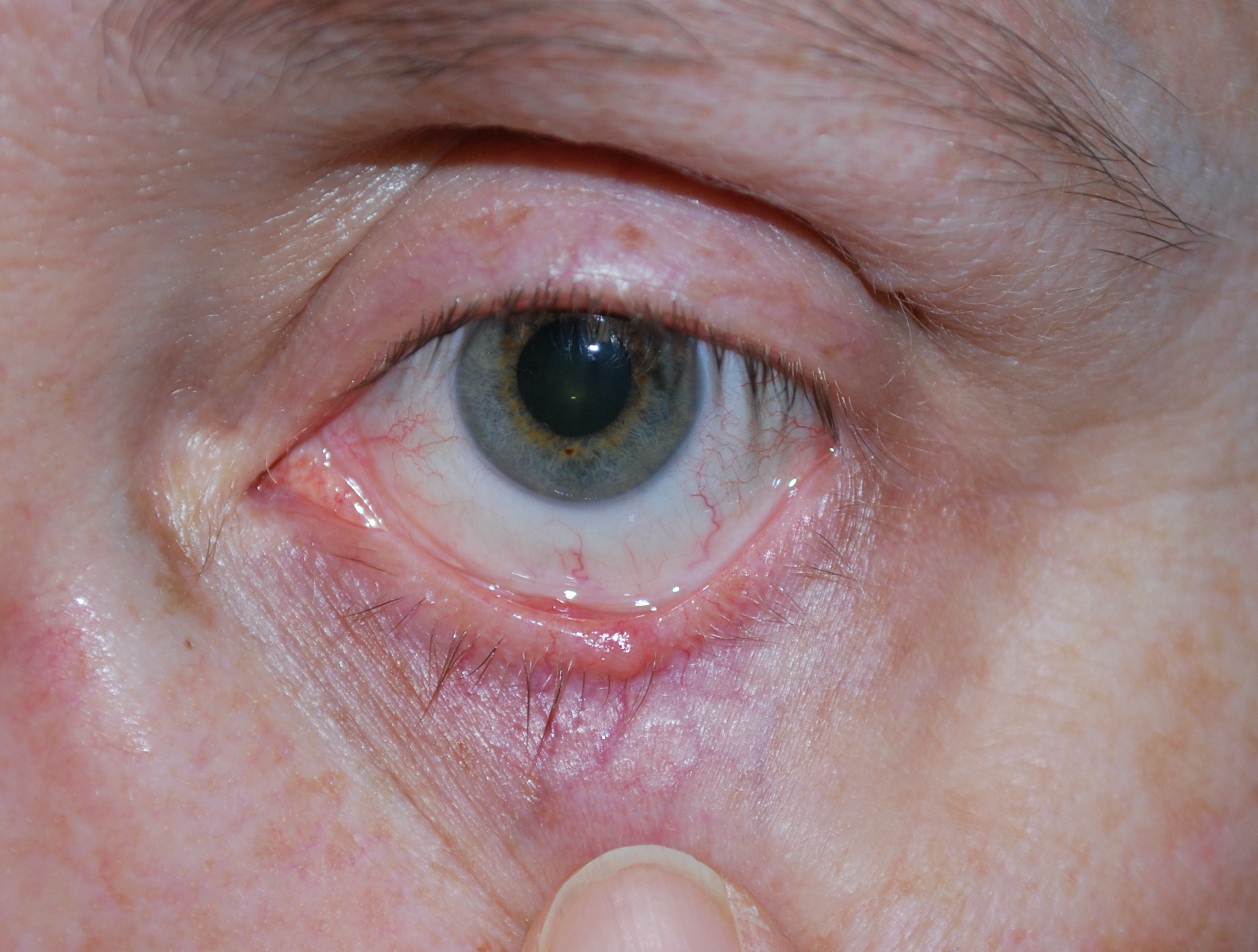
Basal Cell Carcinoma of the left lower eyelid Contributed by Eva Chou
(Click Image to Enlarge)
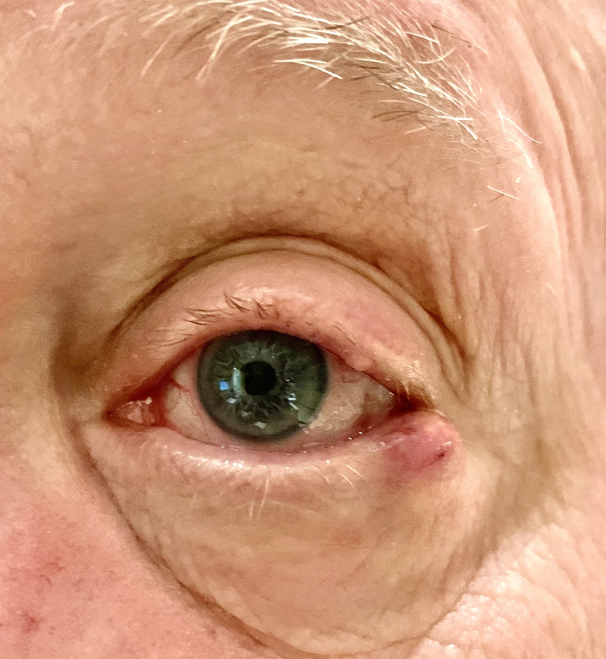
Sebaceous Carcinoma of the lateral left lower eyelid Contributed by Harkaran Rana, DO
(Click Image to Enlarge)
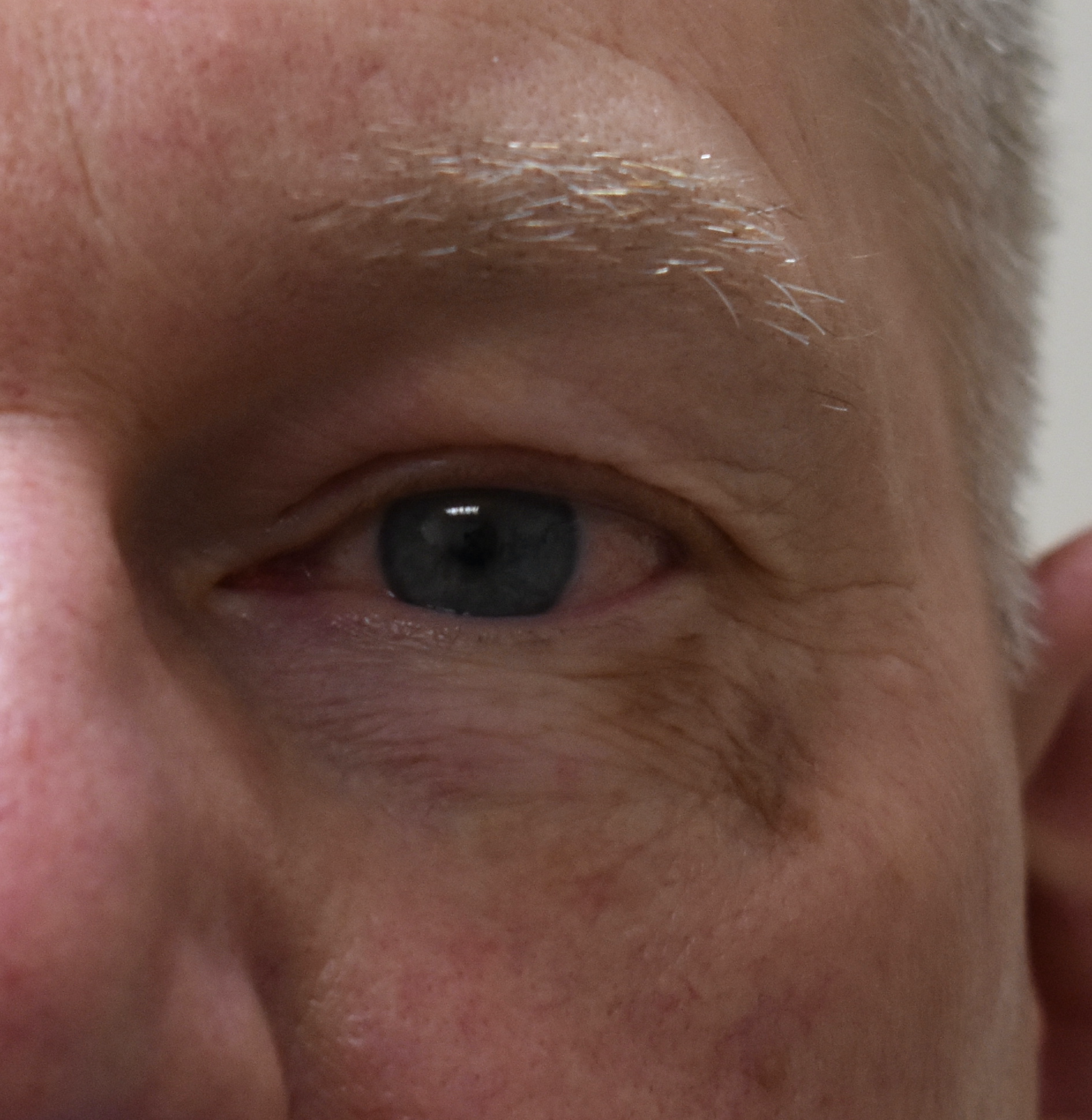
Melanoma in situ involving the left lower eyelid Contributed by Eva Chou
References
Tromme I, Sacré L, Hammouch F, Legrand C, Marot L, Vereecken P, Theate I, van Eeckhout P, Richez P, Baurain JF, Thomas L, Speybroeck N, DEPIMELA study group. Availability of digital dermoscopy in daily practice dramatically reduces the number of excised melanocytic lesions: results from an observational study. The British journal of dermatology. 2012 Oct:167(4):778-86. doi: 10.1111/j.1365-2133.2012.11042.x. Epub 2012 Aug 20 [PubMed PMID: 22564185]
Level 2 (mid-level) evidenceHaenssle HA, Krueger U, Vente C, Thoms KM, Bertsch HP, Zutt M, Rosenberger A, Neumann C, Emmert S. Results from an observational trial: digital epiluminescence microscopy follow-up of atypical nevi increases the sensitivity and the chance of success of conventional dermoscopy in detecting melanoma. The Journal of investigative dermatology. 2006 May:126(5):980-5 [PubMed PMID: 16514414]
Wunderlich K, Suppa M, Gandini S, Lipski J, White JM, Del Marmol V. Risk Factors and Innovations in Risk Assessment for Melanoma, Basal Cell Carcinoma, and Squamous Cell Carcinoma. Cancers. 2024 Feb 29:16(5):. doi: 10.3390/cancers16051016. Epub 2024 Feb 29 [PubMed PMID: 38473375]
Nelson MA, Einspahr JG, Alberts DS, Balfour CA, Wymer JA, Welch KL, Salasche SJ, Bangert JL, Grogan TM, Bozzo PO. Analysis of the p53 gene in human precancerous actinic keratosis lesions and squamous cell cancers. Cancer letters. 1994 Sep 30:85(1):23-9 [PubMed PMID: 7923098]
Level 3 (low-level) evidenceTaguchi M, Watanabe S, Yashima K, Murakami Y, Sekiya T, Ikeda S. Aberrations of the tumor suppressor p53 gene and p53 protein in solar keratosis in human skin. The Journal of investigative dermatology. 1994 Oct:103(4):500-3 [PubMed PMID: 7930674]
Daya-Grosjean L, Sarasin A. The role of UV induced lesions in skin carcinogenesis: an overview of oncogene and tumor suppressor gene modifications in xeroderma pigmentosum skin tumors. Mutation research. 2005 Apr 1:571(1-2):43-56 [PubMed PMID: 15748637]
Level 3 (low-level) evidenceDummer R, Guminksi A, Gutzmer R, Lear JT, Lewis KD, Chang ALS, Combemale P, Dirix L, Kaatz M, Kudchadkar R, Loquai C, Plummer R, Schulze HJ, Stratigos AJ, Trefzer U, Squittieri N, Migden MR. Long-term efficacy and safety of sonidegib in patients with advanced basal cell carcinoma: 42-month analysis of the phase II randomized, double-blind BOLT study. The British journal of dermatology. 2020 Jun:182(6):1369-1378. doi: 10.1111/bjd.18552. Epub 2019 Dec 8 [PubMed PMID: 31545507]
Level 1 (high-level) evidenceSinger JP, Boker A, Metchnikoff C, Binstock M, Boettger R, Golden JA, Glidden DV, Arron ST. High cumulative dose exposure to voriconazole is associated with cutaneous squamous cell carcinoma in lung transplant recipients. The Journal of heart and lung transplantation : the official publication of the International Society for Heart Transplantation. 2012 Jul:31(7):694-9. doi: 10.1016/j.healun.2012.02.033. Epub 2012 Apr 6 [PubMed PMID: 22484291]
Level 2 (mid-level) evidenceAlam M, Brown RN, Silber DH, Mullen GM, Feldman DS, Oren RM, Yancy CW, Cardiac Transplant Research Database Group. Increased incidence and mortality associated with skin cancers after cardiac transplant. American journal of transplantation : official journal of the American Society of Transplantation and the American Society of Transplant Surgeons. 2011 Jul:11(7):1488-97. doi: 10.1111/j.1600-6143.2011.03598.x. Epub [PubMed PMID: 21718441]
Level 2 (mid-level) evidenceLazo de la Vega L, Bick N, Hu K, Rahrig SE, Silva CD, Matayoshi S, Picciarelli P, Wang X, Sugar A, Soong HK, Mian SI, Robinson DR, Chinnaiyan AM, Demirci H, Daniels AB, Worden F, Eberhart CG, Tomlins SA, Rao RC, Harms PW. Invasive squamous cell carcinomas and precursor lesions on UV-exposed epithelia demonstrate concordant genomic complexity in driver genes. Modern pathology : an official journal of the United States and Canadian Academy of Pathology, Inc. 2020 Nov:33(11):2280-2294. doi: 10.1038/s41379-020-0571-7. Epub 2020 May 27 [PubMed PMID: 32461624]
Lee JB, Litzner BR, Vidal CI. Review of the current medical literature and assessment of current utilization patterns regarding mismatch repair protein immunohistochemistry in cutaneous Muir-Torre syndrome-associated neoplasms. Journal of cutaneous pathology. 2017 Nov:44(11):931-937. doi: 10.1111/cup.13010. Epub 2017 Aug 29 [PubMed PMID: 28749576]
Dores GM, Curtis RE, Toro JR, Devesa SS, Fraumeni JF Jr. Incidence of cutaneous sebaceous carcinoma and risk of associated neoplasms: insight into Muir-Torre syndrome. Cancer. 2008 Dec 15:113(12):3372-81. doi: 10.1002/cncr.23963. Epub [PubMed PMID: 18932259]
Eiger-Moscovich M, Eagle RC Jr, Shields CL, Racher H, Lally SE, Silkiss RZ, Shields JA, Milman T. Muir-Torre Syndrome Associated Periocular Sebaceous Neoplasms: Screening Patterns in the Literature and in Clinical Practice. Ocular oncology and pathology. 2020 Aug:6(4):226-237. doi: 10.1159/000504984. Epub 2020 Jan 15 [PubMed PMID: 33005611]
Kyllo RL, Brady KL, Hurst EA. Sebaceous carcinoma: review of the literature. Dermatologic surgery : official publication for American Society for Dermatologic Surgery [et al.]. 2015 Jan:41(1):1-15. doi: 10.1097/DSS.0000000000000152. Epub [PubMed PMID: 25521100]
Owen JL, Kibbi N, Worley B, Kelm RC, Wang JV, Barker CA, Behshad R, Bichakjian CK, Bolotin D, Bordeaux JS, Bradshaw SH, Cartee TV, Chandra S, Cho NL, Choi JN, Council ML, Demirci H, Eisen DB, Esmaeli B, Golda N, Huang CC, Ibrahim SF, Jiang SB, Kim J, Kuzel TM, Lai SY, Lawrence N, Lee EH, Leitenberger JJ, Maher IA, Mann MW, Minkis K, Mittal BB, Nehal KS, Neuhaus IM, Ozog DM, Petersen B, Rotemberg V, Samant S, Samie FH, Servaes S, Shields CL, Shin TM, Sobanko JF, Somani AK, Stebbins WG, Thomas JR, Thomas VD, Tse DT, Waldman AH, Wong MK, Xu YG, Yu SS, Zeitouni NC, Ramsay T, Reynolds KA, Poon E, Alam M. Sebaceous carcinoma: evidence-based clinical practice guidelines. The Lancet. Oncology. 2019 Dec:20(12):e699-e714. doi: 10.1016/S1470-2045(19)30673-4. Epub [PubMed PMID: 31797796]
Level 1 (high-level) evidenceWick MR, Goellner JR, Wolfe JT 3rd, Su WP. Adnexal carcinomas of the skin. II. Extraocular sebaceous carcinomas. Cancer. 1985 Sep 1:56(5):1163-72 [PubMed PMID: 4016704]
Level 3 (low-level) evidenceLanoy E, Dores GM, Madeleine MM, Toro JR, Fraumeni JF Jr, Engels EA. Epidemiology of nonkeratinocytic skin cancers among persons with AIDS in the United States. AIDS (London, England). 2009 Jan 28:23(3):385-93. doi: 10.1097/QAD.0b013e3283213046. Epub [PubMed PMID: 19114864]
Level 2 (mid-level) evidenceLandis MN, Davis CL, Bellus GA, Wolverton SE. Immunosuppression and sebaceous tumors: a confirmed diagnosis of Muir-Torre syndrome unmasked by immunosuppressive therapy. Journal of the American Academy of Dermatology. 2011 Nov:65(5):1054-1058.e1. doi: 10.1016/j.jaad.2010.08.003. Epub 2011 May 6 [PubMed PMID: 21550136]
Level 3 (low-level) evidenceKiyosaki K, Nakada C, Hijiya N, Tsukamoto Y, Matsuura K, Nakatsuka K, Daa T, Yokoyama S, Imaizumi M, Moriyama M. Analysis of p53 mutations and the expression of p53 and p21WAF1/CIP1 protein in 15 cases of sebaceous carcinoma of the eyelid. Investigative ophthalmology & visual science. 2010 Jan:51(1):7-11. doi: 10.1167/iovs.09-4127. Epub 2009 Jul 23 [PubMed PMID: 19628749]
Level 3 (low-level) evidenceBaumüller S, Herwig MC, Mangold E, Holz FC, Loeffler KU. Sebaceous gland carcinoma of the eyelid masquerading as a cutaneous horn in Li--Fraumeni syndrome. The British journal of ophthalmology. 2011 Oct:95(10):1470, 1478. doi: 10.1136/bjo.2009.175158. Epub [PubMed PMID: 20693561]
Level 3 (low-level) evidencePrieto-Granada C, Rodriguez-Waitkus P. Sebaceous Carcinoma of the Eyelid. Cancer control : journal of the Moffitt Cancer Center. 2016 Apr:23(2):126-32 [PubMed PMID: 27218789]
Gandini S, Autier P, Boniol M. Reviews on sun exposure and artificial light and melanoma. Progress in biophysics and molecular biology. 2011 Dec:107(3):362-6. doi: 10.1016/j.pbiomolbio.2011.09.011. Epub 2011 Sep 19 [PubMed PMID: 21958910]
Berwick M, Erdei E, Hay J. Melanoma epidemiology and public health. Dermatologic clinics. 2009 Apr:27(2):205-14, viii. doi: 10.1016/j.det.2008.12.002. Epub [PubMed PMID: 19254665]
van der Leest RJ, Flohil SC, Arends LR, de Vries E, Nijsten T. Risk of subsequent cutaneous malignancy in patients with prior melanoma: a systematic review and meta-analysis. Journal of the European Academy of Dermatology and Venereology : JEADV. 2015 Jun:29(6):1053-62. doi: 10.1111/jdv.12887. Epub 2014 Dec 10 [PubMed PMID: 25491923]
Level 1 (high-level) evidenceMancera N, Smalley KSM, Margo CE. Melanoma of the eyelid and periocular skin: Histopathologic classification and molecular pathology. Survey of ophthalmology. 2019 May-Jun:64(3):272-288. doi: 10.1016/j.survophthal.2018.12.002. Epub 2018 Dec 20 [PubMed PMID: 30578807]
Level 3 (low-level) evidenceSchadendorf D, van Akkooi ACJ, Berking C, Griewank KG, Gutzmer R, Hauschild A, Stang A, Roesch A, Ugurel S. Melanoma. Lancet (London, England). 2018 Sep 15:392(10151):971-984. doi: 10.1016/S0140-6736(18)31559-9. Epub [PubMed PMID: 30238891]
Garcia-Diaz A, Shin DS, Moreno BH, Saco J, Escuin-Ordinas H, Rodriguez GA, Zaretsky JM, Sun L, Hugo W, Wang X, Parisi G, Saus CP, Torrejon DY, Graeber TG, Comin-Anduix B, Hu-Lieskovan S, Damoiseaux R, Lo RS, Ribas A. Interferon Receptor Signaling Pathways Regulating PD-L1 and PD-L2 Expression. Cell reports. 2017 May 9:19(6):1189-1201. doi: 10.1016/j.celrep.2017.04.031. Epub [PubMed PMID: 28494868]
Davies H, Bignell GR, Cox C, Stephens P, Edkins S, Clegg S, Teague J, Woffendin H, Garnett MJ, Bottomley W, Davis N, Dicks E, Ewing R, Floyd Y, Gray K, Hall S, Hawes R, Hughes J, Kosmidou V, Menzies A, Mould C, Parker A, Stevens C, Watt S, Hooper S, Wilson R, Jayatilake H, Gusterson BA, Cooper C, Shipley J, Hargrave D, Pritchard-Jones K, Maitland N, Chenevix-Trench G, Riggins GJ, Bigner DD, Palmieri G, Cossu A, Flanagan A, Nicholson A, Ho JW, Leung SY, Yuen ST, Weber BL, Seigler HF, Darrow TL, Paterson H, Marais R, Marshall CJ, Wooster R, Stratton MR, Futreal PA. Mutations of the BRAF gene in human cancer. Nature. 2002 Jun 27:417(6892):949-54 [PubMed PMID: 12068308]
Level 3 (low-level) evidenceNorth VS, Habib LA, Yoon MK. Merkel cell carcinoma of the eyelid: A review. Survey of ophthalmology. 2019 Sep-Oct:64(5):659-667. doi: 10.1016/j.survophthal.2019.03.002. Epub 2019 Mar 11 [PubMed PMID: 30871952]
Level 3 (low-level) evidenceAmaral T, Leiter U, Garbe C. Merkel cell carcinoma: Epidemiology, pathogenesis, diagnosis and therapy. Reviews in endocrine & metabolic disorders. 2017 Dec:18(4):517-532. doi: 10.1007/s11154-017-9433-0. Epub [PubMed PMID: 28916903]
Feng H, Shuda M, Chang Y, Moore PS. Clonal integration of a polyomavirus in human Merkel cell carcinoma. Science (New York, N.Y.). 2008 Feb 22:319(5866):1096-100. doi: 10.1126/science.1152586. Epub 2008 Jan 17 [PubMed PMID: 18202256]
Shuda M, Chang Y, Moore PS. Merkel cell polyomavirus-positive Merkel cell carcinoma requires viral small T-antigen for cell proliferation. The Journal of investigative dermatology. 2014 May:134(5):1479-1481. doi: 10.1038/jid.2013.483. Epub 2013 Nov 12 [PubMed PMID: 24217011]
Level 3 (low-level) evidencePaulson KG, Tegeder A, Willmes C, Iyer JG, Afanasiev OK, Schrama D, Koba S, Thibodeau R, Nagase K, Simonson WT, Seo A, Koelle DM, Madeleine M, Bhatia S, Nakajima H, Sano S, Hardwick JS, Disis ML, Cleary MA, Becker JC, Nghiem P. Downregulation of MHC-I expression is prevalent but reversible in Merkel cell carcinoma. Cancer immunology research. 2014 Nov:2(11):1071-9. doi: 10.1158/2326-6066.CIR-14-0005. Epub 2014 Aug 12 [PubMed PMID: 25116754]
Becker JC, Stang A, Hausen AZ, Fischer N, DeCaprio JA, Tothill RW, Lyngaa R, Hansen UK, Ritter C, Nghiem P, Bichakjian CK, Ugurel S, Schrama D. Epidemiology, biology and therapy of Merkel cell carcinoma: conclusions from the EU project IMMOMEC. Cancer immunology, immunotherapy : CII. 2018 Mar:67(3):341-351. doi: 10.1007/s00262-017-2099-3. Epub 2017 Nov 30 [PubMed PMID: 29188306]
Lunder EJ, Stern RS. Merkel-cell carcinomas in patients treated with methoxsalen and ultraviolet A radiation. The New England journal of medicine. 1998 Oct 22:339(17):1247-8 [PubMed PMID: 9786759]
Level 3 (low-level) evidenceBaş Z, Sharpe J, Yaghy A, Zhang Q, Shields CL, Hyman L, IRIS Registry Analytic Center Consortium. Prevalence of and Associated Factors for Eyelid Cancer in the American Academy of Ophthalmology Intelligent Research in Sight Registry. Ophthalmology science. 2023 Mar:3(1):100227. doi: 10.1016/j.xops.2022.100227. Epub 2022 Sep 27 [PubMed PMID: 36439695]
Desiato VM, Byun YJ, Nguyen SA, Thiers BH, Day TA. Sebaceous Carcinoma of the Eyelid: A Systematic Review and Meta-Analysis. Dermatologic surgery : official publication for American Society for Dermatologic Surgery [et al.]. 2021 Jan 1:47(1):104-110. doi: 10.1097/DSS.0000000000002660. Epub [PubMed PMID: 33347004]
Level 1 (high-level) evidenceShields JA, Demirci H, Marr BP, Eagle RC Jr, Shields CL. Sebaceous carcinoma of the eyelids: personal experience with 60 cases. Ophthalmology. 2004 Dec:111(12):2151-7 [PubMed PMID: 15582067]
Level 2 (mid-level) evidenceHarvey I, Frankel S, Marks R, Shalom D, Nolan-Farrell M. Non-melanoma skin cancer and solar keratoses. I. Methods and descriptive results of the South Wales Skin Cancer Study. British journal of cancer. 1996 Oct:74(8):1302-7 [PubMed PMID: 8883422]
Marks R, Ponsford MW, Selwood TS, Goodman G, Mason G. Non-melanotic skin cancer and solar keratoses in Victoria. The Medical journal of Australia. 1983 Dec 10-24:2(12):619-22 [PubMed PMID: 6669125]
Flohil SC, van der Leest RJ, Dowlatshahi EA, Hofman A, de Vries E, Nijsten T. Prevalence of actinic keratosis and its risk factors in the general population: the Rotterdam Study. The Journal of investigative dermatology. 2013 Aug:133(8):1971-8. doi: 10.1038/jid.2013.134. Epub 2013 Mar 19 [PubMed PMID: 23510990]
Guorgis G, Anderson CD, Lyth J, Falk M. Actinic Keratosis Diagnosis and Increased Risk of Developing Skin Cancer: A 10-year Cohort Study of 17,651 Patients in Sweden. Acta dermato-venereologica. 2020 Apr 29:100(8):adv00128. doi: 10.2340/00015555-3486. Epub 2020 Apr 29 [PubMed PMID: 32314794]
Saleh GM, Desai P, Collin JR, Ives A, Jones T, Hussain B. Incidence of eyelid basal cell carcinoma in England: 2000-2010. The British journal of ophthalmology. 2017 Feb:101(2):209-212. doi: 10.1136/bjophthalmol-2015-308261. Epub 2016 Apr 29 [PubMed PMID: 27130914]
Kaliki S, Bothra N, Bejjanki KM, Nayak A, Ramappa G, Mohamed A, Dave TV, Ali MJ, Naik MN. Malignant Eyelid Tumors in India: A Study of 536 Asian Indian Patients. Ocular oncology and pathology. 2019 Apr:5(3):210-219. doi: 10.1159/000491549. Epub 2018 Sep 13 [PubMed PMID: 31049330]
Kale SM, Patil SB, Khare N, Math M, Jain A, Jaiswal S. Clinicopathological analysis of eyelid malignancies - A review of 85 cases. Indian journal of plastic surgery : official publication of the Association of Plastic Surgeons of India. 2012 Jan:45(1):22-8. doi: 10.4103/0970-0358.96572. Epub [PubMed PMID: 22754148]
Level 3 (low-level) evidenceBrunetti P, Margo CE, French DD. Incidence of Cutaneous Melanoma of Eyelid Analysis of the Surveillance, Epidemiology, and End Results Database. Ocular oncology and pathology. 2021 Mar:7(1):66-69. doi: 10.1159/000511215. Epub 2020 Nov 17 [PubMed PMID: 33796520]
Olsen CM, Thompson JF, Pandeya N, Whiteman DC. Evaluation of Sex-Specific Incidence of Melanoma. JAMA dermatology. 2020 May 1:156(5):553-560. doi: 10.1001/jamadermatol.2020.0470. Epub [PubMed PMID: 32211827]
Robbins JO, Huck NA, Khosravi P, Torabi SJ, Woodward JA, Kuan EC, Dermarkarian CR. Trends in Demographic, Clinical, Socioeconomic, and Facility-Specific Factors Linked to Eyelid Melanoma Survival: A National Cancer Database Analysis. Ophthalmic plastic and reconstructive surgery. 2025 Jul-Aug 01:41(4):456-464. doi: 10.1097/IOP.0000000000002895. Epub 2025 Jan 6 [PubMed PMID: 39760268]
Oliver JD, Boczar D, Sisti A, Huayllani MT, Restrepo DJ, Spaulding AC, Gabriel E, Bagaria S, Rinker BD, Forte AJ. Eyelid Melanoma in the United States: A National Cancer Database Analysis. The Journal of craniofacial surgery. 2019 Nov-Dec:30(8):2412-2415. doi: 10.1097/SCS.0000000000005673. Epub [PubMed PMID: 31233000]
Lemos B, Nghiem P. Merkel cell carcinoma: more deaths but still no pathway to blame. The Journal of investigative dermatology. 2007 Sep:127(9):2100-3 [PubMed PMID: 17700621]
Albores-Saavedra J, Batich K, Chable-Montero F, Sagy N, Schwartz AM, Henson DE. Merkel cell carcinoma demographics, morphology, and survival based on 3870 cases: a population based study. Journal of cutaneous pathology. 2010 Jan:37(1):20-7. doi: 10.1111/j.1600-0560.2009.01370.x. Epub 2009 Jul 23 [PubMed PMID: 19638070]
Level 3 (low-level) evidenceMerritt H, Sniegowski MC, Esmaeli B. Merkel cell carcinoma of the eyelid and periocular region. Cancers. 2014 May 9:6(2):1128-37. doi: 10.3390/cancers6021128. Epub 2014 May 9 [PubMed PMID: 24821131]
Lemos BD, Storer BE, Iyer JG, Phillips JL, Bichakjian CK, Fang LC, Johnson TM, Liegeois-Kwon NJ, Otley CC, Paulson KG, Ross MI, Yu SS, Zeitouni NC, Byrd DR, Sondak VK, Gershenwald JE, Sober AJ, Nghiem P. Pathologic nodal evaluation improves prognostic accuracy in Merkel cell carcinoma: analysis of 5823 cases as the basis of the first consensus staging system. Journal of the American Academy of Dermatology. 2010 Nov:63(5):751-61. doi: 10.1016/j.jaad.2010.02.056. Epub 2010 Jun 19 [PubMed PMID: 20646783]
Level 3 (low-level) evidenceSmith VA, Camp ER, Lentsch EJ. Merkel cell carcinoma: identification of prognostic factors unique to tumors located in the head and neck based on analysis of SEER data. The Laryngoscope. 2012 Jun:122(6):1283-90. doi: 10.1002/lary.23222. Epub 2012 Apr 20 [PubMed PMID: 22522673]
Level 2 (mid-level) evidenceCohen LM. Lentigo maligna and lentigo maligna melanoma. Journal of the American Academy of Dermatology. 1995 Dec:33(6):923-36; quiz 937-40 [PubMed PMID: 7490362]
Dubow BE, Ackerman AB. Ideas in pathology. Malignant melanoma in situ: the evolution of a concept. Modern pathology : an official journal of the United States and Canadian Academy of Pathology, Inc. 1990 Nov:3(6):734-44 [PubMed PMID: 2263599]
DeWane ME, Kelsey A, Oliviero M, Rabinovitz H, Grant-Kels JM. Melanoma on chronically sun-damaged skin: Lentigo maligna and desmoplastic melanoma. Journal of the American Academy of Dermatology. 2019 Sep:81(3):823-833. doi: 10.1016/j.jaad.2019.03.066. Epub 2019 Mar 28 [PubMed PMID: 30930085]
McKenna JK, Florell SR, Goldman GD, Bowen GM. Lentigo maligna/lentigo maligna melanoma: current state of diagnosis and treatment. Dermatologic surgery : official publication for American Society for Dermatologic Surgery [et al.]. 2006 Apr:32(4):493-504 [PubMed PMID: 16681656]
Weinstock MA, Sober AJ. The risk of progression of lentigo maligna to lentigo maligna melanoma. The British journal of dermatology. 1987 Mar:116(3):303-10 [PubMed PMID: 3567069]
Menzies SW, Liyanarachchi S, Coates E, Smith A, Cooke-Yarborough C, Lo S, Armstrong B, Scolyer RA, Guitera P. Estimated risk of progression of lentigo maligna to lentigo maligna melanoma. Melanoma research. 2020 Apr:30(2):193-197. doi: 10.1097/CMR.0000000000000619. Epub [PubMed PMID: 31095041]
Erickson C, Miller SJ. Treatment options in melanoma in situ: topical and radiation therapy, excision and Mohs surgery. International journal of dermatology. 2010 May:49(5):482-91. doi: 10.1111/j.1365-4632.2010.04423.x. Epub [PubMed PMID: 20534080]
McLeod M, Choudhary S, Giannakakis G, Nouri K. Surgical treatments for lentigo maligna: a review. Dermatologic surgery : official publication for American Society for Dermatologic Surgery [et al.]. 2011 Sep:37(9):1210-28. doi: 10.1111/j.1524-4725.2011.02042.x. Epub 2011 Jun 1 [PubMed PMID: 21631635]
Marsden JR, Fox R, Boota NM, Cook M, Wheatley K, Billingham LJ, Steven NM, NCRI Skin Cancer Clinical Studies Group, the U.K. Dermatology Clinical Trials Network and the LIMIT-1 Collaborative Group. Effect of topical imiquimod as primary treatment for lentigo maligna: the LIMIT-1 study. The British journal of dermatology. 2017 May:176(5):1148-1154. doi: 10.1111/bjd.15112. Epub 2017 Apr 10 [PubMed PMID: 27714781]
Puckett Y, Geist R, Chen CSJ. Lentigo Maligna Melanoma. StatPearls. 2026 Jan:(): [PubMed PMID: 29489150]
Dass SE, Huizenga T, Farshchian M, Mehregan DR. Comparison of SOX-10, HMB-45, and Melan-A in Benign Melanocytic Lesions. Clinical, cosmetic and investigational dermatology. 2021:14():1419-1425. doi: 10.2147/CCID.S333376. Epub 2021 Oct 5 [PubMed PMID: 34675577]
El Shabrawi-Caelen L, Kerl H, Cerroni L. Melan-A: not a helpful marker in distinction between melanoma in situ on sun-damaged skin and pigmented actinic keratosis. The American Journal of dermatopathology. 2004 Oct:26(5):364-6 [PubMed PMID: 15365366]
Sunjaya AP, Sunjaya AF, Tan ST. The Use of BEREP4 Immunohistochemistry Staining for Detection of Basal Cell Carcinoma. Journal of skin cancer. 2017:2017():2692604. doi: 10.1155/2017/2692604. Epub 2017 Dec 31 [PubMed PMID: 29464122]
Sinard JH. Immunohistochemical distinction of ocular sebaceous carcinoma from basal cell and squamous cell carcinoma. Archives of ophthalmology (Chicago, Ill. : 1960). 1999 Jun:117(6):776-83 [PubMed PMID: 10369589]
Joshi UM, Kashani-Sabet M, Kirkwood JM. Cutaneous Melanoma: A Review. JAMA. 2025 Dec 16:334(23):2113-2125. doi: 10.1001/jama.2025.13074. Epub [PubMed PMID: 40853557]
Libson K, Sheridan C, Carr DR, Shahwan KT. Use of Imaging in Cutaneous Squamous Cell Carcinoma to Detect High-Risk Tumor Features, Nodal Metastasis, and Distant Metastasis: A Systematic Review. Dermatologic surgery : official publication for American Society for Dermatologic Surgery [et al.]. 2024 Aug 1:50(8):705-709. doi: 10.1097/DSS.0000000000004191. Epub 2024 Apr 16 [PubMed PMID: 38624106]
Level 1 (high-level) evidenceLagler CN, Freitag SK. Management of periocular actinic keratosis: a review of practice patterns among ophthalmic plastic surgeons. Ophthalmic plastic and reconstructive surgery. 2012 Jul-Aug:28(4):277-81. doi: 10.1097/IOP.0b013e318257f5f2. Epub [PubMed PMID: 22785585]
Kahana A, Worden FP, Elner VM. Vismodegib as eye-sparing adjuvant treatment for orbital basal cell carcinoma. JAMA ophthalmology. 2013 Oct:131(10):1364-6. doi: 10.1001/jamaophthalmol.2013.4430. Epub [PubMed PMID: 23907144]
Level 3 (low-level) evidenceSu MG, Potts LB, Tsai JH. Treatment of periocular basal cell carcinoma with neoadjuvant vismodegib. American journal of ophthalmology case reports. 2020 Sep:19():100755. doi: 10.1016/j.ajoc.2020.100755. Epub 2020 May 23 [PubMed PMID: 32490287]
Level 3 (low-level) evidenceOzolins M, Williams HC, Armstrong SJ, Bath-Hextall FJ. The SINS trial: a randomised controlled trial of excisional surgery versus imiquimod 5% cream for nodular and superficial basal cell carcinoma. Trials. 2010 Apr 21:11():42. doi: 10.1186/1745-6215-11-42. Epub 2010 Apr 21 [PubMed PMID: 20409337]
Level 1 (high-level) evidenceBath-Hextall F, Ozolins M, Armstrong SJ, Colver GB, Perkins W, Miller PS, Williams HC, Surgery versus Imiquimod for Nodular Superficial basal cell carcinoma (SINS) study group. Surgical excision versus imiquimod 5% cream for nodular and superficial basal-cell carcinoma (SINS): a multicentre, non-inferiority, randomised controlled trial. The Lancet. Oncology. 2014 Jan:15(1):96-105. doi: 10.1016/S1470-2045(13)70530-8. Epub 2013 Dec 11 [PubMed PMID: 24332516]
Level 1 (high-level) evidenceWilliams HC, Bath-Hextall F, Ozolins M, Armstrong SJ, Colver GB, Perkins W, Miller PSJ, Surgery Versus Imiquimod for Nodular and Superficial Basal Cell Carcinoma (SINS) Study Group. Surgery Versus 5% Imiquimod for Nodular and Superficial Basal Cell Carcinoma: 5-Year Results of the SINS Randomized Controlled Trial. The Journal of investigative dermatology. 2017 Mar:137(3):614-619. doi: 10.1016/j.jid.2016.10.019. Epub 2016 Dec 5 [PubMed PMID: 27932240]
Level 1 (high-level) evidenceInaba K, Ito Y, Suzuki S, Sekii S, Takahashi K, Kuroda Y, Murakami N, Morota M, Mayahara H, Sumi M, Uno T, Itami J. Results of radical radiotherapy for squamous cell carcinoma of the eyelid. Journal of radiation research. 2013 Nov 1:54(6):1131-7. doi: 10.1093/jrr/rrt069. Epub 2013 Jun 7 [PubMed PMID: 23750022]
Level 2 (mid-level) evidenceYin VT, Pfeiffer ML, Esmaeli B. Targeted therapy for orbital and periocular basal cell carcinoma and squamous cell carcinoma. Ophthalmic plastic and reconstructive surgery. 2013 Mar-Apr:29(2):87-92. doi: 10.1097/IOP.0b013e3182831bf3. Epub [PubMed PMID: 23446297]
Level 3 (low-level) evidenceEl-Sawy T, Sabichi AL, Myers JN, Kies MS, William WN, Glisson BS, Lippman S, Esmaeli B. Epidermal growth factor receptor inhibitors for treatment of orbital squamous cell carcinoma. Archives of ophthalmology (Chicago, Ill. : 1960). 2012 Dec:130(12):1608-11. doi: 10.1001/archophthalmol.2012.2515. Epub [PubMed PMID: 23229707]
Level 3 (low-level) evidenceRoss AH, Kennedy CT, Collins C, Harrad RA. The use of imiquimod in the treatment of periocular tumours. Orbit (Amsterdam, Netherlands). 2010 Apr:29(2):83-7. doi: 10.3109/01676830903294909. Epub [PubMed PMID: 20394545]
Level 3 (low-level) evidenceFreitag SK, Aakalu VK, Tao JP, Wladis EJ, Foster JA, Sobel RK, Yen MT. Sentinel Lymph Node Biopsy for Eyelid and Conjunctival Malignancy: A Report by the American Academy of Ophthalmology. Ophthalmology. 2020 Dec:127(12):1757-1765. doi: 10.1016/j.ophtha.2020.07.031. Epub 2020 Jul 19 [PubMed PMID: 32698034]
Savar A, Ross MI, Prieto VG, Ivan D, Kim S, Esmaeli B. Sentinel lymph node biopsy for ocular adnexal melanoma: experience in 30 patients. Ophthalmology. 2009 Nov:116(11):2217-23. doi: 10.1016/j.ophtha.2009.04.012. Epub 2009 Sep 19 [PubMed PMID: 19766318]
Davis LE, Shalin SC, Tackett AJ. Current state of melanoma diagnosis and treatment. Cancer biology & therapy. 2019:20(11):1366-1379. doi: 10.1080/15384047.2019.1640032. Epub 2019 Aug 1 [PubMed PMID: 31366280]
Ralli M, Botticelli A, Visconti IC, Angeletti D, Fiore M, Marchetti P, Lambiase A, de Vincentiis M, Greco A. Immunotherapy in the Treatment of Metastatic Melanoma: Current Knowledge and Future Directions. Journal of immunology research. 2020:2020():9235638. doi: 10.1155/2020/9235638. Epub 2020 Jun 28 [PubMed PMID: 32671117]
Level 3 (low-level) evidenceMaverakis E, Cornelius LA, Bowen GM, Phan T, Patel FB, Fitzmaurice S, He Y, Burrall B, Duong C, Kloxin AM, Sultani H, Wilken R, Martinez SR, Patel F. Metastatic melanoma - a review of current and future treatment options. Acta dermato-venereologica. 2015 May:95(5):516-24. doi: 10.2340/00015555-2035. Epub [PubMed PMID: 25520039]
Peters GB 3rd, Meyer DR, Shields JA, Custer PL, Rubin PA, Wojno TH, Bersani TA, Tanenbaum M. Management and prognosis of Merkel cell carcinoma of the eyelid. Ophthalmology. 2001 Sep:108(9):1575-9 [PubMed PMID: 11535453]
Sniegowski MC, Warneke CL, Morrison WH, Nasser QJ, Frank SJ, Pfeiffer ML, El-Sawy T, Esmaeli B. Correlation of American Joint Committee on Cancer T category for eyelid carcinoma with outcomes in patients with periocular Merkel cell carcinoma. Ophthalmic plastic and reconstructive surgery. 2014 Nov-Dec:30(6):480-5. doi: 10.1097/IOP.0000000000000153. Epub [PubMed PMID: 24841735]
Level 2 (mid-level) evidenceVoog E, Biron P, Martin JP, Blay JY. Chemotherapy for patients with locally advanced or metastatic Merkel cell carcinoma. Cancer. 1999 Jun 15:85(12):2589-95 [PubMed PMID: 10375107]
Schlaak M, Podewski T, Von Bartenwerffer W, Kreuzberg N, Bangard C, Mauch C, Kurschat P. Induction of durable responses by oral etoposide monochemotherapy in patients with metastatic Merkel cell carcinoma. European journal of dermatology : EJD. 2012 Mar-Apr:22(2):187-91. doi: 10.1684/ejd.2011.1634. Epub [PubMed PMID: 22240092]
Jouary T, Lalanne N, Siberchicot F, Ricard AS, Versapuech J, Lepreux S, Delaunay M, Taieb A. Neoadjuvant polychemotherapy in locally advanced Merkel cell carcinoma. Nature reviews. Clinical oncology. 2009 Sep:6(9):544-8. doi: 10.1038/nrclinonc.2009.109. Epub [PubMed PMID: 19707243]
Level 3 (low-level) evidenceToto V, Colapietra A, Alessandri-Bonetti M, Vincenzi B, Devirgiliis V, Panasiti V, Persichetti P. Upper eyelid Merkel cell carcinoma treated with neoadjuvant chemotherapy and surgical excision. Archives of craniofacial surgery. 2019 Apr:20(2):121-125. doi: 10.7181/acfs.2018.02089. Epub 2019 Apr 20 [PubMed PMID: 31048649]
Garcia GA, Kossler AL. Avelumab as an Emerging Therapy for Eyelid and Periocular Merkel Cell Carcinoma. International ophthalmology clinics. 2020 Spring:60(2):91-102. doi: 10.1097/IIO.0000000000000306. Epub [PubMed PMID: 32205656]
Kokoska ER, Kokoska MS, Collins BT, Stapleton DR, Wade TP. Early aggressive treatment for Merkel cell carcinoma improves outcome. American journal of surgery. 1997 Dec:174(6):688-93 [PubMed PMID: 9409598]
Level 2 (mid-level) evidenceFerreira TA, Pinheiro CF, Saraiva P, Jaarsma-Coes MG, Van Duinen SG, Genders SW, Marinkovic M, Beenakker JM. MR and CT Imaging of the Normal Eyelid and its Application in Eyelid Tumors. Cancers. 2020 Mar 12:12(3):. doi: 10.3390/cancers12030658. Epub 2020 Mar 12 [PubMed PMID: 32178233]
Go CC, Kim DH, Go BC, McGeehan B, Briceño CA. Clinicopathologic Characteristics and Prognostic Factors Impacting Survival in Melanoma of the Eyelid. American journal of ophthalmology. 2022 Feb:234():71-80. doi: 10.1016/j.ajo.2021.07.031. Epub 2021 Jul 31 [PubMed PMID: 34343490]
Quaedvlieg PJ, Tirsi E, Thissen MR, Krekels GA. Actinic keratosis: how to differentiate the good from the bad ones? European journal of dermatology : EJD. 2006 Jul-Aug:16(4):335-9 [PubMed PMID: 16935787]
Level 1 (high-level) evidenceChen GJ, Feldman SR, Williford PM, Hester EJ, Kiang SH, Gill I, Fleischer AB Jr. Clinical diagnosis of actinic keratosis identifies an elderly population at high risk of developing skin cancer. Dermatologic surgery : official publication for American Society for Dermatologic Surgery [et al.]. 2005 Jan:31(1):43-7 [PubMed PMID: 15720095]
Level 2 (mid-level) evidencePhan K, Oh LJ, Goyal S, Rutherford T, Yazdabadi A. Recurrence rates following surgical excision of periocular basal cell carcinomas: systematic review and meta-analysis. The Journal of dermatological treatment. 2020 Sep:31(6):597-601. doi: 10.1080/09546634.2019.1698702. Epub 2019 Dec 4 [PubMed PMID: 31769708]
Level 1 (high-level) evidenceDonaldson MJ, Sullivan TJ, Whitehead KJ, Williamson RM. Squamous cell carcinoma of the eyelids. The British journal of ophthalmology. 2002 Oct:86(10):1161-5 [PubMed PMID: 12234899]
Level 2 (mid-level) evidenceBoniuk M, Zimmerman LE. Sebaceous carcinoma of the eyelid, eyebrow, caruncle, and orbit. Transactions - American Academy of Ophthalmology and Otolaryngology. American Academy of Ophthalmology and Otolaryngology. 1968 Jul-Aug:72(4):619-42 [PubMed PMID: 5706692]
Level 3 (low-level) evidenceRao NA, Hidayat AA, McLean IW, Zimmerman LE. Sebaceous carcinomas of the ocular adnexa: A clinicopathologic study of 104 cases, with five-year follow-up data. Human pathology. 1982 Feb:13(2):113-22 [PubMed PMID: 7076199]
Level 3 (low-level) evidenceDoxanas MT, Green WR. Sebaceous gland carcinoma. Review of 40 cases. Archives of ophthalmology (Chicago, Ill. : 1960). 1984 Feb:102(2):245-9 [PubMed PMID: 6696670]
Level 3 (low-level) evidenceKhan JA, Doane JF, Grove AS Jr. Sebaceous and meibomian carcinomas of the eyelid. Recognition, diagnosis, and management. Ophthalmic plastic and reconstructive surgery. 1991:7(1):61-6 [PubMed PMID: 2018750]
Muqit MM, Roberts F, Lee WR, Kemp E. Improved survival rates in sebaceous carcinoma of the eyelid. Eye (London, England). 2004 Jan:18(1):49-53 [PubMed PMID: 14707966]
Level 2 (mid-level) evidenceParidaens AD, Minassian DC, McCartney AC, Hungerford JL. Prognostic factors in primary malignant melanoma of the conjunctiva: a clinicopathological study of 256 cases. The British journal of ophthalmology. 1994 Apr:78(4):252-9 [PubMed PMID: 8199108]
Level 2 (mid-level) evidenceBecker JC, Stang A, DeCaprio JA, Cerroni L, Lebbé C, Veness M, Nghiem P. Merkel cell carcinoma. Nature reviews. Disease primers. 2017 Oct 26:3():17077. doi: 10.1038/nrdp.2017.77. Epub 2017 Oct 26 [PubMed PMID: 29072302]
Schadendorf D, Lebbé C, Zur Hausen A, Avril MF, Hariharan S, Bharmal M, Becker JC. Merkel cell carcinoma: Epidemiology, prognosis, therapy and unmet medical needs. European journal of cancer (Oxford, England : 1990). 2017 Jan:71():53-69. doi: 10.1016/j.ejca.2016.10.022. Epub 2016 Dec 14 [PubMed PMID: 27984768]
Oestreicher J, Mehta S. Complications of blepharoplasty: prevention and management. Plastic surgery international. 2012:2012():252368. doi: 10.1155/2012/252368. Epub 2012 May 8 [PubMed PMID: 22655191]
Boulos PR, Rubin PA. Cutaneous melanomas of the eyelid. Seminars in ophthalmology. 2006 Jul-Sep:21(3):195-206 [PubMed PMID: 16912018]
Rosen T, Lebwohl MG. Prevalence and awareness of actinic keratosis: barriers and opportunities. Journal of the American Academy of Dermatology. 2013 Jan:68(1 Suppl 1):S2-9. doi: 10.1016/j.jaad.2012.09.052. Epub [PubMed PMID: 23228302]
Roetzheim RG, Pal N, van Durme DJ, Wathington D, Ferrante JM, Gonzalez EC, Krischer JP. Increasing supplies of dermatologists and family physicians are associated with earlier stage of melanoma detection. Journal of the American Academy of Dermatology. 2000 Aug:43(2 Pt 1):211-8 [PubMed PMID: 10906640]
Wehner MR, Linos E, Parvataneni R, Stuart SE, Boscardin WJ, Chren MM. Timing of subsequent new tumors in patients who present with basal cell carcinoma or cutaneous squamous cell carcinoma. JAMA dermatology. 2015 Apr:151(4):382-8. doi: 10.1001/jamadermatol.2014.3307. Epub [PubMed PMID: 25588079]
Level 2 (mid-level) evidence