 Tools
Tools
 Intermittent Exotropia
Introduction
Intermittent exotropia is the most common type of strabismus. This condition is characterized by non-constant exodeviation that predominantly manifests at distance fixation and may progress over a variable period to near fixation. Intermittent exotropia is also known as distance exotropia, divergent squint, periodic exotropia, or exotropia of inattention. Small exophorias are common in newborns, occurring in 60% to 70% of cases, and resolve by 4 to 6 months of age. Intermittent exotropia most commonly presents in childhood.
The typical disease course begins with exophoria, which may progress to intermittent exotropia and eventually to a constant exotropia. However, not all cases are progressive; some may remain stable over time despite lack of treatment, and a few may even improve. In an analysis by von Noorden, 75% of 51 untreated patients with intermittent exotropia demonstrated progression, 9% remained stable, and 16% showed improvement.[1]
Intermittent exotropia is among the most frequently encountered forms of childhood-onset strabismus, characterized by an outward deviation of 1 eye that manifests intermittently under specific conditions, such as fatigue, illness, or inattention. Unlike constant exotropia, in which ocular misalignment is persistent, intermittent exotropia presents as an alternating state of ocular alignment and deviation, reflecting a delicate balance between fusional convergence control and divergent tendencies. The condition occupies a unique position on the strabismic spectrum—bridging phoria and tropia—and its study has long fascinated clinicians and researchers alike due to its complex sensory and motor adaptations, variable clinical course, and nuanced therapeutic decision-making.[2]
From a global perspective, intermittent exotropia represents the most common form of exotropia in both pediatric and adult populations, with an estimated prevalence ranging between 0.5% and 3% depending on geographic and ethnic variations. Studies have consistently reported a higher prevalence in Asian populations, possibly attributed to genetic predisposition, environmental influences, and differing patterns of near work and visual demand. Onset typically occurs in early childhood, between 2 and 6 years of age; however, many patients remain undiagnosed until school age, when caregivers or teachers notice symptoms such as squinting, eye closure in bright light, or intermittent ocular drift. Early recognition and intervention are crucial, as untreated or poorly controlled intermittent exotropia may progress to constant exotropia, loss of stereopsis, and psychosocial difficulties stemming from visible ocular misalignment.[3]
The pathophysiology of intermittent exotropia remains multifactorial, involving intricate interactions among sensory fusion, motor control, accommodative-convergence mechanisms, and neurologic coordination. The fundamental imbalance lies between the tonic divergent forces that naturally separate the visual axes and the fusional convergence mechanisms that maintain binocular alignment. When fusional reserves are strong, ocular alignment is maintained; however, when fatigue, illness, or reduced attention deplete these reserves, the eyes may diverge transiently. Additionally, dissociation during monocular viewing (eg, covering 1 eye or daydreaming) often precipitates a manifest deviation. Bright light has been noted to exacerbate the deviation, possibly due to accommodative relaxation or pupillary constriction, reducing the accommodative stimulus. This phenomenon, often termed photophobia, is characteristic of intermittent exotropia and can serve as a useful diagnostic clue.[4]
Clinically, intermittent exotropia encompasses several subtypes, traditionally classified by their distance-near disparity using the Burian classification: (1) basic type, where the deviation is similar at distance and near; (2) divergence excess type, with a larger deviation at distance than near; (3) simulated divergence excess, where apparent distance dominance normalizes after prolonged occlusion; and (4) convergence insufficiency type, showing greater deviation at near fixation. These subtypes hold diagnostic and therapeutic relevance, as they influence both nonsurgical and surgical treatment strategies. Accurate classification requires comprehensive evaluation under varying fixation conditions, with appropriate occlusion testing to eliminate fusional adaptation.[5]
Symptoms of intermittent exotropia vary widely and depend on the frequency and control of the deviation. Patients may report intermittent diplopia, ocular fatigue, blurred vision, or transient visual confusion, though many children remain asymptomatic due to strong suppression mechanisms. Parents often report occasional outward drifting of the eye, especially when the child is tired, ill, or gazing into the distance. Other common behavioral signs include squinting or closing 1 eye in bright light—a compensatory mechanism to eliminate diplopia—and difficulty maintaining visual focus during reading or sustained tasks. Over time, intermittent exotropia can lead to sensory adaptations, such as suppression scotomas, anomalous retinal correspondence, and a gradual reduction in stereoacuity, underscoring the need for timely evaluation and management.[6]
The diagnostic evaluation of intermittent exotropia relies on detailed clinical assessment using both objective and subjective methods. Key components include measuring deviation with the prism cover test at varying fixation distances, assessing control with standardized scales (eg, the Newcastle Control Score), and evaluating binocular function with tests such as the Worth 4-dot test, Bagolini striated lens test, and stereoacuity testing. Dynamic assessment of control—observing the patient's ability to regain fusion after dissociation—is crucial for grading severity and determining the need for intervention. Ancillary investigations, including synoptophore measurements, ocular motility testing, and evaluation of the accommodative convergence/accommodation (AC/A) ratio, provide valuable information for subtype classification and surgical planning.[7]
Management of intermittent exotropia aims to maintain or restore stable binocular single vision, prevent deterioration to constant exotropia, and achieve satisfactory cosmetic and functional alignment. The treatment approach depends on multiple factors, including the frequency of deviation, control score, degree of suppression, patient age, and psychosocial impact. Nonsurgical options include observation for well-controlled cases; refractive correction, especially in hyperopia or anisometropia; overminus lenses to stimulate accommodative convergence; orthoptic exercises to strengthen fusional control; and occlusion therapy to prevent suppression. Prism therapy may be used in selected cases to temporarily restore single binocular vision.[8]
Surgical intervention is indicated when nonsurgical measures fail, control deteriorates, or the deviation becomes frequent and cosmetically significant. The most commonly performed procedures include bilateral lateral rectus muscle recession for basic and divergence excess types, and unilateral lateral rectus recession with medial rectus resection for unilateral or convergence insufficiency patterns. Surgical outcomes are generally favorable, with success rates of 60% to 80%; however, long-term recurrence remains a challenge, necessitating regular follow-up and, in some cases, reoperation. Innovations such as adjustable sutures, intraoperative alignment monitoring, and customized surgical dosing have enhanced postoperative predictability and outcomes.[7]
Recent research has expanded the understanding of intermittent exotropia beyond traditional motor alignment, emphasizing its impact on quality of life, visual function, and neuroplasticity. Functional magnetic resonance imaging (fMRI) studies have demonstrated altered activity in cortical regions associated with binocular fusion and visual attention, suggesting that intermittent exotropia involves higher-order neural dysregulation in addition to ocular motor imbalance. Additionally, digital and AI-assisted tools have emerged to objectively assess control, deviation magnitude, and treatment outcomes, offering promise for personalized management.[9]
Intermittent exotropia also presents a unique interprofessional challenge. Optimal patient care requires collaboration between ophthalmologists, optometrists, orthoptists, pediatricians, and vision therapists. Ophthalmologists provide diagnostic expertise and determine the need for surgical intervention; optometrists assist with refractive correction and vision therapy; orthoptists play a pivotal role in functional assessment and pre- and postoperative rehabilitation; pediatricians ensure systemic comorbidities are addressed; and counselors support psychosocial adaptation for affected children and families. Such interprofessional collaboration enhances continuity of care, promotes early detection, and improves overall visual outcomes.[9]
From an educational standpoint, understanding intermittent exotropia offers healthcare professionals a rich opportunity to refine their diagnostic acumen, appreciate the subtleties of binocular vision control, and develop patient-centered communication strategies. Early recognition by primary care clinicians and appropriate referral to eye specialists can prevent the progression of intermittent deviations into irreversible sensory adaptations. Moreover, continuous patient education regarding symptoms, adherence to orthoptic therapy, and postoperative follow-up are essential for long-term success.[10]
In conclusion, intermittent exotropia is a multifaceted ocular motility disorder that bridges the interface between sensory and motor control of binocular vision. The clinical course of intermittent exotropia is dynamic, requiring individualized, evidence-based management that integrates clinical observation, patient-centered decision-making, and interprofessional collaboration. As diagnostic technology advances and our understanding of neurovisual integration deepens, the future of management of intermittent exotropia lies in precision-based, multimodal approaches that not only correct ocular alignment but also preserve the integrity of binocular vision and quality of life.[7]
Etiology
Register For Free And Read The Full Article
 Search engine and full access to all medical articles
- 10 free questions in your specialty
- Free CME/CE Activities
- Free daily question in your email
- Save favorite articles to your dashboard
- Emails offering discounts
Learn more about a Subscription to StatPearls Point-of-Care
Etiology
The etiology of intermittent exotropia is not very clearly defined. However, several theories have been proposed by different researchers to explain the underlying pathogenesis.[8]
- Innervational factors: Duane proposed that exodeviations occur secondary to an innervational imbalance that disrupts the reciprocal relationship between active convergence and divergence mechanisms.[11]
- Mechanical factors: Bielschowsky proposed that the abnormal resting position associated with exodeviations contributes to their development. This abnormal position is determined by anatomic and mechanical factors such as the orientation of the orbit, shape, and size of the orbits and globes, volume and viscosity of retrobulbar tissue, insertion, and functioning of the eye muscles, length, elasticity, and the anatomical and structural arrangement of fascias and ligaments of the orbits.[12]
- Fusion faculty: Any obstacle that disrupts binocular vision can lead to eye deviation. A defect of the fusion faculty has been suggested as the essential cause of squint. An inadequate fusion faculty of the eyes leads to an unstable state of equilibrium, and the eyes deviate inwards or outwards even on slight provocation.[13]
- AC/A ratio: Cooper and Medow proposed the role of a high accommodative convergence/accommodation ratio in intermittent exotropias.[14] Kushner later found that approximately 60% of patients with true divergence exotropias had a high AC/A ratio, and 40% had a normal AC/A ratio.[15]
- Refractive errors: Underlying uncorrected refractive errors have been postulated as a mechanism for exodeviations. In uncorrected myopes, less than normal accommodative effort is needed for near vision. This decreased accommodative convergence has been described as the underlying cause for increased exodeviations among myopes.[16] Similarly, in uncorrected high hyperopes, clear vision is unattainable even with maximum accommodative accommodation, or leads to asthenopic symptoms.[17] Consequently, the convergence mechanism becomes underactive, leading a low AC/A ratio.
Anisomyopia and anisometropia result in unclear retinal images, which function as an obstacle to binocular fusion and thus facilitate suppression thereby predisposing individuals to exodeviations.[18]
- Hemiretinal suppression: Knapp and Jampolsky postulated bilateral, bitemporal hemiretinal suppression as the underlying mechanism that leads to progression from exophoria to intermittent exotropia.[19][20]
Intermittent exotropia is a multifactorial condition resulting from a complex interplay between motor, sensory, anatomical, and neurological factors that disrupt binocular alignment control. The condition reflects a dynamic imbalance between divergent fusional forces that pull the eyes outward and convergent fusional reserves that maintain binocular alignment. Etiologically, it is not caused by a single abnormality but by a spectrum of deficits involving neuromuscular control, accommodation–convergence relationships, refractive status, and higher-order sensory processing.[3]
Motor Mechanisms
Motor etiologies account for the imbalance between convergence and divergence mechanisms.
- Weakness of fusional convergence is the most common contributing factor. The patient maintains ocular alignment only with excessive effort, resulting in intermittent breakdown of fusion.
- Excess tonic divergence increases the natural outward resting position of the eyes.
- An abnormal AC/A ratio can alter the relationship between focusing and convergence, producing distance-near disparity.[7]
Table 1. Motor Mechanisms Implicated in Intermittent Exotropia
|
Mechanism |
Description |
Clinical Correlation |
|
Weak fusional convergence |
Inadequate convergence effort to overcome divergence |
Poor control, frequent drift in fatigue/bright light |
|
Excess tonic divergence |
Baseline exodeviation due to increased tonic divergence tone |
Constant outward drift tendency |
|
Abnormal AC/A ratio |
Reduced or variable coupling of accommodation and convergence |
Distance or near-type exotropia |
|
Lateral rectus overaction |
Enhanced lateral rectus tone or contracture |
Prominent divergence in distance fixation |
|
Medial rectus weakness |
Inadequate convergence drive or mechanical restriction |
Convergence insufficiency type intermittent exotropia |
Sensory Mechanisms
Disruption of binocular sensory input can lead to loss of fusion and intermittent ocular misalignment.
- Blur or anisometropia reduces the quality of fusion, particularly during visual fatigue.
- Suppression and anomalous retinal correspondence may develop to avoid diplopia, perpetuating the deviation.
- Loss of stereopsis impairs fusion maintenance and may allow the deviation to increase in magnitude over time.[3]
Table 2. Sensory Factors Associated with Intermittent Exotropia
|
Factor |
Mechanism |
Effect on Alignment |
|
Anisometropia |
Unequal image clarity impairs fusion |
Reduces convergence effort |
|
Suppression |
Cortical inhibition of 1 eye's input |
Prevents diplopia but weakens fusion |
|
Low stereoacuity |
Defective depth perception |
Inability to sustain binocular alignment |
|
Early-onset sensory deprivation |
Conditions such as congenital cataract and corneal opacity |
Permanent fusion loss leading to exodeviation |
Anatomical and Orbital Factors
Anatomic predispositions may influence ocular alignment and orbital mechanics.
- A longer orbital axis and divergent orbital walls, commonly observed in Asian populations, increase the tendency toward divergence.
- A high interpupillary distance may increase the tendency toward a divergent resting posture.
- Connective tissue laxity or age-related changes (in adults) reduce extraocular muscle stability, contributing to intermittent or consecutive exotropia.[4]
Table 3. Anatomical Risk Factors
|
Anatomical Factor |
Description |
Associated Type of Intermittent Exotropia |
|
Wide interpupillary distance |
Enhances divergent alignment tendency |
Basic and divergence excess |
|
Lateral rectus pulley instability |
Alters the extraocular muscle vector |
Variable and unpredictable deviations |
|
Long axial globe/orbit ratio |
Alters ocular motility mechanics |
Large-angle intermittent exotropia |
|
Connective tissue laxity (adult-onset) |
Leads to the decompensation of phoria |
Intermittent to constant transition |
Neurological and Central Integration Factors
Neural control of binocular vision requires continuous coordination between vergence centers, oculomotor nuclei, and visual cortical areas. Disruption or delayed maturation of these pathways can precipitate intermittent exotropia.
- Functional MRI studies reveal aberrant activation in the frontal eye fields, posterior parietal cortex, and cerebellum, regions involved in vergence control.
- Immature or deficient neural adaptation mechanisms can prevent stable fusion during visual stress.
- Genetic factors play a role; familial clustering of intermittent exotropia supports a heritable predisposition, though specific loci remain under investigation.[21]
Table 4. Neural and Genetic Associations
|
Mechanism |
Proposed Pathway |
Evidence/Example |
|
Cortical vergence control deficit |
Impaired frontal eye field or cerebellar modulation |
fMRI showing reduced activation in vergence tasks |
|
Neurodevelopmental delay |
Delayed maturation of binocular fusion centers |
Common in early-onset intermittent exotropia |
|
Familial tendency |
Genetic susceptibility with variable expression |
Approximately 30%-40% positive family history |
Environmental and Behavioural Factors
Environmental and behavioural triggers often precipitate the breakdown of fusion in individuals predisposed to intermittent exotropia.
- Visual fatigue, illness, stress, and prolonged distance fixation increase the likelihood of ocular drift.
- Excessive screen time and near-work imbalance may contribute to altered accommodative-convergence coordination, especially in children.
- Psychological factors, such as anxiety or attention disorders, may exacerbate control deterioration by affecting cortical suppression control.[22]
Acquired and Secondary Causes
Intermittent exotropia may also develop secondarily to other ocular or systemic conditions:
- Postoperative decompensation following esotropia correction.
- Posttraumatic orbital or cranial nerve injury disrupts fusional pathways.
- Systemic disorders, such as thyroid eye disease and myasthenia gravis, impair the balance of the extraocular muscles.
- Neurological insults, such as intracranial lesions affecting midbrain vergence centers.[23]
Table 5. Secondary Causes of Intermittent Exotropia Frontal Eye Field
|
Category |
Examples |
Mechanism |
|
Post-surgical |
Post-esotropia correction |
Overcorrection or reduced convergence tone |
|
Neurological |
Midbrain lesions and demyelinating diseases |
Disruption of vergence control nuclei |
|
Myogenic |
Myasthenia gravis and thyroid myopathy |
Extraocular imbalance |
|
Traumatic |
Orbital wall fractures |
Altered ocular mechanics |
|
Refractive or accommodative |
High hyperopia and anisometropia |
Reduced accommodative convergence |
Epidemiology
The majority of cases of exodeviations start shortly after birth. In a study of 472 patients with intermittent exotropia, 204 had the deviation from birth, 16 developed the deviation around 6 months of age, and 72 developed it between 6 and 12 months of age.[24] A study from China reported a prevalence of intermittent exotropia of 3.24% in the study population.[25]
Most studies describe a preponderance of female patients in exotropia. In a 10-year study from the United States, 205 children were diagnosed with exotropia, with an annual incidence of 64.1 per 100,000 patients among children aged 19 or younger.[26] Among these, 86% had either an intermittent exotropia, an underlying convergence insufficiency, or a central nervous system pathology with an exotropia.
Intermittent exotropia is the most prevalent form of exodeviation and among the most common types of childhood strabismus worldwide. This condition represents a spectrum of disorders characterized by intermittent outward deviation of 1 eye, often manifesting during periods of fatigue, illness, or inattention. Epidemiological studies demonstrate considerable variability in prevalence due to differences in study populations, diagnostic criteria, and ethnic composition.[8]
Global Prevalence
The global prevalence of intermittent exotropia is estimated to range from 0.5% to 3.5% in population-based studies. In Asian populations, particularly in East and Southeast Asia, the prevalence is substantially higher, ranging from 2.5% to 3.5%, compared to approximately 0.9% to 1.5% in Western populations.
- In a large-scale study from China, Wu et al (2018, Investigative Ophthalmology & Visual Science) reported a prevalence of 24% for intermittent exotropia among school-aged children, making it the predominant type of strabismus in the cohort.
- Similarly, in a Korean national survey, Kim et al (2019, PLoS One) found that intermittent exotropia accounted for nearly 60% of all diagnosed strabismus cases, underscoring its regional predominance.
- In contrast, data from the United States (Cotter et al, 2020, Ophthalmology) indicate an overall prevalence of strabismus of approximately 5%, with exotropia accounting for 25% to 35% of cases, of which half are intermittent.[2]
Age of Onset and Distribution
The majority of intermittent exotropia cases manifest in early childhood, though onset may vary from infancy to early school age.
- In a classic longitudinal study by von Noorden and Campos (2002), of 472 patients with intermittent exotropia, 204 (43%) were found to have the deviation from birth, 16 (3%) developed it by 6 months, and 72 (15%) between 6 and 12 months of age.
- The typical diagnostic peak occurs between 2 and 6 years, correlating with the developmental phase of binocular control and the increased visual demands of preschool children.
- Adult-onset intermittent exotropia is less common but may occur secondary to decompensated exophoria, loss of sensory fusion, or neuromuscular fatigue.[3]
Sex Distribution
Several extensive cohort studies have demonstrated a slight female predominance in intermittent exotropia, though this may reflect referral bias or sociocultural factors influencing health-seeking behavior.
- In a 10-year population-based study conducted in Minnesota, USA (Govindan et al, Ophthalmology, 2005), which included 205 children with exotropia, 54% were female, indicating a modest sex disparity.
- Similar findings were observed in East Asian populations, where females accounted for approximately 55% to 60% of cases (Hatt et al, 2017, Eye).Although the biological basis of this gender difference remains uncertain, hormonal or connective tissue influences on extraocular muscle tone have been postulated.[4]
Incidence and Population-Based Data
The annual incidence of childhood exotropia in the United States is estimated at 64.1 per 100,000 children aged 19 or younger, according to the Rochester Epidemiology Project. Of these, nearly 86% were classified as intermittent types or had convergence insufficiency components.
- By age 19, the cumulative incidence of intermittent exotropia was 1%, higher than that for constant exotropia (0.6%).
- Importantly, intermittent exotropia is often diagnosed earlier but progresses more slowly, underscoring the value of long-term follow-up.[21]
Ethnic and Geographic Variations
Ethnicity plays a significant role in the epidemiological distribution of intermittent exotropia.
- Asian and African-American children exhibit a higher prevalence of exotropia, whereas Caucasian and Hispanic populations show a relative predominance of esotropia.
- In Singapore and Hong Kong, studies (Fu et al, British Journal of Ophthalmology, 2021) reported intermittent exotropia as the leading cause of pediatric strabismus, accounting for 70% to 80% of diagnosed cases.
- Conversely, in Scandinavian populations, esotropia remains slightly more common, reflecting possible environmental or genetic protective factors.[27]
Socioeconomic and Environmental Influences
Environmental factors such as increased near work, screen exposure, and urban lifestyle have been proposed as modern risk modifiers. Rapid shifts in visual demand (near-to-distance) may challenge the maintenance of fusion in predisposed children. Additionally, prematurity, low birth weight, and family history are recognized risk enhancers, likely due to delayed maturation of oculomotor control mechanisms.
- Family studies have identified a 30% to 40% positive family history of strabismus among intermittent exotropia patients, suggesting polygenic inheritance with incomplete penetrance.
- Associations with systemic or developmental disorders, such as Down syndrome, craniofacial anomalies, and neuromuscular dysfunction, further support a multifactorial etiology.[28]
Natural Course and Progression
Epidemiological follow-up studies suggest that intermittent exotropia may progress over time.
- Hatt et al (2014, American Journal of Ophthalmology) reported that 40% of untreated intermittent exotropia cases showed worsening control or conversion to constant exotropia within 3 to 5 years.
- Conversely, a small proportion (10%-15%) demonstrate spontaneous improvement, particularly in younger patients with strong fusional convergence and near stereoacuity. These findings highlight the variable natural history and reinforce the need for individualized monitoring and early intervention.[29]
Table 6. Global Epidemiology of Intermittent Exotropia
|
Region/Study |
Prevalence/Incidence |
Age Group Studied |
Sex Ratio (F:M) |
Key Findings |
|
China (Wu et al, 2018, IOVS) |
3.24% |
School-aged children |
1.1:1 |
The most common form of strabismus |
|
South Korea (Kim et al, 2019, PLoS One) |
2.7% |
Nationwide pediatric data |
1.2:1 |
Exotropia > Esotropia (2.5:1 ratio) |
|
United States of America (Govindan et al, 2005, Ophthalmology) |
64.1/100,000 annual incidence |
19 or younger |
1.17:1 |
86% were intermittent or convergence types |
|
United Kingdom (Hatt et al, 2017, Eye) |
0.9% |
Pediatric cohort |
1.05:1 |
Higher prevalence of intermittent versus constant exotropia |
|
Singapore (Fu et al, 2021, Br J Ophthalmol) |
2.9% |
Children aged 5-12 |
1.3:1 |
Accounts for 75% of all strabismus cases |
Pathophysiology
Intermittent exotropia is a dynamic dysfunction of the binocular visual system characterized by periodic loss of ocular alignment due to an imbalance between the mechanisms of divergence and convergence. The hallmark of the condition is the alternation between phases of fusion (orthophoria) and deviation (exotropia), reflecting a delicate equilibrium between motor control, sensory fusion, and higher-order cortical modulation. The disorder is not static; its manifestation fluctuates with attention, fatigue, illumination, and emotional state, highlighting the interplay between voluntary and reflexive control of eye alignment.[3]
Motor Control Imbalance
The failure of fusional convergence to counteract tonic divergence is the most fundamental pathophysiologic mechanism.
- Tonic divergence reflects the resting position of the eyes in the absence of fusion stimuli. In intermittent exotropia, this divergent resting tone is exaggerated, predisposing the eyes to drift outward when fusional convergence is weakened.
- Fusional convergence acts as the opposing force maintaining alignment. Reduced amplitude or fatigability of fusional convergence results in intermittent breakdown.
- The AC/A ratio determines the extent to which accommodation contributes to convergence. A low AC/A ratio leads to distance-type exotropia (greater at distance than at near), whereas a high AC/A ratio predisposes to convergence-type exotropia.[3]
Mechanistic sequence:
- Normal fusion maintains alignment.
- Fatigue, inattention, or monocular occlusion interrupts fusion.
- The eye drifts outward to its tonic divergent position.
- When attention is refocused, convergence re-engages to restore binocular vision.[4]
This cyclical phenomenon underlies the term intermittent, as the deviation alternates between controlled and manifest states.[2]
Sensory Fusion Dysfunction
Sensory fusion—the cortical process by which the 2 retinal images are combined into a single percept—is essential to maintain ocular alignment. In intermittent exotropia, defective or unstable sensory fusion contributes significantly to pathogenesis.
- When 1 eye drifts outward, the brain receives disparate foveal images, leading to diplopia or visual confusion.
- To avoid diplopia, the visual cortex suppresses the image from the deviated eye, leading to intermittent suppression scotomas.
- With prolonged suppression, the brain may adapt via anomalous retinal correspondence—a cortical remapping in which non-foveal retinal points are paired to restore a single visual perception despite misalignment.
- Over time, these sensory adaptations weaken fusional drives and perpetuate the deviation.
Loss of fusion also explains why stereoacuity decreases in proportion to the frequency and magnitude of the deviation. Early-onset or long-standing intermittent exotropia can cause permanent deficits in depth perception, particularly fine stereopsis.[7]
Neural and Cortical Control Mechanisms
Binocular coordination depends on an intricate neural network extending from the ocular motor nuclei in the brainstem to cortical and cerebellar centers. Modern neuroimaging studies have demonstrated several neural correlates of intermittent exotropia:
- Frontal eye fields: Responsible for initiating and sustaining vergence eye movements. fMRI shows reduced activation during convergence tasks in patients with intermittent exotropia.
- Cerebellar vermis and fastigial nucleus: Modulate vergence accuracy and adaptation. Dysfunction here leads to sluggish recovery of alignment after dissociation.
- Parietal and occipital cortices: Involved in sensory fusion and stereopsis integration. Disruption of binocular neurons in these regions correlates with decreased depth perception.
- Brainstem (supraoculomotor area and mesencephalic reticular formation): Houses near-response neurons coordinating accommodation, convergence, and pupillary constriction. Impaired signaling results in delayed or insufficient convergence.
Thus, intermittent exotropia represents not only a peripheral ocular motility disorder but also a central vergence control dysfunction—a mismatch between neural convergence demand and ocular motor output.[30]
Role of Accommodation and Vergence Adaptation
Accommodation and convergence are linked by the AC/A ratio, which determines the amount of convergence that accompanies each accommodation step. In intermittent exotropia:
- A low AC/A ratio limits convergence during near tasks, causing distance exotropia (divergence excess type).
- Conversely, a high AC/A ratio can produce convergence excess patterns, though less commonly.
- Adaptive mechanisms, such as vergence adaptation (a sustained increase in convergence following repetitive demands), are weaker in intermittent exotropia, leading to rapid fusion fatigue.
Over time, chronic reliance on accommodative convergence can lead to asthenopia and near blur. Orthoptic training aims to strengthen this adaptive loop.[31]
Photophobia and Bright Light Phenomenon
A unique and diagnostically important feature of intermittent exotropia is the bright-light squinting reflex, in which patients close 1 eye in bright light. Several mechanisms have been proposed to explain this phenomenon:
- Bright light induces pupillary constriction, reducing accommodative drive and convergence, thereby unmasking the exodeviation.
- Increased retinal illumination may enhance suppression of the deviating eye's image.
- Alternatively, squinting may serve as a compensatory mechanism to restore binocular fusion.
This phenomenon underscores the delicate coupling of sensory input and motor alignment in intermittent exotropia.[32]
Plasticity and Progression
The intermittent nature of exotropia reflects the plastic equilibrium between motor and sensory adaptation:
- In the early stages, strong fusional reserves allow rapid recovery of alignment.
- With increasing frequency or duration of deviation, suppression deepens, stereoacuity deteriorates, and neural adaptation shifts toward a constant tropia.
- The progression from intermittent to constant exotropia reflects decompensation of sensory-motor balance, indicating a failure of cortical fusion mechanisms.
Functional neuroimaging confirms that this transition correlates with reduced inter-hemispheric connectivity between visual cortices and diminished activity in binocular neurons.[33]
Integrative Mechanistic Model
Intermittent exotropia emerges from a dynamic triad of interacting mechanisms.
- Motor imbalance: Excess tonic divergence with weak fusional convergence.
- Sensory adaptation: Suppression and anomalous retinal correspondence.
- Central dysregulation: Abnormal cortical and cerebellar modulation of vergence control.
These processes exist on a continuum, varying among individuals and evolving over time. The degree of neural adaptability and fusional reserve determines whether the deviation remains intermittent or progresses to a constant misalignment.[21]
Table 7. Pathophysiological Mechanisms in Intermittent Exotropia
|
Component |
Primary Mechanism |
Clinical Correlate |
Supporting Evidence |
|
Motor control |
Weak convergence and high tonic divergence |
Intermittent drift and fatigue-related loss of control |
Prism cover testing and AC/A ratio |
|
Sensory fusion |
Suppression and anomalous retinal correspondence |
Diplopia suppression and reduced stereopsis |
Worth 4-dot test and Bagolini striated lens test |
|
Cortical modulation |
Reduced frontal eye field, cerebellar, and occipital activity |
Fluctuating alignment and delayed fusion recovery |
fMRI studies (Yan et al, Front Hum Neurosci, 2022) |
|
Accommodation linkage |
Low AC/A ratio and weak adaptive convergence |
Distance-type exotropia |
Synoptophore assessment |
|
Environmental modulation |
Light, fatigue, and stress |
Bright light eye closure |
Clinical observation |
Histopathology
Unlike inflammatory or degenerative disorders, intermittent exotropia does not exhibit frank pathological tissue damage but rather subtle histological and ultrastructural alterations within the extraocular muscles and their neuromuscular control apparatus. These findings are consistent with a chronic adaptive process involving altered muscle fiber composition, innervation density, and cytoskeletal remodelling, reflecting the abnormal vergence dynamics characteristic of the disorder.[2]
Extraocular Muscle Morphology
Histologic evaluation of resected or recessed lateral and medial rectus muscles from patients undergoing surgery for intermittent exotropia reveals several reproducible patterns:
- Muscle fiber size variation: There is mild-to-moderate variation in muscle fiber diameter, suggesting a mixture of atrophic and hypertrophic fibers secondary to chronic disuse or compensatory overaction.
- The lateral rectus often demonstrates relative hypertrophy (from chronic divergent activity).
- The medial rectus may show relative thinning and reduced cross-sectional area, indicating reduced convergent load.
- Increased connective tissue content: Endomysial and perimysial connective tissue proliferation is commonly observed, consistent with fibrotic remodeling due to long-standing imbalance of use.
- Altered mitochondrial distribution: Electron microscopy studies (Lennerstrand et al, Acta Ophthalmol, 2014) show irregular mitochondrial clustering and loss of organized sarcomere alignment, reflecting metabolic adaptation to chronic low-frequency activity.[21]
Muscle Fiber Typing and Enzymatic Studies
Histochemical analyses demonstrate an altered ratio of fast-twitch and slow-twitch fibers, which correlates with the sustained vergence effort required in intermittent exotropia.
Table 8. Comparative Histochemical Features of the Lateral and Medial Rectus Muscles
|
Muscle Type |
Predominant Fiber Type |
Histochemical Findings |
Functional Correlation |
|
Lateral rectus |
Type II (fast-twitch glycolytic) |
Increased ATPase and succinate dehydrogenase activity |
Rapid divergent saccades |
|
Medial rectus |
Type I (slow oxidative) |
Reduced oxidative enzyme activity in intermittent exotropia |
Fatigable convergence control |
These findings indicate a functional denervation-reinnervation pattern, suggesting motor unit remodelling due to fluctuating neural drive.
Neuromuscular Junction and Innervation Changes
Ultrastructural and immunohistochemical studies have revealed:
- Enlarged neuromuscular junctions with increased postsynaptic folding in both medial and lateral recti, implying compensatory synaptic reinforcement in response to fluctuating motor tone.
- Sprouting of terminal axons and increased acetylcholinesterase activity in medial rectus samples, possibly reflecting adaptive reinnervation.
- Evidence of partial denervation has been reported in some specimens, characterized by reduced synaptic vesicle density and focal Schwann cell proliferation (Anderson et al, Invest Ophthalmol Vis Sci, 2016).
Collectively, these changes suggest neurogenic remodelling secondary to chronic dysinnervation rather than primary myopathy.[34]
Myonuclear and Sarcomeric Alterations
High-resolution electron microscopy demonstrates:
- Z-line streaming and sarcomere disarray, particularly in fatigued medial rectus fibers.
- Increased satellite cell density, reflecting ongoing low-grade regeneration.
- Nuclear centralization in a subset of fibers, a marker of metabolic adaptation rather than degeneration.
These structural alterations are consistent with sustained submaximal load and repetitive vergence effort observed in patients with intermittent exotropia attempting fusion maintenance.[9]
Cerebellar and Cortical Correlates
Histopathologic evidence from animal and postmortem human studies reveals changes not only in ocular muscles but also in central control circuits:
- Reduced synaptic density in the supraoculomotor area and mesencephalic reticular formation, regions responsible for near response control.
- Cerebellar vermis Purkinje cell loss and dendritic thinning have been reported in chronic cases of strabismus (Tusa et al, Cereb Cortex, 2018), suggesting aberrant cerebellar plasticity.
- In cortical regions, particularly the frontal eye fields and layer IV of the visual cortex, studies have shown reduced binocularity in cortical neurons, correlating with loss of fine stereopsis.
These central structural changes underpin the failure of binocular integration and the progressive suppression mechanisms characteristic of intermittent exotropia.[35]
Immunohistochemical Markers
Immunostaining of extraocular muscle biopsies from patients with intermittent exotropia reveals distinct molecular profiles.
- Upregulation of MyoD and myogenin in medial rectus samples—markers of continuous regenerative activity.
- Altered expression of slow myosin heavy chain isoforms, indicating phenotypic plasticity of muscle fibers adapting to divergent stress.
- Reduced expression of neurotrophic factors such as BDNF and NT-4 in the lateral rectus, potentially contributing to altered motor neuron input.
Such findings support the view that intermittent exotropia involves chronic, low-grade neuromuscular remodelling rather than degenerative pathology.[3]
Table 9. Histopathologic Features of Intermittent Exotropia
|
Tissue/Region |
Key Findings |
Interpretation |
|
Medial rectus |
Fiber thinning and reduced oxidative enzyme activity |
Reduced convergence load |
|
Lateral rectus |
Hypertrophy and high ATPase activity |
Divergence dominance |
|
Neuromuscular junction |
Enlarged endplates and synaptic remodelling |
Adaptive reinnervation |
|
Connective tissue |
Endomysial fibrosis |
Chronic strain response |
|
Cerebellum |
Purkinje cell atrophy and dendritic thinning |
Loss of adaptive vergence modulation |
|
Visual cortex |
Reduced binocular neuron density |
Defective sensory fusion |
|
Immunohistochemistry |
MyoD, myogenin ↑; BDNF ↓ |
Muscle plasticity with reduced trophic support |
Toxicokinetics
Unlike inflammatory or degenerative disorders, intermittent exotropia does not exhibit frank pathological tissue damage but rather subtle histological and ultrastructural alterations within the extraocular muscles and their neuromuscular control apparatus. These findings are consistent with a chronic adaptive process involving altered muscle fiber composition, innervation density, and cytoskeletal remodelling, reflecting the abnormal vergence dynamics characteristic of the disorder.
Extraocular Muscle Morphology
Histologic evaluation of resected or recessed lateral and medial rectus muscles from patients undergoing surgery for intermittent exotropia reveals several reproducible patterns.
- Muscle fiber size variation: There is mild to moderate variation in muscle fiber diameter, suggesting a mixture of atrophic and hypertrophic fibers secondary to chronic disuse or compensatory overaction.
- The lateral rectus often demonstrates relative hypertrophy from chronic divergent activity.
- The medial rectus may show relative thinning and reduced cross-sectional area, indicating reduced convergent load.
- Increased connective tissue content: Endomysial and perimysial connective tissue proliferation is commonly observed, consistent with fibrotic remodeling due to long-standing imbalance of use.
- Altered mitochondrial distribution: Electron microscopy studies (Lennerstrand et al, Acta Ophthalmol, 2014) show irregular mitochondrial clustering and loss of organized sarcomere alignment, reflecting metabolic adaptation to chronic low-frequency activity.[2]
Muscle Fiber Typing and Enzymatic Studies
Histochemical analyses demonstrate an altered ratio of fast-twitch and slow-twitch fibers, which correlates with the sustained vergence effort required in intermittent exotropia (see Table. Comparative Histochemical Features of the Lateral and Medial Rectus Muscles).[3]
These findings indicate a functional denervation-reinnervation pattern, suggesting motor unit remodelling due to fluctuating neural drive.
Neuromuscular Junction and Innervation Changes
Ultrastructural and immunohistochemical studies have revealed:
- Enlarged neuromuscular junctions with increased postsynaptic folding in both medial and lateral recti, implying compensatory synaptic reinforcement in response to fluctuating motor tone.
- Sprouting of terminal axons and increased acetylcholinesterase activity in medial rectus samples, possibly reflecting adaptive reinnervation.
- Evidence of partial denervation has been reported in some specimens, characterized by reduced synaptic vesicle density and focal Schwann cell proliferation (Anderson et al, Invest Ophthalmol Vis Sci, 2016).
Collectively, these changes suggest neurogenic remodelling secondary to chronic dysinnervation rather than primary myopathy.[21]
Myonuclear and Sarcomeric Alterations
High-resolution electron microscopy demonstrates:
- Z-line streaming and sarcomere disarray, particularly in fatigued medial rectus fibers.
- Increased satellite cell density, reflecting ongoing low-grade regeneration.
- Nuclear centralization in a subset of fibers, a marker of metabolic adaptation rather than degeneration.
These structural alterations are consistent with sustained submaximal loading and repetitive vergence effort observed in patients with intermittent exotropia attempting to maintain fusion.[4]
Cerebellar and Cortical Correlates
Histopathologic evidence from animal and post-mortem human studies reveals changes not only in ocular muscles but also in central control circuits:
- Reduced synaptic density in the supraoculomotor area and mesencephalic reticular formation, regions responsible for near response control.
- Cerebellar vermis Purkinje cell loss and dendritic thinning have been reported in chronic cases of strabismus (Tusa et al, Cereb Cortex, 2018), suggesting aberrant cerebellar plasticity.
- In cortical regions, particularly the frontal eye fields and layer IV of the visual cortex, studies have shown reduced binocularity in cortical neurons, correlating with loss of fine stereopsis.
These central structural changes underpin the failure of binocular integration and the progressive suppression mechanisms characteristic of intermittent exotropia.[4]
Immunohistochemical Markers
Immunostaining in extraocular muscle biopsies from intermittent exotropia cases demonstrates distinctive molecular profiles:
- Upregulation of MyoD and myogenin in medial rectus samples—markers of continuous regenerative activity.
- Altered expression of slow myosin heavy chain isoforms, indicating phenotypic plasticity of muscle fibers adapting to divergent stress.
- Reduced expression of neurotrophic factors such as BDNF and NT-4 in the lateral rectus, potentially contributing to altered motor neuron input.
Such findings support the view that intermittent exotropia involves chronic, low-grade neuromuscular remodelling rather than degenerative pathology (see Table. Histopathologic Features of Intermittent Exotropia).[36]
History and Physical
Patients with intermittent exotropia are often asymptomatic. Parents reporting that the child closes 1 eye in bright light or occasionally shows a deviation, particularly when 44 daydreaming or physically tired, is the most common presentation. Most patients are asymptomatic, which is related to a well-developed suppression mechanism. In addition, patients often exhibit normal retinal correspondence when the eyes are aligned, but abnormal retinal correspondence on sensory testing when 1 eye deviates.
Patients with intermittent exotropia may report a variety of symptoms.
- Transient diplopia: Occasionally, patients may complain of intermittent binocular horizontal double vision or discomfort associated with eye deviation.
- Asthenopic symptoms: These symptoms are experienced in the initial phases, when fusion begins to break down and the eyes deviate from the ortho position. Patients may complain of eyestrain, blurring, headache, and difficulty with prolonged reading.
- Diplophotophobia: Closure of 1 eye in bright sunlight. Bright sunlight dazzles the retina, disrupting the fusion and thus causing the deviation to manifest.[37]
- Micropsia: This can occur as a result of accommodative convergence to control the deviation.
Classification of Intermittent Exotropia
Burian classified intermittent exotropia into 4 groups based on distance and near deviations.[38] The differences are based on underlying fusional convergence and divergence mechanisms.
- Basic type: When the difference between the distance deviation and near deviation is less than 10 prism diopter (PD). These patients have normal fusional, accommodation, AC/A ratio, and proximal convergence.
- True divergence excess: After a patch test, the distance deviation exceeds the near deviation by at least 10 PD. These patients may have a high or normal AC/A ratio. Patients with a high AC/A ratio are at risk of overcorrection if surgery is performed based on the distance deviation.
- Convergence insufficiency: The deviation for near exceeds the deviation for distance by at least 10 PD.
- Pseudo-divergence excess: When distance deviation exceeds the near deviation by more than 10 PD, but after the monocular patch test, the difference between distance and near deviation decreases to less than 10 PD. The patch test disrupts the associated higher tonic fusional convergence in these patients; thus, an increase in near deviation is observed after the test, bringing it closer to distance deviation values. This phenomenon has been described by Kushner as tenacious proximal fusion.[39][40][39] Kushner modified this classification and expanded it to include categories such as tenacious proximal fusion, high AC/A ratio, low AC/A ratio, proximal convergence, and pseudo-convergence insufficiency.[15]
Intermittent exotropia is one of the most common forms of childhood-onset strabismus. It is characterized by a periodic outward deviation of 1 eye that alternates with periods of orthotropia (normal alignment). The condition reflects a delicate imbalance between fusional convergence and tonic divergence, and its manifestations evolve with age, fatigue, attention, and sensory adaptation.
Age of Onset
- Most cases are first noticed between 6 months and 6 years of age.
- Parents often report noticing the eye drifting outward during daydreaming, illness, fatigue, or exposure to bright light.
- In adults, intermittent exotropia may represent decompensation of a previously controlled exophoria or, less commonly, a sequela of trauma or neurologic disease.[2]
Chief Complaints
- Intermittent outward deviation of 1 eye, particularly noticeable when the patient is tired or inattentive.
- Asthenopic symptoms, such as eye strain, headaches, or blurred vision, occur after prolonged near work.
- Photophobia or bright light closure of 1 eye (sunlight squint reflex)—a hallmark of intermittent exotropia.
- Intermittent diplopia, particularly for distance fixation or after fatigue.
- Parents or teachers may report that the child loses focus or does not look directly at others at times.[21]
History of Onset and Frequency
- The deviation may initially occur only occasionally, with its frequency and duration gradually increasing over time.
- Episodes become more frequent during illness, stress, or fatigue, and less frequent when the patient is alert and focused.
- History often reveals that the patient can voluntarily realign the eyes or pull them in, suggesting preserved fusional control.[8]
Family and Personal History
- A positive family history of strabismus or refractive errors (particularly myopia) is common.
- Birth and developmental history should be reviewed to exclude neurological, perinatal, or sensory causes of secondary exotropia.
- Systemic illnesses, such as thyroid dysfunction and connective tissue disorders, are typically absent in idiopathic cases but must be ruled out.[41]
Visual Function Symptoms
- Variable binocular vision—good alignment at times, but with intermittent suppression during episodes.
- Difficulty maintaining single vision during prolonged distance viewing (eg, watching television and classroom board viewing).
- Some adults may report reduced depth perception or stereopsis, especially during decompensated phases.[42]
Associated Factors
- Symptoms often worsen with fatigue, emotional stress, fever, or reduced illumination.
- Near work typically improves alignment in most patients, except in the convergence insufficiency subtype.
- No pain, redness, or ocular surface symptoms are typically reported, distinguishing it from inflammatory or neuro-ophthalmic causes.[43]
Physical Examination
A comprehensive ophthalmic and orthoptic examination is essential to confirm the diagnosis, classify the subtype, and determine the degree of control.
General Observations
- The deviation is often intermittent and variable.
- When the patient is alert and fixating, ocular alignment appears normal.
- On inattention or covering 1 eye, the deviating eye drifts outward.[44]
Cover-Uncover Test
- Intermittent exotropia becomes manifest when 1 eye is covered (loss of fusion).
- On uncovering, the eye refixates with a fast, corrective inward movement (fusional convergence), confirming the intermittent nature.
- The speed and accuracy of refixation indicate the strength of fusional control.[3]
Alternate Cover Test
- Reveals the full latent deviation (total exodeviation), which is typically larger than the manifest angle.
- Prism bars are used to quantify the deviation in PD at distance (6 m) and near (33 cm).
- The deviation is typically greater at a distance than near in divergence excess type and greater at near in convergence insufficiency type.[4]
Assessment of Control
Clinical control is graded based on the Newcastle or Mayo Intermittent Exotropia Control Scales.
Table 10. Grading of Control in Intermittent Exotropia
|
Control Grade |
Clinical Description |
|
Good control |
Deviation breaks only after cover; recovers without blinking or refixation |
|
Fair control |
Requires blinking or refixation to regain fusion |
|
Poor control |
Exotropia presents spontaneously; fusion is rarely achieved |
Ocular Motility and Versions
- Ocular motility is full in all directions unless a restrictive or paralytic cause is present.
- Overaction of the lateral rectus or mild inferior oblique overaction may be observed in some patients.
- Convergence amplitude is assessed using the near point of convergence. A remote near point of convergence indicates convergence insufficiency.[7]
Sensory Examination
- Stereopsis (depth perception) is tested with the Tittmus or Randot stereotests.
- During control, stereoacuity may be normal, but it reduces markedly during decompensation.
- The Worth 4-dot test or the Bagolini striated lens test reveals suppression of the deviating eye during manifest exotropia.
- Anomalous retinal correspondence may be detected on synoptophore testing in long-standing cases.[45]
Refractive Evaluation
- Cycloplegic refraction is essential.
- Myopia is commonly associated with spectacle correction, and spectacle correction may improve fusion.
- Hyperopia or anisometropia can destabilize binocular control.
Neurologic and Systemic Evaluation
- Pupils, extraocular motility, and neurological examination are normal in primary intermittent exotropia.
- In atypical or adult-onset cases, neuroimaging is warranted to exclude cranial nerve palsies, orbital masses, or myasthenia gravis.[7]
Table 11. Typical Clinical Subtypes on Examination
Table 12. Red Flag Findings Suggesting Secondary Exotropia
|
Finding |
Possible Cause |
|
Limitations of adduction |
Lateral rectus fibrosis or cranial nerve III palsy |
|
Variable deviation with ptosis |
Myasthenia gravis |
|
Sensory exotropia with reduced vision |
Unilateral visual loss |
|
Sudden onset in adults |
Neurologic lesion or trauma |
Key Diagnostic Indicators
- Intermittent outward deviation increases with fatigue, attention lapse, or distance fixation.
- Bright light squinting (closing 1 eye outdoors) is highly characteristic.
- Preserved fusion and stereoacuity in early stages.
- Progressive cases show reduced sensory fusion, increased deviation angle, and constant exotropia.
Table 13. History and Examination Correlates in Intermittent Exotropia
|
Feature |
Typical Findings |
Clinical Significance |
|
Age of onset |
1-6 years |
Early-onset; better fusion potential |
|
Main complaint |
Eye drifting outward |
Characteristic symptom |
|
Triggering factors |
Fatigue, bright light, and illness |
Indicative of intermittent control |
|
Diplopia |
Intermittent and transient |
Sensory suppression adaptation |
|
Stereoacuity |
Reduced during decompensation |
Functional severity marker |
|
Deviation type |
Exotropia is greater at distance |
Suggests divergence excess subtype |
Evaluation
Intermittent exotropia is a clinical diagnosis and does not need any specific laboratory or radiographic tests. A thorough assessment of control and precise measurement of the deviation are essential. These patients typically have bilateral good vision and freely alternating fixation. However, patients with strabismic amblyopia might show a fixation pattern. The deviations are typically comitant, and ocular movements are full and free. A complete sensory and motor examination should be performed and deviations measured for near, distance, and all 9 diagnostic gazes. This approach is useful for monitoring the progression or deterioration of exotropia during follow-up visits. The basic evaluation can be categorized as follows:[21]
Subjective Methods
The control of exodeviation is assessed using Newcastle Scoring and is divided into Home control and Office control. Scoring is assigned to each patient on a scale of 0 to 9, with 0 indicating the best control and 9 the worst.[46][47]
Home control: Exodeviation noticed
- Never—0
- <50% of the time when the child is awake, appears for distance only—1
- >50% of the time when the child is awake, appears for distance only—2
- Squinting observed for distance as well as for near fixation—3 [4]
Office control: While fixating at a distance
- Manifests only after cover test, and re-fixates without need for blink (good)—0
- Blinks or re-fixates after the cover test (fair)—1
- Exotropia remains manifested after the cover test, and no recovery happens even with blinking—2
- Manifests exotropia spontaneously (poor)—3
Office control: While fixating at near
- Manifests only after cover test, and re-fixates without need for blink (good)—0
- Blinks or re-fixates after the cover test (fair)—1
- Exotropia remains manifested after the cover test, and no recovery happens even with blinking—2
- Manifests exotropia spontaneously (poor)—3
Total score = Home control + Office control (distance) + Office control (near)
Objective Methods
- Distance stereoacuity: Distance stereoacuity provides an objective assessment of the control of deviation and serves as a significant indicator of the deterioration of fusion. Normal distance stereoacuity indicates good control with little or no suppression.[48][49]
- Near stereoacuity: Near stereoacuity does not correlate well with the degree of control in intermittent exotropia [50] and plays a limited role in determining the patient's treatement.
- Measuring the angle of deviation: Patients with intermittent exotropia need a prolonged alternate cover test to break the tenacious proximal fusion and reveal full deviation. A patch test is advised if there is a significant difference in near and distance deviations.[51]
- Nine gaze measurements: Intermittent exotropia may be associated with other eye movement anomalies, mainly overaction of the inferior oblique muscle and lateral incomitance (a decrease in the amount of exodeviation on side gaze).[52]
- Patch test: The patch test is indicated when there is a near-distance deviation disparity. This test is used to dissociate tonic fusional convergence and helps differentiate true divergence excess from pseudo-divergence excess. The patch is applied to 1 eye for 30 minutes, and measurements are repeated after removing the patch without allowing the eyes to fuse in between.
- Lens gradient method: This test is used to identify patients with true divergence excess exodeviations, as indicated by a high AC/A ratio. This test is performed in patients who have a disparity between distance and near deviation, with distance deviation exceeding near deviation by greater than or equal to 10 prism dioptres after the patch test. After the patch test with eyes still dissociated, the measurements are repeated for near with a +3 D add. If the near deviation increases by greater than or equal to 20 PD for near after the lens gradient method, a diagnosis of true divergence excess intermittent exotropia with a high AC/A ratio is made. The importance of this test lies in the fact that these patients also have distance-near disparity post-surgical correction and need bifocal spectacles to control consecutive esotropia for near.[53]
- Far distance measurement: This method involves assessing deviation by asking the patient to fixate at a far distance rather than at 6 m. The test helps uncover the full deviation by reducing near-convergence. A prospective randomized trial showed that 86% of patients who underwent surgery for the maximum angle of deviation had a satisfactory outcome, compared to 62% in the group operated for standard deviations measured with a 6-m target deviation.[54]
The evaluation of intermittent exotropia is comprehensive and interprofessional, focusing on quantifying the magnitude of deviation, assessing binocular sensory function, and determining the degree of control. Ancillary investigations, such as neuroimaging or laboratory tests, are reserved for atypical or secondary cases.
Clinical and Orthoptic Evaluation
Visual acuity testing:
- Monocular and binocular visual acuity should be assessed using age-appropriate methods, such as Lea symbols, HOTV, or Snellen.
- Equal acuity in both eyes supports a diagnosis of primary intermittent exotropia, whereas unilateral reduction suggests sensory exotropia.
- Refractive correction should be prescribed before further evaluation to avoid overestimating the deviation.
Refraction:
- Cycloplegic refraction is mandatory for all pediatric patients (atropine 1% or cyclopentolate 1%).
- Correcting myopia, hyperopia, or anisometropia often improves fusional control and should precede surgical planning.[55]
Ocular Alignment Assessment
Cover tests:
- Cover-uncover test: Detects manifest deviation (tropia).
- Alternate cover test: Quantifies the total deviation (latent + manifest).
- These tests are performed at distance (6 m) and near (33 cm) fixation to identify the subtype of intermittent exotropia.[56]
Table 14. Subtypes of Intermittent Exotropia Based on Distance-Near Deviations
|
Type |
Distance-Near Relationship |
Diagnostic Implication |
|
Basic intermittent exotropia |
Equal deviation |
Normal AC/A ratio |
|
Divergence excess |
>10 PD greater at distance |
Suggests distance fusion weakness |
|
Convergence insufficiency |
>10 PD greater at near |
Suggests near vergence weakness |
Prism and alternate cover test:
- Used to measure deviation magnitude in PD with base-in prisms until neutralization.
- Both horizontal and vertical components are recorded.
- Measurement at primary, upgaze, downgaze, and lateral gazes identifies pattern deviations (A- or V-pattern exotropia).
Control assessment (fusion control score):
- Quantifies frequency of deviation using Newcastle Control Score or Mayo Clinic Scale.[57]
Table 15. Fusion Control Assessment Using Newcastle Control Score
|
Score |
Description |
|
0-2 |
Good control; deviation only after the cover test |
|
3-4 |
Moderate; requires blink or refixation |
|
5 |
Poor, spontaneous exotropia is present most of the time |
Sensory Function Testing
Stereoacuity:
- Tested using Randot, Titmus, or Frisby stereotests.
- Normal or near-normal stereopsis confirms intermittent rather than constant exotropia.
- Worsening stereoacuity over time is a marker for decompensation and surgical indication.
Worth 4-dot test:
- Performed at a distance (6 m) and near (33 cm).
- Detects suppression or diplopia under dissociative conditions.
- Alternate suppression patterns at different distances may indicate a divergence excess subtype.[58]
Bagolini striated lens test:
- Assesses retinal correspondence and fusion stability in natural viewing.
- Helps differentiate normal retinal correspondence from anomalous correspondence in long-standing cases.[59]
Synoptophore Evaluation
- Measures the objective angle of deviation, fusion ranges, and sensory adaptation.
- Provides key data on simultaneous perception, fusion amplitude, and stereopsis under controlled conditions.[60]
Table 16. Convergence and Vergence Assessment in Intermittent Exotropia
|
Test |
Method |
Interpretation |
|
Near point of convergence |
Move the target from a distance to near until diplopia occurs |
Remote near point of convergence >10 cm indicates convergence weakness |
|
Positive fusional vergence |
Base-out prisms at near until diplopia |
Low Positive fusional vergence → Convergence insufficiency |
|
Negative fusional vergence |
Base-in prisms |
Helps assess divergence capacity |
These tests are essential for identifying vergence insufficiency or excess, which guide both nonsurgical therapy (orthoptic exercises) and surgical strategy (muscle selection and dosage).
Evaluation of ocular motility:
- Perform versions and deductions in all gaze positions to exclude restrictive or paralytic causes.
- Assess lateral rectus overaction, inferior oblique overaction, and pattern strabismus (A/V patterns).
- Forced duction testing may be performed intraoperatively to confirm the absence of mechanical restriction.[61]
Sensory Suppression and diplopia testing:
- The after-image test or double Maddox rod differentiates between true suppression and anomalous retinal correspondence.
- Diplopia charting may be performed pre- and postoperatively in adults to evaluate fusional potential.
Ancillary and Specialized Investigations
Neuroimaging (indications):
Although imaging is not routinely required, MRI or computed tomography orbit/brain is indicated when:
- Onset is sudden or associated with neurologic symptoms.
- There is restricted ocular motility or incongruent findings.
- Secondary causes such as orbital fracture, mass lesion, or cranial nerve palsy are suspected.[62]
Laboratory tests:
- Generally not required for primary intermittent exotropia.
- Thyroid function tests, acetylcholine receptor antibodies, or myasthenia workup may be considered in atypical presentations.
Table 17. Electrophysiologic and Imaging Studies (Research/Advanced Centers)
|
Modality |
Purpose |
Clinical Relevance |
|
Binocular visual evoked potentials |
Evaluate cortical binocular interaction |
Detects suppression asymmetry |
|
fMRI |
Maps cortical eye movement and fusion centers |
Research tool for pathophysiologic correlation |
|
Oculomotor tracking (EyeLink and Nystagmus Labs) |
Measures saccadic latency and vergence dynamics |
Objective measure of fusional stability |
Alignment with Guidelines
American Association for Pediatric Ophthalmology and Strabismus (AAPOS, 2023):
- Mandatory baseline tests: Visual acuity, cycloplegic refraction, cover tests at distance and near, stereoacuity, and control assessment.
- Surgical indications include deteriorating control, increasing angle, or loss of stereopsis.[63]
American Academy of Ophthalmology (AAO, Preferred Practice Pattern 2022):
- Recommends quantitative measurement of deviation and documentation of fusion control at each visit.
- Advocates for nonsurgical management (orthoptic therapy and refractive correction) before surgery.
Royal College of Ophthalmologists (RCOphth, UK, 2022):
- Advises routine orthoptic evaluation and sensory testing in all children with suspected intermittent exotropia.
- MRI is recommended for acute or atypical presentations.[64]
Table 18. Diagnostic Tests and Their Purpose
|
Test |
Purpose |
Diagnostic Contribution |
|
Visual acuity and refraction |
Identify amblyopia or refractive error |
Baseline vision, rule out sensory causes |
|
Cover tests |
Detect and quantify deviation |
Essential for diagnosis |
|
Prism cover test |
Measure deviation magnitude |
Guides the surgical dose |
|
Stereoacuity |
Assess binocularity |
Marker of control |
|
Worth 4-dot test/Bagolini striated lens test |
Detect suppression or diplopia |
Sensory adaptation |
|
Near point of convergence |
Evaluate convergence amplitude |
Identifies convergence insufficiency |
|
Synoptophore |
Quantify motor/sensory fusion |
Pre- and postoperative planning |
|
MRI/CT |
Exclude secondary causes |
For atypical or acquired cases |
Treatment / Management
Management of intermittent exotropia varies from observation to nonsurgical or surgical intervention based on the patient's deviation, control, and complaints. A prospective observational study of 183 children aged 3 to 10 with intermittent exotropia found that the probability of deterioration at 3 years (defined as constant exotropia or decline in stereopsis) was 15%.[65] Another retrospective study of patients aged 5 to 25 showed that without surgery, the angle of deviation remained stable in 58%, improved in 19%, and worsened in 23%.[66](B2)
Nonsurgical Treatment
The goal of nonsurgical management is to promote binocular vision by eliminating suppression, facilitating recognition of double vision when the eyes are misaligned, and building fusional reserves to control exodeviation. This approach may be preferred in patients with small deviations (<20 PD), in very young patients in whom accurate measurements cannot be obtained, or in cases where surgical overcorrection could lead to amblyopia or loss of fixation.[67] Additionally, patients with a high AC/A ratio may respond to nonsurgical interventions. The various nonsurgical management options include:
- Correction of refractive error: Uncorrected refractive errors can impair fusion and thus lead to manifest deviations. Cycloplegic refraction should be performed in all patients, and a trial of corrective lenses is advised. This approach is particularly beneficial in myopic patients, who might regain their control with refractive correction alone.[68] (B3)
- Orthoptics: These exercises may be used to improve the control of the deviation. The aim is to make the patient aware of the manifest deviation. Convergence exercises are helpful in patients with a remote near the point of convergence or who demonstrate poor fusional convergence amplitudes. Active anti-suppression and diplopia awareness techniques are useful in patients with suppression.
- Overcorrecting minus lenses: This is based on the principle of stimulating accommodative convergence, thereby reducing exodeviation.[69][70] (B3)
- Part-time occlusion: This approach is a passive anti-suppression technique, particularly useful for very young children. Alternate eye occlusion should be advised in patients with equal fixation patterns. Although this method may improve control of the deviation, long-term outcomes remain inadequately studied.[71] A multicenter, randomized, controlled trial assessed the role of patching in children aged 3 to 10 years with intermittent exotropia.[72] Children were randomized to observation or 6 months of 3-hour daily patching. At 6 months, the rate of deterioration was low in both groups, suggesting that both observation and patching are reasonable management options. (A1)
- Prismotherapy: The conventional approach uses a prism base to enhance bifoveal stimulation. Large numbers of prisms are often required, which may degrade visual quality and reduce compliance.[73]]
Surgical Treatment
Indications for surgery include the preservation or restoration of binocular function and the preservation of cosmesis. One of the essential indications for surgical intervention in intermittent exotropia is an increased frequency or duration of tropia since this indicates deteriorating fusional control. Signs of progression of intermittent exotropia include the following:
- Gradual loss of fusional control noticed by the increasing frequency of the manifest phase of squint
- Development of secondary convergence insufficiency
- Increase in the size of basic deviation
- Development of suppression
- Decrease in stereoacuity
The different surgical approaches include the following:
- Unilateral medial rectus muscle resection combined with a lateral rectus muscle recession
- Bilateral lateral rectus muscle recessions
The choice of surgery depends on the surgeon's preference. Few support bilateral symmetric surgery to avoid horizontal incomitance and to prevent palpebral fissure narrowing, which can be associated with horizontal rectus muscle resections. Few authors have advocated bilateral lateral rectus recessions as superior in patients with true divergence excess intermittent exotropia. Most surgeons prefer to operate on the most significant distance deviation that can be documented repeatedly.[54](A1)
- Lateral incomitance: Patients with preoperative lateral incomitance are likely to require overcorrection after surgery.[74] Thus, reducing the amount of recession is recommended, particularly when the lateral gaze deviation is 50% less than the deviation in the primary position.
- A- and V-patterns: Intermittent exotropia may be associated with inferior or superior oblique overactions, resulting in A- and V-patterns. In patients with inferior oblique overaction and a significant V-pattern, the inferior oblique weakening should be considered at the time of the horizontal muscle surgery. If significant superior oblique overaction and an A-pattern are present, either an infra placement of the lateral rectus muscles or a superior oblique weakening procedure should be considered. Small vertical deviations without significant patterns can generally be ignored, as vertical phorias of less than 8 PD typically resolve only following horizontal muscle surgery.
Botulinum toxin injection is also an option for intermittent exotropia, though it has not been much explored. A nonrandomized, case-controlled study among children aged 3 to 144 months with intermittent exotropia showed results similar to surgical intervention. These children received 2.5 units of botulinum toxin injected into each lateral rectus muscle. Results showed that 69% of patients were orthophoric 12 to 44 months following the Botox injection.[75](B2)
Differential Diagnosis
The differential diagnosis of intermittent exotropia includes all other forms of exotropia, which can be distinguished based on history and clinical examination. These include:
- Constant exotropia: Typically appears within the first 6 months of life, remains persistent, and does not resolve spontaneously.
- Sensory exotropia: Commonly observed in patients with poor visual function in 1 eye and typically develops in an older child or an adult as the eye with defective vision gradually drifts.
- Consecutive exotropia: Refers to exotropia developing in a previously esotropic eye, most often due to surgical overcorrection, but it may also occur spontaneously in eyes with poor vision.
- Duane's retraction syndrome: Characterized by variable limitation of adduction or abduction and can present as exotropia, esotropia, or orthotropia. Ocular movements, changes in palpebral fissure, and, if needed, electromyography can help differentiate this condition.[21]
Table 19. Differential Diagnosis for Intermittent Exotropia
|
Condition |
Key Clinical Features |
Differentiating Features from Intermittent Exotropia |
Diagnostic Tests/Clues |
|
Constant exotropia |
Constant outward deviation of 1 eye, present at all times |
No periods of fusion; deviation does not vary with fatigue or attention |
Cover test shows constant tropia; no recovery with effort |
|
Congenital exotropia |
Large-angle exotropia from birth or early infancy; often associated with neurological disorders |
Present since birth and not intermittent; poor fusion potential |
Onset <6 months; poor response to alternate cover test; abnormal neuroimaging may be present |
|
Sensory exotropia |
In cases of unilateral poor vision or amblyopia, the eye drifts outward due to sensory deprivation |
Loss of fusion from decreased visual input; vision is markedly asymmetric |
Visual acuity testing shows unilateral reduction; fundus examination reveals primary ocular pathology |
|
Divergence excess exotropia |
Exodeviation is greater at distance than near; normal near fusion |
True divergence excess maintains good near-control; it may mimic an intermittent type |
Distance-near disparity >10 PD; normal AC/A ratio; confirmed with occlusion test |
|
Convergence insufficiency |
Exodeviation is greater at the near point than at the distance; the near point of convergence is remote |
Exotropia primarily during near work; no distance deviation |
Near point of convergence >10 cm; difficulty maintaining near fusion |
|
Pseudoexotropia |
Apparent outward deviation without true misalignment due to facial or anatomical factors |
No true ocular deviation; corneal reflex symmetric |
Hirschberg test normal; no deviation on cover-uncover test |
|
Decompensated exophoria |
Latent deviation that becomes manifest intermittently due to fatigue or illness |
Smaller deviation angle; preserved fusion at times; often symptomatic with diplopia |
Prism cover test shows smaller deviation (<10-15 PD); intermittent diplopia complaints |
|
Cranial nerve III palsy (partial) |
Outward deviation due to paresis of the medial rectus; associated ptosis or anisocoria |
Limited adduction, ptosis, and possible pupil involvement distinguish it from intermittent exotropia |
Ocular motility testing; MRI of the brain if new-onset or associated neurological signs |
|
Thyroid eye disease |
Restrictive myopathy causes variable exotropia or esotropia |
Associated proptosis, lid retraction, diplopia in certain gazes; adults affected |
Orbital imaging (CT/MRI); thyroid function tests |
|
Mechanical exotropia (orbital fracture/mass) |
Restriction of extraocular movements; often post-trauma or orbital pathology |
Limitations of ocular motility, diplopia, enophthalmos, and restricted ductions |
Forced duction test positive; computed tomography orbit shows mechanical restriction |
|
Neuromuscular junction disorders (eg, myasthenia gravis) |
Variable ptosis, diplopia, and fatigable weakness |
Fluctuating alignment but not specific to exodeviation; may mimic intermittent tropia |
Ice pack test; repetitive nerve stimulation; acetylcholine receptor antibody testing |
|
Retinal slip or suppression disorders |
Poor sensory fusion secondary to retinal or cortical issues |
Intermittent fusion failure unrelated to muscle imbalance |
Worth 4-dot test or Bagolini striated lens test show abnormal suppression |
Clinical Pearls
- Always differentiate intermittent exotropia from sensory and paralytic causes to avoid unnecessary surgery.
- A detailed cover-uncover test at both distance and near, along with fusional control scoring, is key to diagnosis.
- Pseudoexotropia must be ruled out before labeling as true exotropia.
- Neuroimaging is warranted in atypical cases (acute onset, asymmetry, and restricted movement).[4]
Pertinent Studies and Ongoing Trials
Intermittent exotropia has been extensively studied through prospective randomized trials and long-term cohort studies, particularly under the Pediatric Eye Disease Investigator Group (PEDIG) and other national and international collaborations. Evidence from these studies underpins modern management—balancing nonsurgical therapy, observation, and surgical timing.[2]
Table 20. Natural History and Progression Studies
|
Study/Author |
Design and Population |
Key Findings |
Implications |
|
Holmes et al, 2019 (PEDIG, Ophthalmology) |
Prospective 3-year study (183 children, 3-10 years) |
Only 15% showed deterioration (loss of stereoacuity or constant exotropia) without intervention |
Supports initial observation for well-controlled cases [76] |
|
Hatt et al, 2015 (Am J Ophthalmol) |
Prospective cohort, 5-year follow-up |
About one-third progressed to constant exotropia or required surgery |
Early signs of control loss predict surgical requirement [77] |
|
Richard & Parks, 2013 (J AAPOS) |
Retrospective, 15-year review |
Younger onset and poor near control correlated with faster deterioration |
Highlights the importance of age-based monitoring frequency [78] |
Abbreviation: AAPOS, American Association for Pediatric Ophthalmology and Strabismus.
Summary
Most intermittent exotropia cases remain stable for several years, but poor control, earlier age of onset, and reduced stereoacuity predict progression, which forms the basis for modern follow-up intervals (every 6–12 months).
Table 21. Nonsurgical and Orthoptic Therapy Trials
|
Study/Author |
Therapeutic Approach |
Key Findings |
Clinical Implications |
|
PEDIG 2019 (Ophthalmology) |
Part-time patching versus observation |
Patching modestly improved control but was not significantly different at 6 months |
Observation is acceptable for a well-controlled intermittent exotropia [76] |
|
PEDIG 2021 (JAMA Ophthalmology) |
Overminus lenses versus observation |
Overminus lenses improved control at 12 months; mild myopic shift reported |
Short-term benefit; requires cautious monitoring [79] |
|
Kim et al, 2020 (Br J Ophthalmol) |
Convergence exercises and overminus correction |
Combined therapy improved fusional vergence and near control |
Supports multimodal conservative therapy in early stages |
Abbreviation: PEDIG, Pediatric Eye Disease Investigator Group.
Summary
International guidelines (AAO, AAPOS, and RCOphth) recommend initial nonsurgical management—including part-time patching, orthoptic therapy, or overminus lenses—before surgical consideration, particularly in mild-to-moderate intermittent exotropia.
Treatment Planning
The treatment of intermittent exotropia is highly individualized and depends on patient age, deviation magnitude, fusional control, stereoacuity, and psychosocial impact. The primary aim is to maintain or restore binocular single vision while preventing the progression to constant exotropia or amblyopia. Modern management integrates nonsurgical, orthoptic, and surgical strategies, guided by periodic assessment using standardized control scales and stereoacuity tests.[2]
Table 22. General Principles of Treatment Planning
|
Goal |
Therapeutic Focus |
|
Maintain binocular function |
Preserve stereopsis and fusion |
|
Improve control of deviation |
Enhance convergence and fusional reserves |
|
Prevent sensory suppression |
Early correction of refractive or amblyopic components |
|
Restore cosmesis and psychosocial comfort |
Timely surgical alignment |
|
Minimize recurrence |
Postoperative orthoptic and visual therapy |
Stepwise Management Approach
Observation and monitoring:
- Indications: Well-controlled, small-angle intermittent exotropia (<15 PD) with good stereopsis.
- Protocol:
- Review every 6-12 months with assessment of control, stereoacuity, and deviation magnitude.
- Educate parents to observe for increased frequency or constant outward drift.
- Rationale: Studies (eg, Holmes et al, PEDIG 2019) show that only 15% of well-controlled cases deteriorate over 3 years without treatment.[3]
Table 23. Nonsurgical/Optical Management
|
Approach |
Mechanism |
Indications |
Comments |
|
Refractive correction |
Improves fusion by eliminating blur and accommodative strain |
All patients |
Correct myopia, hyperopia, and anisometropia |
|
Overminus lenses (−2.00 to −3.00 D) |
Stimulates accommodative convergence |
Moderate intermittent exotropia with poor control at a distance |
Effective in children <10 years; monitor for myopic shift |
|
Part-time patching (2-3 h/d) |
Reduces suppression and promotes alternation |
Early, alternating intermittent exotropia |
Limited long-term benefit per PEDIG (2019) |
|
Prisms (base-in) |
Aids fusion in mild convergence insufficiency type |
Small residual deviation |
Rarely long-term; often adjunctive |
|
Orthoptic/vision therapy |
Strengthens fusional convergence and near point |
Convergence insufficiency type: postoperative rehabilitation |
Effective with motivated older children/adults Pharmacological aids (adjunctive) |
Abbreviation: PEDIG, Pediatric Eye Disease Investigator Group.
- Low-dose miotics (eg, echothiophate iodide): Rarely used; may improve accommodative convergence but are limited due to adverse effects (iris cysts and cataract risk).
- Botulinum toxin: Occasionally used in select cases of recurrent exotropia post-surgery or in poor surgical candidates.
Table 24. Indications for Surgery
|
Indication |
Clinical Marker |
|
Progressive loss of control |
Frequent exotropia (>50% of waking hours) |
|
Constant deviation |
Loss of the intermittent phase |
|
Decreasing stereoacuity |
Drop in Titmus/randot threshold >2 levels |
|
Increasing deviation |
>25-30 PD on repeated measurements |
|
Psychosocial distress |
Cosmesis or self-image issues |
According to AAPOS and AAO Preferred Practice Pattern (2022), surgery is recommended in the above situations.
Timing of surgery is critical—earlier intervention before suppression and stereoacuity loss yields better long-term outcomes.
Table 25. Surgical Treatment Options
|
Procedure |
Typical Indications |
Advantages/Key Notes |
|
Bilateral lateral rectus recession |
Basic type intermittent exotropia; symmetrical deviation |
Predictable, lower recurrence |
|
Unilateral recession-resection |
Small-to-moderate unilateral deviation |
Useful when deviation dominance exists |
|
Medial rectus resection (bilateral) |
Convergence insufficiency type |
Improves near fusion |
|
Adjustable suture technique |
Adults and cooperative adolescents |
Allows postoperative fine-tuning |
|
Reoperation (for recurrence) |
Residual or recurrent exotropia >15 PD |
Often, medial rectus resection or additional LR recession |
Target surgical correction: Typically 2 to 4 PD overcorrection postoperatively to account for postoperative exodrift.
Postoperative Management
- Follow-up schedule:
- Day 1 → Week 1 → Month 1 → 3, 6, and 12 months
- Long-term review annually
- Assess:
- Alignment, fusion stability, and suppression testing
- Refractive status and near point of convergence
- Manage residual exotropia:
- Orthoptic therapy
- Prism adaptation for small-angle residual deviation
- Secondary surgery if >15 PD recurrence
- Nonsurgical rehabilitation and vision therapy
Modern digital orthoptic platforms (eg, virtual reality and dichoptic fusion exercises) are emerging as effective adjuncts.
- Studies (EXO-VR Trial, NCT05123928) show improved vergence amplitude and sensory fusion after 6-8 weeks of supervised virtual reality therapy.
- Amblyopia therapy may be combined when fixation preference or sensory suppression develops.[80]
Algorithm for Treatment Planning
- Step 1: Evaluate control, deviation, and stereoacuity
- Step 2: Correct refractive error and initiate observation or orthoptic therapy
- Step 3: In cases of progression or poor control, consider optical therapy (overminus and prisms)
- Step 4: With persistent deterioration or social/cosmetic concerns, plan surgery (bilateral lateral rectus or recession-resection)
- Step 5: Implement postoperative orthoptic training to stabilize fusion [81]
Table 26. International Guidelines for the Management of Intermittent Exotropia
|
Guideline Body |
Key Recommendations |
|
AAPOS (2023) |
Observation or overminus therapy in mild cases; surgery for deteriorating control or stereoacuity loss |
|
AAO PPP (2022) |
Prioritize nonsurgical management before surgery; individualized planning |
|
RCOphth (UK, 2022) |
Include sensory assessment preoperatively; early surgical intervention yields better functional results |
|
PEDIG Protocol (2019) |
Observation is safe in a well-controlled intermittent exotropia; overminus effective short-term |
Abbreviation: AAPOS, American Association for Pediatric Ophthalmology and Strabismus; AAO PPP, American Academy of Ophthalmology Preferred Practice Pattern; RCOphth, Royal College of Ophthalmologists; PEDIG, Pediatric Eye Disease Investigator Group.
Prognosis and Long-Term Planning
- Success rates: 70%-80% long-term motor alignment after surgery.
- Recurrence rate: 20%-30% over 5-10 years, often managed with reoperation or orthoptic reinforcement.
- Functional prognosis: Better if stereoacuity and fusion are maintained preoperatively.
Predictors of Favourable Outcome
- Age <10 years at surgery
- Good preoperative control
- Absence of suppression or amblyopia [82]
Toxicity and Adverse Effect Management
Although the management of intermittent exotropia is generally safe and non-toxic, complications may arise from pharmacological interventions, optical therapies, and surgical procedures. Awareness and early recognition of these adverse effects are essential to ensure optimal visual outcomes and patient safety.
Table 27. Adverse Effects of Nonsurgical Therapy
|
Modality |
Potential Adverse Effect |
Mechanism |
Management Strategy |
|
Overminus lenses |
Myopic shift |
Prolonged accommodative stimulation induces axial elongation |
Use minimal minus power (−1.00 to −2.00 D); monitor refraction every 6-12 months |
|
Part-time occlusion therapy |
Occlusion amblyopia (rare) |
Excessive patching of the dominant eye in young children |
Limit patching to 2-3 h/d; alternate eye patching |
|
Prism therapy |
Asthenopia and adaptation |
Overuse leads to prismatic dependency and fatigue |
Gradual reduction or discontinuation; orthoptic reinforcement |
|
Vision therapy (orthoptics) |
Headache, diplopia, and fatigue |
Overexertion of convergence or vergence training |
Adjust session duration; include rest breaks |
|
Pharmacologic miotics (eg, echothiophate iodide) |
Iris cysts, lens opacities, and brow ache |
Chronic cholinergic stimulation and ciliary spasm |
Rarely used now; discontinue the drug and monitor the anterior segment |
|
Botulinum toxin injection (rarely used) |
Temporary ptosis and vertical deviation |
Diffusion to the levator or vertical rectus |
Usually self-limiting within 3-4 months; supportive care only |
Summary
Nonsurgical complications are typically dose- or duration-dependent and reversible. Adherence to appropriate dosing, monitoring, and counseling ensures safe long-term therapy.[83]
Surgical Complications
Surgical correction, while highly effective, carries inherent risks that may affect ocular motility, alignment, and cosmesis.
Table 28. Early Postoperative Complications
|
Complication |
Etiology |
Prevention/Management |
|
Chemosis and conjunctival edema |
Excessive manipulation or cautery |
Cold compresses, topical steroids, and lubrication |
|
Subconjunctival hemorrhage |
Rupture of small vessels |
Resolves spontaneously; reassurance |
|
Corneal epithelial defect |
Poor protection during surgery |
Lubricating drops, antibiotic ointment, and patching |
|
Overcorrection (esotropia) |
Excessive recession or inadequate resection |
Usually temporary; may need prisms or botulinum injection if persistent |
|
Diplopia |
Sensory mismatch or overcorrection |
Observation (common transiently); persistent cases may need prism therapy |
|
Suture granuloma/infection |
Reaction to suture material or contamination |
Topical antibiotic-steroid combination; excision if chronic |
Table 29. Late Postoperative Complications
|
Complication |
Mechanism |
Management |
|
Recurrent exotropia |
Slipped muscle, stretched scar, and inadequate correction |
Orthoptic therapy, reoperation (medial rectus strengthening) |
|
Consecutive esotropia |
Excessive initial correction |
Prisms, botulinum injection, or surgical adjustment |
|
Scarring and conjunctival cysts |
Post-surgical fibrosis |
Excision or steroid drops if symptomatic |
|
Anterior segment ischemia (rare) |
Multiple rectus surgeries |
Staged procedures; avoid simultaneous surgery on >2 rectus muscles per eye |
|
Scleral perforation |
Deep scleral passes during surgery |
Immediate retinal evaluation, cryotherapy or laser if needed |
Table 30. Pharmacologic Toxicity (Anesthesia and Adjuncts)
|
Agent/Step |
Possible Toxicity |
Precaution/Management |
|
Local anesthetics (lidocaine and bupivacaine) |
Retrobulbar hemorrhage and cardiac arrhythmia |
Minimal volume, avoid intravascular injection |
|
General anesthesia |
Oculocardiac reflex (bradycardia) |
Atropine prophylaxis; intraoperative monitoring |
|
Topical antibiotics/steroids post-surgery |
Steroid-induced intraocular pressure rise |
Short course with monitoring at follow-up |
Neuro-Optometric and Psychosocial Sequelae
- Asthenopia or diplopia may persist post-surgery due to delayed sensory adaptation and can be managed with orthoptic re-education and fusion exercises.
- Psychological distress or dissatisfaction from cosmetic asymmetry can occur, underscoring the importance of thorough preoperative counseling and realistic goal-setting.[2]
Table 31. Toxicity Monitoring and Prevention Protocol
|
Phase |
Recommended Monitoring |
Intervention if Abnormal |
|
Pre-treatment |
Cycloplegic refraction and baseline stereoacuity |
Optimize optical correction before therapy |
|
During therapy |
Control scores, ocular alignment, and refraction every 6-12 months |
Reduce or stop overminus/patching if amblyopia risk |
|
Post-surgery |
Alignment, motility, anterior segment, and intraocular pressure |
Manage inflammation; detect recurrence early |
Adverse Event Prevention Strategies
- Meticulous surgical planning: Avoid overcorrection, account for exodrift tendency.
- Gradual optical adjustments: Prevent induced refractive changes.
- Postoperative orthoptic rehabilitation: Stabilizes fusion and reduces recurrence.
- Regular follow-up: Early detection of late complications such as fibrosis or recurrence.
Key Takeaways
- Most adverse effects in intermittent exotropia management are preventable and transient.
- Overminus-induced myopia and postoperative recurrence remain the most common issues.
- Use evidence-based protocols, such as AAPOS and AAO, for dosing, surgical extent, and monitoring frequency.
- Patient education, especially for parents in pediatric cases, is vital to minimize risks and enhance adherence.[2]
Staging
Unlike malignant or progressive structural diseases, intermittent exotropia is staged according to functional control, deviation magnitude, and sensory status rather than anatomical progression. Staging helps determine disease severity, prognosis, and timing of intervention. Multiple validated scoring systems—such as the Newcastle Control Score and the Office-based Control Scale (PEDIG)—allow clinicians to objectively document disease progression and guide surgical decision-making.[2]
Functional Staging Based on Control
The degree of control—how frequently and easily the eye deviates—is the most practical measure of intermittent exotropia severity.
Table 32. Clinical Classification (Functional Staging)
|
Stage |
Clinical Description |
Typical Features |
|
Stage I: Latent phase (Exophoria) |
Eyes aligned under binocular viewing; deviation appears only when fusion is disrupted |
No manifest exotropia; excellent control |
|
Stage II: Intermittent phase (compensated intermittent exotropia) |
Occasional outward drift; regains alignment easily |
Flicks of exotropia under fatigue or distance fixation |
|
Stage III: Intermittent phase (decompensating intermittent exotropia) |
Deviation manifests frequently; slower recovery or requires blinking to realign |
Partial control; suppression areas begin to form |
|
Stage IV: Constant phase (decompensated) |
Constant exotropia without recovery |
Loss of fusion, suppression, or amblyopia may develop |
Interpretation:
Progression from stage I to IV indicates worsening fusional control and sensory adaptation. Stages III to IV typically warrant surgical consideration.[2]
Table 33. Quantitative Staging of Intermittent Exotropia Using Office-Based Control Scale (PEDIG, 2009)
|
Grade |
Control Description |
|
0 |
No exotropia unless dissociated (phoria only) |
|
1 |
Exotropia <5 s after dissociation; recovers without blink |
|
2 |
Exotropia appears <5 s after dissociation; requires a blink to realign |
|
3 |
Exotropia >5 s after dissociation; recovers with effort |
|
4 |
Exotropia remains >5 s; recovery slow or incomplete |
|
5 |
Constant exotropia without fusion |
Abbreviation: PEDIG, Pediatric Eye Disease Investigator Group.
Graded separately for distance and near.
Clinical use:
- Widely used in clinical trials (eg, PEDIG 2019).
- Grades ≥3 suggest poor control and often predict progression or the need for surgery.[3]
Table 34. Newcastle Control Score (Haggerty et al, 2004) for Intermittent Exotropia
|
Component |
Score Range |
Description |
|
Home control |
0-3 |
Frequency of outward drift noticed by parents |
|
Clinic control (distance and near) |
0-3 each |
Time taken for recovery post-cover test |
|
Total Score |
0-9 |
Higher = worse control |
Combines home- and clinic-based control assessments.
Interpretation:
- 0-3: Good control → Observe
- 4-6: Moderate control → Nonsurgical/orthoptic therapy
- 7-9: Poor control → Consider surgical alignment [4]
Table 35. Staging by Angle of Deviation
|
Stage |
Deviation Magnitude (PD) |
Typical Clinical Implication |
|
Mild |
<15 PD |
Often phoric; monitor |
|
Moderate |
15-25 PD |
Eligible for nonsurgical management |
|
Severe |
>25-30 PD |
Poor fusional reserves; surgery is likely required |
Distance-near disparity also defines subtypes:
- Basic intermittent exotropia: Equal at distance and near (most common)
- Divergence excess: Greater at distance (>10 PD difference)
- Convergence insufficiency: Greater at near (>10 PD difference)
- Pseudo-divergence excess: Disparity disappears after prolonged occlusion[84]
Table 36. Sensory Staging (Binocular Function)
|
Sensory Level |
Findings |
Implication |
|
Normal sensory fusion |
Stereopsis intact (Titmus/Randot >60″) |
Good prognosis with conservative therapy |
|
Suppression scotoma (intermittent) |
Fusion is disrupted during the tropic phase |
Moderate control; consider early surgery |
|
Constant suppression |
Absence of fusion responses |
Poor prognosis for binocular recovery |
|
Anomalous retinal correspondence |
Sensory adaptation with abnormal fusion |
Advanced stage; surgical realignment essential |
Table 37. Combined Functional-Sensory-Quantitative Staging Table
|
Stage |
Functional Control |
Angle of Deviation (PD) |
Stereoacuity |
Suggested Management |
|
Stage I: Latent (phoric) |
Excellent (PEDIG 0-1) |
<15 |
Normal |
Observation |
|
Stage II: Mild intermittent |
Good-Moderate (PEDIG 2-3; NCS ≤3) |
15-25 |
Slightly reduced |
Orthoptic therapy ± overminus |
|
Stage III: Decompensating intermittent |
Poor (PEDIG 4; NCS 4-6) |
25-35 |
Reduced (100-400″) |
Optical therapy; consider surgery |
|
Stage IV: Constant exotropia |
Lost (PEDIG 5; NCS ≥7) |
>35 |
Suppression/anomalous retinal correspondence |
Surgery ± postoperative fusion therapy |
Abbreviations: NCS, Newcastle Control Score; PEDIG, Pediatric Eye Disease Investigator Group.
Modern Digital and AI-Based Staging Approaches
Recent innovations employ computer vision algorithms and eye-tracking systems for objective quantification of control and fusional latency:
- AI-based intermittent exotropia Grading Systems (Zhang et al, 2023, Front Med Technol) use real-time gaze tracking to assess frequency and recovery duration, correlating strongly with PEDIG grading (r > 0.90).
- Virtual reality (EXO-VR Trial, 2024) incorporates digital control scoring and stereoacuity tracking, promising more precise staging and therapy adjustment.
Clinical Relevance
- Early stages (I-II): Managed conservatively; aim to maintain binocular stability.
- Intermediate stages (III): Indicate sensory adaptation; timely intervention prevents decompensation.
- Advanced stage (IV): Requires surgical correction and postoperative visual rehabilitation to regain functional fusion.[85]
Prognosis
There is a lack of a standard definition for surgical success in patients with intermittent exotropia. The varied treatment approaches, differences in intervention timing, and the paucity of long-term follow-up further compound the ambiguity. Most studies defined surgical success as a residual misalignment of less than or equal to 10 PD. Various studies with varying follow-up durations have reported success rates ranging from 0% to 100%. The success rates reported in the literature range from 50% to 80%, with follow-up durations of 6 months to 5 years; longer follow-ups tend to show lower success rates. Recent studies have also reported variable success rates in all types of intermittent exotropia, around 40% to 70%.[86][87][88]
Kushner compared the surgical results for different degrees of intermittent exotropia, surgical procedure, and amount of surgical dosage and drew the following conclusions:[54]
- Patients with high AC/A are at high risk of developing consecutive esotropia at near.
- Patients with tenacious proximal fusion have a better chance of surgical success.
- The surgery in patients with intermittent exotropia should be planned based on the maximum deviation documented consistently for the patient.[2]
The overall prognosis of intermittent exotropia is generally favorable when recognized early and appropriately managed. Most patients retain excellent binocular function and normal visual acuity, especially with timely optical, orthoptic, or surgical intervention. However, the natural history of intermittent exotropia is variable—some patients remain stable for years, whereas others experience progressive deterioration in control and stereoacuity.
Natural Course and Long-Term Outcomes
- Spontaneous improvement: A subset of children, particularly those with small-angle deviations (<15 PD) and good fusional reserves, may show stable or improved control without intervention.
- Progression to constant exotropia: Approximately 15% to 25% of untreated cases show worsening of deviation over 3 to 5 years, with gradual loss of intermittent control (Holmes et al, PEDIG Study, Ophthalmology 2019).
- Stable control: Approximately 60% to 70% of patients maintain stable control with nonsurgical measures such as overminus correction or orthoptic therapy.
Thus, the course of intermittent exotropia reflects a dynamic balance between fusional divergence and convergence mechanisms, influenced by age, refractive correction, and neurological adaptation.[3]
Table 38. Prognostic Determinants
|
Prognostic Factor |
Favorable Indicators |
Unfavorable Indicators |
|
Age at onset |
Onset >2 years (allows normal binocular development) |
Onset <1 year (risk of sensory adaptation) |
|
Frequency of deviation |
Rare, short-lasting episodes |
Frequent or prolonged exotropia |
|
Control |
Good/fair control (PEDIG 0-2; NCS ≤3) |
Poor control (PEDIG ≥4; NCS ≥6) |
|
Angle of deviation |
<25 PD |
>30 PD or increasing angle |
|
Stereoacuity |
Normal or near-normal |
Loss of fine stereopsis (>200 arcsec) |
|
Response to overminus/orthoptics |
Good fusional response |
Nonresponders, suppression, or anomalous retinal correspondence |
|
Postoperative status |
Mild early overcorrection (2-4 PD esotropia) |
Immediate postoperative exodrift or undercorrection |
Abbreviations: NCS, Newcastle Control Score; PEDIG, Pediatric Eye Disease Investigator Group.
Interpretation:
The presence of good preoperative control and preserved stereopsis is the strongest predictor of long-term alignment success.
Table 39. Postsurgical Prognosis
|
Parameter |
Evidence-Based Outcome |
|
Initial motor success rate |
75%-90% (Ying et al, Br J Ophthalmol 2020) |
|
Long-term stability (≥5 years) |
65%-75% maintain alignment within 10 PD of orthotropia |
|
Recurrence rate |
20%-35% after 5-10 years, often gradual exodrift |
|
Overcorrection (esotropia) |
5%-10%; typically resolves in 4-6 weeks |
|
Sensory recovery (stereopsis) |
>80% retain or regain fusion after successful alignment |
Surgical outcomes in intermittent exotropia are highly successful, primarily when performed at the appropriate functional stage.
Children undergoing surgery before 10 with good fusional amplitudes achieve the best sensory and motor outcomes. Adults may require longer adaptation periods but generally achieve good cosmetic and functional outcomes.
Impact of Nonsurgical Management
- Overminus therapy: Yields temporary improvement in control and alignment, especially in children younger than 10; however, myopic shift of approximately 0.25 D/year is reported with prolonged use (>2 years).
- Vision therapy: Demonstrates significant improvement in near fusional amplitudes and control scores in convergence insufficiency-type intermittent exotropia.
- Observation protocols (PEDIG 2019): Indicate that stable cases can safely be followed up every 6 to 12 months with minimal risk of deterioration.[3]
Sensory Prognosis
- Stereopsis Preservation:
- Maintained in over 85% to 90% of well-controlled or surgically aligned patients.
- Loss of fusion indicates a poor prognosis for binocular recovery if exotropia becomes constant.
- Suppression and anomalous retinal correspondence:
- These adaptations occur in long-standing or neglected intermittent exotropia, especially when the deviation exceeds 30 PD or control is lost.
- If not addressed early in childhood, these changes may become irreversible.[89]
Table 40. Recurrence and Long-Term Follow-Up
|
Timing of Recurrence |
Typical Cause |
Management Strategy |
|
Early (<6 months) |
Undercorrection, slipped muscle |
Reoperation if >15 PD residual |
|
Intermediate (6-24 months) |
Inadequate fusion reinforcement |
Orthoptic therapy or prism correction |
|
Late (>2 years) |
Sensory deterioration or growth-related orbital changes |
Secondary surgery or botulinum therapy |
Predictors of recurrence:
- Younger age at surgery (<4 years)
- Preoperative angle >35 PD
- Poor postoperative fusional amplitude
- Lack of orthoptic follow-up
Quality-of-Life and Psychosocial Outcomes
- Studies reveal that intermittent exotropia significantly affects psychosocial well-being, self-esteem, and peer interactions in school-age children.
- Successful surgical alignment and fusion restoration markedly improve quality-of-life indices (IXTQ scores improved by 40% to 50% post-surgery).
- Adults also report enhanced social confidence and occupational satisfaction following correction.[3]
Table 41. Prognosis Summary Table
|
Aspect |
Good Prognosis |
Poor Prognosis |
|
Control |
Stable, intermittent |
Frequent/constant deviation |
|
Stereoacuity |
Preserved fusion |
Suppression/anomalous retinal correspondence |
|
Management response |
Responsive to orthoptics/surgery |
Resistant, recurrent exodrift |
|
Age and timing |
Early detection and correction |
Late, untreated progression |
|
Psychosocial outcome |
Improved quality of life |
Persistent self-image concerns |
Modern Predictive and AI-Based Prognostic Tools
- AI-assisted predictive modeling using preoperative computed tomography and gaze-tracking parameters can estimate postoperative stability with >90% accuracy.
- Machine learning algorithms integrating control scale, age, and stereoacuity are being developed to stratify recurrence risk and personalize treatment follow-up schedules.
Key Prognostic Takeaways
- Early diagnosis, active monitoring, and appropriate intervention yield the best long-term outcomes.
- The majority (70%-80%) of patients achieve satisfactory ocular alignment and functional binocular vision.
- Lifelong follow-up is recommended, as exodrift and sensory adaptation may occur even years after successful alignment.
- Prognosis worsens with delayed treatment, poor control, or sensory adaptation.[90]
Complications
Complications associated with surgical correction are similar to those of any squint surgery and can be broadly categorized as anesthesia-related and surgical, which may occur intraoperatively or postoperatively.
Anesthesia-Related Complications
- Oculocardiac reflex
- Malignant hyperthermia
- Cardiac arrest
- Hepatic porphyria
- Succinylcholine-induced apnoea
Surgical Complications
Intraoperative:
- Hemorrhage
- Lost or slipped muscle
- Inadvertent injury to surrounding structures
- Globe perforation
- Wrong muscle or wrong eye surgery
Postoperative:
- Diplopia
- Monofixation syndrome
- Loss of stereopsis
- Suture reaction
- Conjunctival granuloma
- Anterior segment ischemia
- Retinal detachment
- Under or overcorrections
- Adhesive syndrome [91]
Table 42. Comprehensive Complications of Intermittent Exotropia Management
|
Category |
Complication |
Description/Mechanism |
Clinical Impact |
Management/Prevention Strategy |
|
Surgical complications |
Overcorrection (esotropia) |
Excessive medial rectus tightening or excessive lateral rectus recession leading to postoperative esodeviation. |
Diplopia, suppression, or amblyopia in children |
Mild cases resolve spontaneously; persistent cases require prisms or secondary surgery |
|
Undercorrection/residual exotropia |
Incomplete alignment due to inadequate recession, muscle slippage, or poor control. |
Persistence or recurrence of exodeviation, cosmetic dissatisfaction |
Revision surgery or vision therapy; strict postoperative orthoptic follow-up |
|
|
Consecutive esotropia (late) |
Occurs weeks to months after surgery due to fibrosis or scarring. |
Diplopia at distance and near; asthenopia |
Prisms, botulinum toxin injection, or surgical adjustment |
|
|
Recurrence of exotropia |
Progressive loss of control or slippage of the recessed muscle; higher risk in divergence excess type. |
Reduced binocular function and stereopsis |
Long-term follow-up; postoperative orthoptic therapy to maintain fusion |
|
|
Infection/suture granuloma |
Secondary bacterial contamination or foreign body reaction. |
Local redness, discharge, discomfort |
Topical antibiotics or excision of the granuloma if persistent |
|
|
Anterior segment ischemia (rare) |
Compromised anterior ciliary circulation after multi-muscle surgery. |
Corneal edema, uveitis, decreased vision |
Avoid multi-muscle surgery in elderly or vascular patients; staged procedures |
|
| Nonsurgical complications |
Loss of fusion/suppression |
Deterioration of sensory fusion due to prolonged misalignment |
Decreased stereoacuity, amblyopia in children |
Orthoptic exercises; early surgical intervention if control is poor |
|
Diplopia |
Occurs postoperatively due to altered sensory adaptation |
Visual confusion, distress |
Temporary patching or prism therapy; adaptation improves over weeks |
|
|
Asthenopia (eye strain) |
Excessive convergence effort in the intermittent phase |
Headache, fatigue during near tasks |
Over-minus lenses or orthoptic therapy; ergonomic advice |
|
|
Psychosocial distress |
Cosmetic concern due to visible eye deviation, especially in adolescents |
Low self-esteem, social withdrawal |
Psychological counseling; timely correction for cosmetic rehabilitation |
|
| Long-term/functional complications |
Loss of stereopsis |
Poor fusion post-surgery or recurrence leading to loss of depth perception. |
Difficulty in fine motor activities, impaired spatial judgment |
Postoperative fusion training; early intervention |
|
Monofixation syndrome |
Small residual deviation with suppression scotoma and limited stereopsis |
Asymptomatic or mild loss of binocularity |
Observation: therapy is not usually needed |
|
|
Convergence insufficiency (post-surgery) |
Secondary to altered muscle dynamics or inadequate convergence training |
Diplopia during near work, reading difficulty |
Convergence exercises, base-in prisms |
|
|
Sensory adaptation failure |
Inability to reestablish normal binocular vision after alignment |
Diplopia and visual confusion persist |
Gradual orthoptic therapy; avoid early re-surgery |
Table 43. Summary of Major Complications
|
Complication Type |
Incidence (%) |
Severity |
Notes |
|
Overcorrection |
10%-20% |
Mild to moderate |
May regress spontaneously in 2-3 weeks |
|
Undercorrection / Recurrence |
15%-30% |
Moderate |
Commonest late complication |
|
Diplopia |
5%-10% |
Mild |
Usually transient |
|
Infection/granuloma |
<2% |
Mild |
Managed conservatively |
|
Psychosocial distress |
Variable |
Moderate |
Requires counseling support |
Clinical Pearls
- Transient overcorrection is desirable postoperatively to reduce the risk of long-term recurrence.
- Postoperative orthoptic rehabilitation is essential to maintain binocular stability.
- Early identification of recurrence within the first 6 to 12 months is associated with better outcomes.
- Counseling and visual therapy play a crucial role in patient satisfaction and compliance.[2]
Postoperative and Rehabilitation Care
Postoperative treatment depends on the alignment of the eyes. The patient may report diplopia; the eyes may be in the ortho position, or may have residual exodeviations or consecutive esotropia.
- Orthotropia: In young patients, a small esotropia of 8 to 10 PD is ideal, as the eyes tend to deviate outward postoperatively. In patients with orthotropia, immediate postoperative orthoptic exercises should be recommended to strengthen positive fusional convergence and facilitate control of the newly established bifoveal single vision.
- Consecutive esotropia: A residual esotropia of up to 10 PD is ideal. Even patients with up to 20 PD of residual esotropia may resolve over time. Nonsurgical measures should be tried for at least 1 month, as spontaneous resolution is likely.[3]
Children
There is a high risk of monocular fixation syndrome and amblyopia suppression. Therefore, the following measures are advocated within 2 weeks post-surgery.
- Refraction using cycloplegics should be performed, and any hypermetropia should be fully corrected.
- Bifocals: If there is greater near deviation present post-surgery.
- Occlusion therapy: Part-time alternate eye patching or monocular patching based on the fixation pattern.
- Prismotherapy: Fresnel prisms can be advised to fully correct the deviation and maintain bifoveal fixation.
A decision for repeat surgery should be taken if the child remains overcorrected by more than 15 PD, despite the nonsurgical measures.[4]
Adults
In patients with a visually mature system, if overcorrection exceeds 20 PD, nonsurgical measures may be tried after a 6- to 8-week watch period. As explained above, the same options of nonsurgical treatment in the form of hypermetropic correction, bifocals, or prismotherapy might be considered. Any decision regarding repeat surgery should be made only after 6 months post-surgery.
Residual Exotropia
- Small residual exotropia (15-18 PD): These patients can be treated with nonconservative measures.
- Optical correction: A full correction should be given for any underlying myopic refraction.
- Cycloplegics: Patients with hypermetropia or emmetropia may be started on 1% cyclopentolate eye drops twice daily to promote accommodative convergence.
- Orthoptic exercises: Anti-suppression exercises or fusional exercises should be continued till alignment is obtained.
- Prismotherapy: Base-in prisms equal to fully neutralizing the deviation may be helpful to prevent diplopia and maintain binocular fusion.
- Large residual exotropia (15-18 PD): Patients with a large residual exotropia in the first postoperative week probably require additional surgery. However, it is generally recommended to wait for 8 to 12 weeks before re-operating for the residual exotropia. If the primary surgery was bilateral lateral rectus recession of 6 mm or less, re-recession of the lateral rectus might be planned. If the primary recession exceeded 6 mm, bilateral medial rectus resections with a conservative approach might be planned, as overcorrection is common after resecting against a large recession.[92]
The postoperative phase in the management of intermittent exotropia is as critical as the surgical correction itself, with a focus on maintaining ocular alignment, restoring fusion, and preventing recurrence. Rehabilitation extends beyond wound healing to re-establish coordinated binocular vision and enhance quality of life through interprofessional follow-up.
Immediate Postoperative Phase (Day 0-7)
Goals: Control inflammation, prevent infection, and promote optimal healing.
Table 43. Immediate Postoperative Care for Intermittent Exotropia
|
Parameter |
Recommended Care |
Purpose |
|
Topical therapy |
Combination of antibiotic-steroid drops (eg, moxifloxacin + prednisolone acetate 4 times per day for 1 week, taper gradually) |
Prevent infection and reduce inflammation |
|
Lubrication |
Artificial tears 3-4 times per day |
Prevent irritation and dryness |
|
Pain management |
Oral nonsteroidal anti-inflammatory drugs or acetaminophen |
Relieve postoperative discomfort |
|
Eye protection |
Use a protective eye shield at night for 5-7 days |
Prevent accidental rubbing or trauma |
|
Activity restriction |
Avoid swimming, strenuous activities, and contact sports for 2 weeks |
Reduce risk of wound dehiscence or infection |
|
Follow-up |
postoperative day 1, week 1, and week 3 visits |
Early identification of misalignment or complications |
Expected findings:
Mild conjunctival congestion, chemosis, or foreign-body sensation are common and usually resolve within a week.
Early Recovery Phase (Weeks 2-6)
This stage focuses on alignment, stabilization, and re-education of binocular vision.
Table 44. Early Recovery Care for Intermittent Exotropia
|
Aspect |
Rehabilitation Focus |
|
Alignment monitoring |
Record deviation angle and control score at distance and near. Mild esotropia (≤6 PD) immediately post-surgery is desirable to prevent late exodrift |
|
Fusion stimulation |
Begin convergence and divergence exercises after 2-3 weeks (eg, pencil push-ups and Brock string) |
|
Prism therapy |
Temporary base-in prisms can be used for residual exotropia or diplopia |
|
Vision therapy |
Computer-based or virtual reality–assisted fusion exercises enhance sensory re-adaptation |
|
Refraction assessment |
Perform cycloplegic refraction at 4-6 weeks to address latent hyperopia or accommodative spasm |
Orthoptic and Sensory Rehabilitation
Orthoptic therapy plays a pivotal role in long-term success by improving fusional reserves and maintaining binocular stability.
Convergence Training
- Begin 3-4 weeks post-surgery under orthoptist supervision.
- Exercises: Pencil push-ups, stereograms, base-out prism jumps, and accommodative facility drills.
- Duration: 15-20 minutes daily for 8-12 weeks.
Binocular Fusion Reinforcement
- Regular testing with Worth 4-dot and Titmus stereopsis.
- Suppression managed with alternate occlusion therapy or dichoptic stimulation.[93]
Digital Rehabilitation Tools
- Software-based programs such as HTS iNet, RevitalVision, or Oculus-based virtual reality fusion games improve compliance, especially in children and adolescents.
Late Postoperative Phase (After 3 Months)
The focus shifts to long-term stability and the prevention of recurrence.
Table 45. Late Postoperative Care for Intermittent Exotropia
|
Follow-Up Interval |
Clinical Objective |
|
3 months |
Assess ocular alignment and stereoacuity |
|
6 months |
Evaluate for exodrift (>10 PD warrants early intervention) |
|
1 year and annually thereafter |
Monitor for sensory adaptation or recurrence |
Residual or recurrent exotropia (<15 PD) may be managed with:
- Orthoptic therapy continuation
- Prism correction
- Botulinum toxin injection in the antagonist muscle (temporary alignment restoration)
Table 46. Management of Postoperative Complications
|
Complication |
Typical Timing |
Management Approach |
|
Transient overcorrection (esotropia) |
1-2 weeks |
Alternate patching or base-out prisms; usually self-resolving |
|
Persistent undercorrection/exodrift |
3-6 months |
Fusion therapy or reoperation if >15 PD |
|
Slipped or lost muscle |
Immediate to early |
Surgical re-exploration |
|
Conjunctival cyst/granuloma |
2-4 weeks |
Topical steroids or excision if persistent |
Table 47. Interprofessional Rehabilitation Care in Intermittent Exotropia
|
Team Member |
Role |
|
Strabismus surgeon |
Oversees postoperative alignment and surgical outcomes |
|
Orthoptist/vision Therapist |
Conducts structured vergence and fusion therapy |
|
Optometrist |
Performs refraction, prism adjustments, and optical monitoring |
|
Pediatric ophthalmologist |
Manages amblyopia and fusion development in children |
|
Ophthalmic nurse/Counselor |
Ensures compliance with medications, hygiene, and follow-up visits |
Optimal recovery involves collaboration across multiple specialties.
Patient and Family Counseling
- Educate about the importance of follow-up to prevent recurrence.
- Emphasize that mild early overcorrection is intentional and transient.
- Encourage daily home-based fusion exercises and adherence to spectacle correction.
- Discuss the psychosocial benefits of improved cosmesis and visual comfort.
Long-Term Rehabilitation Goals
- Maintain ocular alignment within 10 PD of orthotropia.
- Preserve or restore normal stereopsis (>60 arcsec).
- Prevent recurrence through regular orthoptic follow-up.
- Ensure sustained visual efficiency and psychosocial well-being.[21]
Consultations
Management of intermittent exotropia often requires an interprofessional approach, especially in complex or refractory cases. Effective coordination among ophthalmic subspecialists, orthoptists, paediatricians, and rehabilitation specialists ensures comprehensive care that addresses both visual function and psychosocial well-being.
Ophthalmology and Strabismus Specialist
- Primary consultation: The paediatric or strabismus ophthalmologist is the central consultant responsible for diagnosis, classification, and determining the need for nonsurgical or surgical intervention.
- Role:
- Perform a detailed binocular vision assessment.
- Plan individualized therapy (optical, orthoptic, or surgical).
- Evaluate postoperative alignment and manage recurrences.[94]
Orthoptist/Vision Therapy Specialist
- Role:
- Assess fusional amplitudes, control scores, and stereoacuity using standardized tools (eg, Worth 4-dot test or Titmus test).
- Conduct structured orthoptic exercises—convergence, anti-suppression, and fusion therapy.
- Use computerized or virtual reality–based training to enhance compliance in paediatric patients.
- Referral indication:
- Poor control or suppression before surgery.
- Postoperative fusion rehabilitation and recurrence prevention.[95]
Optometrist
- Role:
- Evaluate and correct refractive errors, such as hyperopia, myopia, or anisometropia.
- Prescribe overminus lenses to improve control of small-angle deviations.
- Monitor the accommodative-convergence relationship and prescribe prism correction for residual deviations.
- Referral indication:
- Inconsistent visual acuity, accommodative dysfunction, or need for optical correction optimization.[96]
Paediatrician/Developmental Specialist
- Role:
- Screen for neurological or systemic conditions associated with secondary exotropia, such as cerebral palsy, developmental delay, or craniofacial anomalies.
- Coordinate interprofessional input in syndromic or neurodevelopmental cases.
- Referral indication:
- Atypical ocular motility or suspected systemic associations.[97]
Neuro-Ophthalmologist
- Role:
- Evaluate cases with sudden-onset or paralytic exotropia where neurological etiology is suspected.
- Assess for cranial nerve palsies, orbital pathology, or intracranial lesions.
- Referral indication:
- Unexplained ocular motility limitation.
- Associated neurological signs or acquired exotropia in adults.[98]
Oculoplastic Surgeon (If Indicated)
- Role:
- Evaluate mechanical causes of exotropia, including orbital fracture, fibrosis, and thyroid eye disease.
- Assist in combined surgical management for orbital pathology–associated deviations.[99]
Psychologist/Vision Rehabilitation Counsellor
- Role:
- Address psychosocial effects, self-esteem issues, and social stigma associated with strabismus, especially in children and adolescents.
- Provide counseling for coping strategies and postoperative adaptation.
- Referral indication:
- Children showing emotional distress or reduced participation due to ocular misalignment.[100]
Occupational/Educational Therapist
- Role:
- Support children with reading difficulties, hand-eye coordination issues, or academic underperformance due to intermittent diplopia or eye strain.
- Implement adaptive strategies in classroom environments.[101]
Low Vision/Rehabilitation Specialist (Selective)
- Role:
- Rarely required but beneficial for patients with coexisting amblyopia or sensory deficits.
- Aid in visual efficiency training and ergonomic optimization.[102]
Table 48. Interprofessional Team Coordination in Intermittent Exotropia
|
Professional |
Key Responsibilities |
|
Ophthalmologist/surgeon |
Lead treatment planning and follow-up |
|
Orthoptist/vision therapist |
Fusion and convergence training |
|
Optometrist |
Optical correction, prism management |
|
Paediatrician |
Systemic evaluation and coordination |
|
Neuro-ophthalmologist |
Rule out neurological causes |
|
Psychologist/counsellor |
Address psychosocial impact |
|
Rehabilitation specialist |
Support adaptive visual training |
A team-based approach ensures holistic management of intermittent exotropia—integrating optical correction, surgical precision, fusion therapy, and psychosocial rehabilitation. This interprofessional collaboration enhances patient satisfaction, functional recovery, and long-term ocular stability.
Patients with intermittent exotropia require a detailed evaluation by clinicians, orthoptists, and ophthalmic technicians. The healthcare team should discuss the possible underlying pathogenesis of the condition and help parents make an informed decision about management. The team should play an active role by ensuring treatment compliance through patching or orthoptics exercises. These patients should be closely followed by the healthcare team, which also helps with better decision-making when surgical intervention is needed.[2]
The team members should be well-versed in postoperative care instructions and the correct administration of eye drops. They should explain to the patient/parents the red-flag symptoms/signs during the follow-up, which should be addressed at the earliest possible opportunity. Each team member should understand their role in the patient's treatment, evaluate the patient at each visit, discuss the future treatment plan, and ensure compliance and regular follow-ups.[3]
The management of intermittent exotropia requires coordinated interprofessional collaboration across ophthalmology, optometry, orthoptics, pediatrics, and rehabilitation disciplines. Effective teamwork ensures accurate diagnosis, individualized treatment planning, optimized visual outcomes, and holistic psychosocial support for patients and families.[103]
Table 49. Interprofessional Team Structure and Responsibilities in Intermittent Exotropia Management
|
Team Member |
Primary Role and Contribution |
|
Ophthalmologist/strabismus surgeon |
Leads the diagnostic evaluation, determines surgical and nonsurgical management, and coordinates long-term follow-up. |
|
Orthoptist/vision therapist |
Conducts binocular function testing (control scores, fusion amplitudes, stereopsis) and administers orthoptic/vision therapy programs to strengthen convergence and fusion postoperatively. |
|
Optometrist |
Evaluates and corrects refractive errors, prescribes overminus or prism glasses, and monitors accommodative-convergence balance. |
|
Pediatrician/family clinician |
Screens for associated neurological or developmental abnormalities; coordinates systemic evaluation if secondary causes of exotropia are suspected. |
|
Nurses/ophthalmic technicians |
Support pre- and postoperative care, assist in patient education, monitor visual acuity and ocular motility during clinic visits, and ensure continuity of care. |
|
Psychologist/counselor |
Provides psychosocial and behavioral support, particularly for pediatric patients with self-image concerns or poor compliance with therapy. |
|
Educators/rehabilitation specialists |
Identify academic or functional challenges related to eye strain, diplopia, or poor concentration; help in school-based adaptations. |
Skills and Communication Strategies
Effective interprofessional communication is essential for coordinating diagnostics, ensuring consistent therapeutic protocols, and maintaining patient safety.
- Standardized assessment tools such as the Newcastle Control Score and Prism Cover Test results should be documented in a shared electronic health record.
- Case discussions between ophthalmologists, optometrists, and orthoptists improve decision-making for surgery timing and postoperative care.
- Regular team huddles or virtual meetings help track progress, identify compliance barriers, and adjust therapy plans collaboratively.
- Family-centered counseling ensures that caregivers are informed and engaged in home-based exercises, follow-up schedules, and lifestyle modifications.[104]
Ethical and Professional Responsibilities
- The team should maintain ethical transparency regarding treatment options, risks, expected outcomes, and realistic visual goals.
- Informed consent should include discussions about surgical alternatives, possible recurrence, and the need for ongoing therapy.
- Special attention must be given to pediatric autonomy, ensuring that children are appropriately engaged and reassured throughout the treatment process.
- Cultural sensitivity is vital when counseling families from diverse backgrounds, especially regarding cosmetic and social concerns.[105]
Care Coordination and Workflow Integration
- Establish referral pathways among pediatricians, optometrists, and ophthalmologists to facilitate early detection of intermittent exotropia.
- Implement shared management protocols that combine refractive correction, orthoptic therapy, and surgical evaluation to avoid duplication and enhance efficiency.
- Use interdisciplinary care plans with task delegation to improve continuity and reduce communication errors.
- Tele-ophthalmology platforms can facilitate follow-up and remote monitoring in underserved regions, ensuring equitable access to care.[106]
Enhancing Patient-Centered Outcomes
- Integrating medical and psychosocial perspectives promotes comprehensive care that prioritizes both visual and emotional well-being.
- A structured team approach improves:
- Binocular function and stereopsis preservation
- Long-term surgical success and alignment stability
- Patient satisfaction and compliance
- Early intervention for recurrence through vigilant team monitoring[4]
Team-Based Quality and Safety Practices
- Checklist-driven preoperative assessments ensure safety and reduce surgical errors.
- Postoperative review protocols by both surgeon and orthoptist allow early identification of over- or undercorrections.
- Regular interdisciplinary morbidity and outcome audits foster accountability and continuous improvement.
- Education and training workshops for nurses, residents, and technicians reinforce understanding of intermittent exotropia evaluation and management pathways.[107]
Table 50. Collaborative Workflow and Roles in Intermittent Exotropia Management
|
Stage |
Lead Professional |
Supporting Roles |
Outcome Goal |
|
Screening and early detection |
Pediatrician |
Optometrist |
Timely referral to ophthalmology |
|
Diagnosis and evaluation |
Ophthalmologist |
Orthoptist and nurse |
Accurate deviation quantification |
|
Nonsurgical therapy |
Orthoptist |
Optometrist |
Improved control and fusion |
|
Surgical planning and execution |
Ophthalmologist |
Nurse and Anesthetist |
Safe and precise realignment |
|
Postoperative rehabilitation |
Orthoptist |
Counselor |
Sustained alignment, improved quality of life |
|
Long-term follow-up |
Interprofessional |
Family and Pediatrician |
Stable binocular function and patient confidence |
Key Takeaway
Optimal outcomes in intermittent exotropia depend not solely on surgical success but on the synergistic functioning of an interprofessional team that combines surgical precision, optical correction, vision therapy, and psychosocial care. Collaborative teamwork ensures long-term binocular stability, enhanced patient safety, and improved quality of life.[2]
Deterrence and Patient Education
Educating patients and actively involving parents/guardians in decision-making and management are important. In young patients with good control and small-angle deviations, an observation alone might be sufficient. At times, patching with close observation or orthoptics exercises might be needed. The management plan must be discussed with parents, and their involvement is mandatory to ensure that children adhere to the treatment correctly.[2]
Parents should be informed that orthoptics exercises may help delay surgery. The need for regular follow-ups and parental involvement to assess home control should be well discussed. A detailed discussion of the risks and benefits associated with different management options should be conducted. Parents should also be counseled on the potential impact of strabismus on the child's psychological health and its effect on social behavior and education.[3]
Intermittent exotropia is a chronic, fluctuating ocular misalignment that can progress if left unrecognized and untreated early. Comprehensive patient and family education is essential to improve adherence, reduce anxiety, and ensure long-term visual and psychosocial outcomes. Effective counseling begins at diagnosis and continues through orthoptic training, surgery, and postoperative follow-up.[4]
Early Recognition and Parental Awareness
Parents should be educated about:
- Early signs: Outward eye deviation, frequent eye rubbing, squinting in bright light, or closing 1 eye while reading.
- Intermittent nature: Deviation may appear occasionally, especially during fatigue, illness, or daydreaming, and may not always indicate worsening.
- Importance of timely evaluation: Delays in assessment can result in reduced binocular function and amblyopia.
- Key message: Early detection enables nonsurgical intervention and improves sensory outcomes.[108]
Understanding the Condition
Explain in simple terms:
- The deviation results from a temporary loss of fusion control rather than a structural defect.
- It can be managed with exercises, lenses, or prisms, and surgery may be needed only when control deteriorates.
- Many children can maintain normal vision if appropriately monitored.
Encourage parents to view intermittent exotropia as a treatable visual coordination disorder rather than a cosmetic defect.[109]
Adherence to Follow-Up and Treatment
Educate families on:
- The importance of regular ophthalmologic follow-ups, even if the eyes appear aligned.
- Compliance with orthoptic therapy and refractive correction to maintain fusion and prevent recurrence.
- Post-surgical orthoptic rehabilitation and vision therapy are vital to sustain alignment.
Provide a follow-up schedule (eg, every 3-6 months) and emphasize long-term commitment.[110]
Table 51. Lifestyle and Environmental Recommendations for Intermittent Exotropia
|
Aspect |
Recommendations |
|
Visual hygiene |
Encourage reading breaks (20-20-20 rule) to prevent visual fatigue. |
|
Outdoor play |
Outdoor activity helps improve distance fusion control and overall eye coordination. |
|
Screen time |
Limit prolonged gadget use and ensure appropriate working distances. |
|
Lighting |
Ensure adequate illumination during near tasks to reduce strain. |
Encourage balance between near and distance visual activities to prevent decompensation.
Postoperative Patient Education
After surgical correction, patients and families should be counseled that:
- Mild early overcorrection (esotropia) is common and often temporary.
- Postoperative redness or swelling resolves within 2 to 3 weeks.
- Orthoptic exercises and compliance with follow-up enhance long-term stability.
- Signs of recurrence (eye drift, frequent closure of 1 eye) should prompt early re-evaluation.[86]
Provide written postoperative instructions and contact details for urgent concerns.
Psychosocial Support
- Children and adolescents with visible ocular deviation may experience embarrassment, bullying, or social withdrawal.
- Counseling helps families address emotional aspects and prevent self-esteem issues.
- In older patients, psychological reassurance is vital, as it explains that surgery is safe and effective and improves both vision and appearance.
- Referral to psychological or school counseling services may be beneficial in cases of severe anxiety or adjustment issues.
Prevention of Recurrence or Progression
Key preventive strategies include:
- Consistent spectacle wear and binocular therapy.
- Avoiding excessive visual fatigue.
- Prompt correction of refractive errors and amblyopia.
- Regular monitoring during growth spurts or puberty, when control may fluctuate.
Parental engagement in daily eye exercise routines ensures sustained results.[111]
Table 52. Family and Caregiver Roles in the Management of Intermittent Exotropia
|
Family Role |
Educational Points |
|
Observation |
Note frequency, duration, and triggers of deviation episodes. |
|
Motivation |
Encourage daily compliance with orthoptic exercises. |
|
Reinforcement |
Praise improvements to enhance a child’s confidence and participation. |
|
Communication |
Maintain coordination with the ophthalmologist and orthoptist for follow-up updates. |
Key Counseling Messages
- Intermittent exotropia is manageable and often correctable with timely care.
- Surgery is not the first step—behavioral, optical, and orthoptic approaches are vital early interventions.
- Postoperative care determines long-term alignment success.
- Ongoing collaboration between family and eye care professionals yields the best outcomes.[112]
Pearls and Other Issues
Intermittent exotropia represents one of the most dynamic and clinically variable forms of strabismus, with management outcomes heavily influenced by early detection, accurate quantification, and individualized therapy. Recognizing subtle deterioration in control and implementing timely intervention remain key to preserving binocular vision and preventing psychosocial distress.
Clinical Pearls
- Early recognition and follow-up are critical: Children with intermittent outward deviation may show good control initially but can progress rapidly to constant exotropia if untreated. Regular assessment of control scores and sensory fusion is essential.
- Over-minus lenses remain a valuable first-line option: Mild overcorrection can stimulate accommodative convergence and improve control in young, compliant patients. It is particularly effective in those with small to moderate deviations.
- Post-surgical small esotropia is favorable: A transient overcorrection immediately after surgery (up to 6 PD) predicts better long-term stability and minimizes late exodrift.
- Fusion recovery exercises are mandatory: Postoperative orthoptic therapy enhances binocular function and reduces recurrence rates. Modern digital/virtual reality–based tools improve engagement and compliance.
- Psychosocial impact should never be underestimated: Even mild exotropia can affect self-image and social interactions, particularly in school-aged children and adolescents. Addressing aesthetic and emotional aspects improves overall quality of life.
- Consider refractive contribution: Hyperopia, anisometropia, or astigmatism can reduce fusional reserve; correcting these errors can improve control before surgical planning.[2]
Table 53. Common Pitfalls and Preventive Strategies in Intermittent Exotropia Management
|
Pitfall |
Impact |
How to Avoid |
|
Underestimating control loss |
May delay timely intervention and worsen binocular outcomes |
Use standardized scales (eg, Newcastle Control Score) during every visit |
|
Performing surgery too early |
Risk of overcorrection and loss of fusion potential |
Reserve for cases with poor control, large angle (>25 PD), or symptomatic diplopia |
|
Ignoring postoperative orthoptic therapy |
Increased recurrence of exodeviation |
Incorporate structured fusion training post-surgery |
|
Inadequate parent counseling |
Poor compliance and unrealistic expectations |
Provide clear education on the long-term nature of intermittent exotropia |
|
Neglecting psychological evaluation |
Child anxiety, social withdrawal, or poor self-esteem |
Integrate counseling or behavioral therapy as needed |
Prevention and Long-Term Maintenance
- Regular monitoring: 6- to 12-month reviews are essential even after apparent stabilization.
- Strengthening fusional amplitudes: Encourage convergence exercises and balance near-distance activities.
- Parental vigilance: Parents should record episodes of deviation and triggers to guide clinical assessment.
- Controlled screen use: Reduce excessive near tasks and prolonged digital exposure in children.
- Early intervention: Address amblyopia, refractive errors, and sensory deficits to preserve binocular vision.[113]
Practical Insights from Literature
- Predictors of surgical success: Better outcomes are observed with smaller preoperative deviations (<35 PD), good preoperative stereoacuity, and younger age (<8 years) at the time of intervention.
- Recurrence patterns: Most recurrences occur within 6 to 12 months; ongoing orthoptic support reduces relapse rates.
- Role of botulinum toxin: Temporary chemodenervation of the lateral rectus is a promising adjunct in select recurrent or small-angle deviations.[4]
Table 54. Pearls for Interprofessional Collaboration
|
Professional |
Contribution |
|
Strabismus surgeon |
Designs surgical plan and manages postoperative alignment |
|
Orthoptist/vision therapist |
Conducts binocular fusion training and follow-up exercises |
|
Optometrist |
Manages refractive correction and prism adjustments |
|
Pediatrician |
Screens for systemic or neurological associations |
|
Counselor/psychologist |
Addresses anxiety and self-image issues |
|
School health programs |
Early detection and referral for alignment anomalies |
Interprofessional synergy leads to better compliance, visual outcomes, and holistic rehabilitation.
Key Takeaway
Intermittent exotropia is not merely an ocular deviation—it is a dynamic interplay of neurosensory adaptation, motor control, and psychosocial balance. Successful management hinges on timely diagnosis, preservation of fusion, and family engagement at every stage of care.[114]
Enhancing Healthcare Team Outcomes
Intermittent exotropia represents a common form of childhood strabismus characterized by an outward deviation that varies with fatigue, attention, and illness. Accurate diagnosis requires a detailed assessment of control, refractive status, binocular function, and subtype classification. Management ranges from observation and refractive correction to orthoptic therapy and surgery, with treatment tailored to the magnitude of deviation, the level of control, and the functional impact. Ongoing follow-up is essential because progression, loss of control, and psychosocial concerns may influence the timing of intervention.
High-quality care depends on coordinated interprofessional collaboration among ophthalmologists, optometrists, orthoptists, pediatricians, nurses, technicians, counselors, and rehabilitation specialists. Clinicians and advanced practitioners refine diagnostic accuracy, integrate standardized tools, and determine appropriate nonsurgical or surgical strategies. Orthoptists support binocular function testing and therapy, whereas nurses and technicians ensure continuity of care through postoperative care, medication guidance, and monitoring for red-flag symptoms. Pharmacists reinforce safe drop administration, and behavioral professionals address adherence challenges. Clear communication, shared documentation, and family-centered counseling strengthen decision-making, enhance treatment adherence, and promote stable alignment, patient safety, and long-term visual outcomes.
Media
(Click Image to Enlarge)
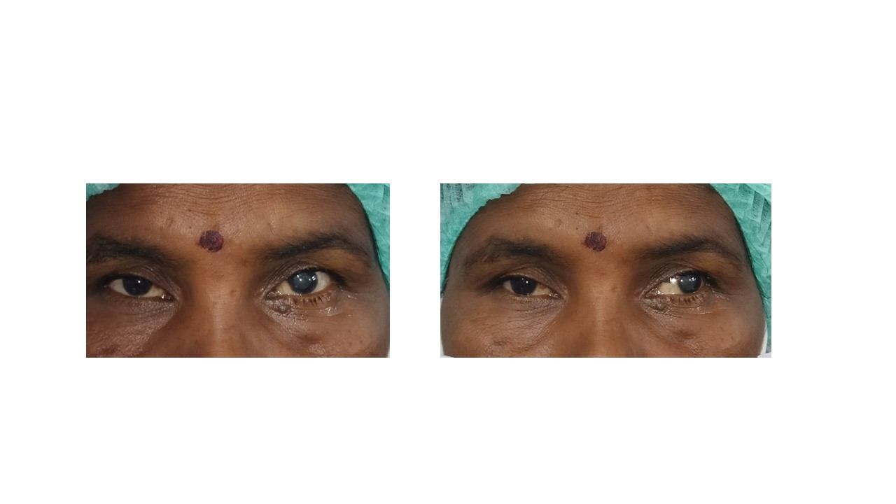
Orthotropia and Exotropia. The image shows a patient depicting orthotropia at near and exotropia after the fusion is suspended using the alternate cover test, suggestive of intermittent exotropia.
Contributed by K Kaur, MBBS, DNB, FPOS, FICO, MRCS Ed, MNAMS
(Click Video to Play)
Intermittent Exotropia. The video demonstrates a 5-year-old female patient with intermittent exotropia, showing good control as evidenced by rapid refixation during the alternate cover test.
Contributed by K Kaur, MBBS, DNB, FPOS, FICO, MRCS Ed, MNAMS
References
Tang W, He B, Luo J, Deng Z, Wang X, Duan X. Effect of the Control Ability on Stereopsis Recovery of Intermittent Exotropia in Children. Journal of pediatric ophthalmology and strabismus. 2021 Nov-Dec:58(6):350-354. doi: 10.3928/01913913-20210615-02. Epub 2021 Aug 26 [PubMed PMID: 34435904]
Yang Y, Lou J, Hou J, Tan S, Chang S, Yuan Y, Yu X. Corrective surgery improves visual motor and visual perception skills in children with intermittent exotropia. Optometry and vision science : official publication of the American Academy of Optometry. 2025 Nov 1:102(11):699-706. doi: 10.1097/OPX.0000000000002305. Epub 2025 Oct 15 [PubMed PMID: 41212121]
Swelam A, Ghaith KAA, Hegab M, Ghaith S, Al Khathami A, Milisic S. Combined bilateral lateral rectus recession and medial rectus Faden procedure in true divergence excess exotropia: a case report. Journal of surgical case reports. 2025 Nov:2025(11):rjaf868. doi: 10.1093/jscr/rjaf868. Epub 2025 Nov 5 [PubMed PMID: 41209665]
Level 3 (low-level) evidenceJost RM, Kumar K, Dao LM, Luu B, Alex AA, Koritala BA, Cheng-Patel C, Kelly KR, Birch EE. Everyday Consequences of Childhood Intermittent Exotropia. Journal of binocular vision and ocular motility. 2025 Nov 6:():1-12. doi: 10.1080/2576117X.2025.2579357. Epub 2025 Nov 6 [PubMed PMID: 41199138]
Ehrenberg M, Dotan G, Friling R, Palevski D, Elbaz U, Sternfeld A. Surgical Outcomes of Patients With Mild to Moderate Exodeviation With and Without Combined Superior Oblique Palsy. Journal of pediatric ophthalmology and strabismus. 2025 Nov 5:():1-6. doi: 10.3928/01913913-20250929-01. Epub 2025 Nov 5 [PubMed PMID: 41190567]
Phanphruk W, Hennein L, Hunter DG. Strabismus Surgery in Orthophoric Patients with Abduction Limitation. Clinical ophthalmology (Auckland, N.Z.). 2025:19():3991-3999. doi: 10.2147/OPTH.S543647. Epub 2025 Oct 29 [PubMed PMID: 41185761]
Yu J, Zhou Y. Clinical Analysis of the Effects of Different Types of Contact Lenses on the Correction of Myopia With Small-Angle Strabismus and Visual Function. Eye & contact lens. 2025 Dec 1:51(12):553-561. doi: 10.1097/ICL.0000000000001229. Epub 2025 Oct 31 [PubMed PMID: 41172279]
K Mohammad N, Ali Rajab I. Types of refractive errors in a sample of Iraqi children with Intermittent exotropia. F1000Research. 2024:13():1318. doi: 10.12688/f1000research.156932.5. Epub 2025 Jul 22 [PubMed PMID: 41000504]
Level 2 (mid-level) evidenceNishanth S, M S, S V, Kumaramanickavel G, Gijs M, Berendschot TTJM, Bauer NJC. A case of unilateral progression of myopia after strabismus surgery. Journal of AAPOS : the official publication of the American Association for Pediatric Ophthalmology and Strabismus. 2025 Dec:29(6):104677. doi: 10.1016/j.jaapos.2025.104677. Epub 2025 Oct 23 [PubMed PMID: 41138974]
Level 3 (low-level) evidenceYang Y, Hou J, Huang H, Wei C, Chen J, Wu T, Liu J, Deng D, Ling S, Yang W, Yan J, Chang S, Yu X. Reading ability in children with intermittent exotropia and its association with binocular visual function and visual information processing capability. BMC ophthalmology. 2025 Sep 1:25(1):497. doi: 10.1186/s12886-025-04326-y. Epub 2025 Sep 1 [PubMed PMID: 40890623]
Lee JY, Park KA, Oh SY. Lateral rectus muscle recession for intermittent exotropia with anomalous head position in type 1 Duane's retraction syndrome. Graefe's archive for clinical and experimental ophthalmology = Albrecht von Graefes Archiv fur klinische und experimentelle Ophthalmologie. 2018 Dec:256(12):2467-2471. doi: 10.1007/s00417-018-4079-5. Epub 2018 Jul 30 [PubMed PMID: 30062561]
Wu Y, Peng T, Zhou J, Xu M, Gao Y, Zhou J, Hou F, Yu X. A Survey of Clinical Opinions and Preferences on the Non-surgical Management of Intermittent Exotropia in China. Journal of binocular vision and ocular motility. 2021 Oct-Dec:71(4):167-174 [PubMed PMID: 34449280]
Level 3 (low-level) evidenceBui Quoc E, Milleret C. Origins of strabismus and loss of binocular vision. Frontiers in integrative neuroscience. 2014:8():71. doi: 10.3389/fnint.2014.00071. Epub 2014 Sep 25 [PubMed PMID: 25309358]
Cooper J. Intermittent exotropia of the divergence excess type. Journal of the American Optometric Association. 1977 Oct:48(10):1261-73 [PubMed PMID: 908827]
Kushner BJ, Morton GV. Distance/near differences in intermittent exotropia. Archives of ophthalmology (Chicago, Ill. : 1960). 1998 Apr:116(4):478-86 [PubMed PMID: 9565045]
Kekunnaya R, Chandrasekharan A, Sachdeva V. Management of Strabismus in Myopes. Middle East African journal of ophthalmology. 2015 Jul-Sep:22(3):298-306. doi: 10.4103/0974-9233.159728. Epub [PubMed PMID: 26180467]
von Noorden GK, Avilla CW. Accommodative convergence in hypermetropia. American journal of ophthalmology. 1990 Sep 15:110(3):287-92 [PubMed PMID: 2396654]
JAMPOLSKY A, FLOM BC, WEYMOUTH FW, MOSES LE. Unequal corrected visual acuity as related to anisometropia. A.M.A. archives of ophthalmology. 1955 Dec:54(6):893-905 [PubMed PMID: 13268145]
KNAPP P. Intermittent exotropia: evaluation and therapy. The American orthoptic journal. 1953 Sep:3():27-33 [PubMed PMID: 13104828]
JAMPOLSKY A. Differential diagnostic characteristics of intermittent exotropia and true exophoria. The American orthoptic journal. 1954 Jun:4():48-55 [PubMed PMID: 13180847]
Tınkır Kayıtmazbatır E, Güler HA, Acar Duyan Ş, Bozkurt Oflaz A, Bozkurt B. Visual Health in Autism Spectrum Disorder: Screening Outcomes, Clinical Associations, and Service Gaps. Medicina (Kaunas, Lithuania). 2025 Oct 1:61(10):. doi: 10.3390/medicina61101779. Epub 2025 Oct 1 [PubMed PMID: 41155763]
Feng K, Jing F. [Tuina for intermittent exotropia: a case report]. Zhongguo zhen jiu = Chinese acupuncture & moxibustion. 2025 Sep 12:45(9):1218-1220. doi: 10.13703/j.0255-2930.20250331-0008. Epub 2025 Jul 7 [PubMed PMID: 40954050]
Level 3 (low-level) evidenceVatkar V, Aher P, Gore S, Toshniwal S, Gollamudi S. Unilateral Oil Droplet Cataract in a Child: Importance of Early Detection and Intervention. Annals of African medicine. 2025 Aug 26:():. doi: 10.4103/aam.aam_249_25. Epub 2025 Aug 26 [PubMed PMID: 40874469]
Audren F. [Intermittent exotropia]. Journal francais d'ophtalmologie. 2019 Nov:42(9):1007-1019. doi: 10.1016/j.jfo.2018.12.031. Epub 2019 Jul 10 [PubMed PMID: 31301849]
Pan CW, Zhu H, Yu JJ, Ding H, Bai J, Chen J, Yu RB, Liu H. Epidemiology of Intermittent Exotropia in Preschool Children in China. Optometry and vision science : official publication of the American Academy of Optometry. 2016 Jan:93(1):57-62. doi: 10.1097/OPX.0000000000000754. Epub [PubMed PMID: 26583796]
Govindan M, Mohney BG, Diehl NN, Burke JP. Incidence and types of childhood exotropia: a population-based study. Ophthalmology. 2005 Jan:112(1):104-8 [PubMed PMID: 15629828]
Level 2 (mid-level) evidenceMcKean-Cowdin R, Cotter SA, Tarczy-Hornoch K, Wen G, Kim J, Borchert M, Varma R, Multi-Ethnic Pediatric Eye Disease Study Group. Prevalence of amblyopia or strabismus in asian and non-Hispanic white preschool children: multi-ethnic pediatric eye disease study. Ophthalmology. 2013 Oct:120(10):2117-24. doi: 10.1016/j.ophtha.2013.03.001. Epub 2013 May 19 [PubMed PMID: 23697956]
Biswas S, El Kareh A, Qureshi M, Lee DMX, Sun CH, Lam JSH, Saw SM, Najjar RP. The influence of the environment and lifestyle on myopia. Journal of physiological anthropology. 2024 Jan 31:43(1):7. doi: 10.1186/s40101-024-00354-7. Epub 2024 Jan 31 [PubMed PMID: 38297353]
Cotter SA, Mohney BG, Chandler DL, Holmes JM, Wallace DK, Melia BM, Wu R, Kraker RT, Superstein R, Crouch ER, Paysse EA, Pediatric Eye Disease Investigator Group. Three-year observation of children 12 to 35 months old with untreated intermittent exotropia. Ophthalmic & physiological optics : the journal of the British College of Ophthalmic Opticians (Optometrists). 2020 Mar:40(2):202-215. doi: 10.1111/opo.12668. Epub [PubMed PMID: 32202318]
Zhou M, Shi Q, Li H, Su M, Zhang H, Hong J, Wang X, Wan X, Fu J, Liu Z. Altered static and dynamic functional connectivity in childhood basic-type intermittent exotropia. Brain communications. 2025:7(5):fcaf358. doi: 10.1093/braincomms/fcaf358. Epub 2025 Sep 15 [PubMed PMID: 41040847]
Horwood AM, Riddell PM. Evidence that convergence rather than accommodation controls intermittent distance exotropia. Acta ophthalmologica. 2012 Mar:90(2):e109-17. doi: 10.1111/j.1755-3768.2011.02313.x. Epub 2012 Jan 26 [PubMed PMID: 22280437]
Oh BL, Suh SY, Choung HK, Kim SJ. Squinting and photophobia in intermittent exotropia. Optometry and vision science : official publication of the American Academy of Optometry. 2014 May:91(5):533-9. doi: 10.1097/OPX.0000000000000251. Epub [PubMed PMID: 24727823]
Level 2 (mid-level) evidenceLiu P, Fu J, Zhang R, Chu H. Initial postoperative plasticity as a predictor of mid-term stereoacuity outcome after surgery for intermittent exotropia. BMC ophthalmology. 2023 May 15:23(1):213. doi: 10.1186/s12886-023-02958-6. Epub 2023 May 15 [PubMed PMID: 37189107]
Ansari AAA, Sharma P, Khateeb EW, Bhombal F, Lad S, Jain V, Usmani A, Jain A, Abdal MO. Effectiveness of binocular vision therapy in managing patients with intermittent exotropia. Indian journal of ophthalmology. 2025 Oct 1:73(10):1422-1425. doi: 10.4103/IJO.IJO_724_25. Epub 2025 Sep 25 [PubMed PMID: 40995896]
Yang Y, Lou J, Tan S, Hou J, Huang H, Liu J, Chen J, Wu T, Sang X, Wang R, Hou F, Chang S, Yu X, Deng D. Surgical Realignment Reverses Contrast Sensitivity Deficits in Children With Intermittent Exotropia: One-Year Results of a Cohort Study. Investigative ophthalmology & visual science. 2025 Aug 1:66(11):71. doi: 10.1167/iovs.66.11.71. Epub [PubMed PMID: 40874696]
Tuohy JL, Byer BJ, Royer S, Keller C, Nagai-Singer MA, Regan DP, Seguin B. Evaluation of Myogenin and MyoD1 as Immunohistochemical Markers of Canine Rhabdomyosarcoma. Veterinary pathology. 2021 May:58(3):516-526. doi: 10.1177/0300985820988146. Epub 2021 Mar 11 [PubMed PMID: 33691532]
Kelkar JA, Gopal S, Shah RB, Kelkar AS. Intermittent exotropia: Surgical treatment strategies. Indian journal of ophthalmology. 2015 Jul:63(7):566-9. doi: 10.4103/0301-4738.167109. Epub [PubMed PMID: 26458472]
BURIAN HM, SPIVEY BE. THE SURGICAL MANAGEMENT OF EXODEVIATIONS. American journal of ophthalmology. 1965 Apr:59():603-20 [PubMed PMID: 14270998]
Wang X, Wang F, Xi S, Jiang C, Liu Y, Wen W, Zhao C. Short-Term Near Stereoacuity Improvements Following Favorable Surgical Alignment in Exotropic and Esotropic Patients. Seminars in ophthalmology. 2022 Feb 17:37(2):177-186. doi: 10.1080/08820538.2021.1946093. Epub 2021 Jul 5 [PubMed PMID: 34223803]
Sprunger DT, Lambert SR, Hercinovic A, Morse CL, Repka MX, Hutchinson AK, Cruz OA, Wallace DK, American Academy of Ophthalmology Preferred Practice Pattern Pediatric Ophthalmology/Strabismus Panel. Esotropia and Exotropia Preferred Practice Pattern®. Ophthalmology. 2023 Mar:130(3):P179-P221. doi: 10.1016/j.ophtha.2022.11.002. Epub 2022 Dec 14 [PubMed PMID: 36526451]
Aurell E, Norrsell K. A longitudinal study of children with a family history of strabismus: factors determining the incidence of strabismus. The British journal of ophthalmology. 1990 Oct:74(10):589-94 [PubMed PMID: 2285681]
Sverdlichenko I, Mandelcorn MS, Issashar Leibovitzh G, Mandelcorn ED, Markowitz SN, Tarita-Nistor L. Binocular visual function and fixational control in patients with macular disease: A review. Ophthalmic & physiological optics : the journal of the British College of Ophthalmic Opticians (Optometrists). 2022 Mar:42(2):258-271. doi: 10.1111/opo.12925. Epub 2021 Dec 4 [PubMed PMID: 34862635]
Goering M, Drennan KB, Moshirfar M. Convergence Insufficiency. StatPearls. 2026 Jan:(): [PubMed PMID: 32119277]
Yadav S, Tandon R. Comprehensive eye examination: what does it mean? Community eye health. 2019:32(107):S1-S4 [PubMed PMID: 32123482]
O'Connor AR, Tidbury LP. Stereopsis: are we assessing it in enough depth? Clinical & experimental optometry. 2018 Jul:101(4):485-494. doi: 10.1111/cxo.12655. Epub 2018 Jan 27 [PubMed PMID: 29377291]
Haggerty H, Richardson S, Hrisos S, Strong NP, Clarke MP. The Newcastle Control Score: a new method of grading the severity of intermittent distance exotropia. The British journal of ophthalmology. 2004 Feb:88(2):233-5 [PubMed PMID: 14736781]
Level 2 (mid-level) evidenceBuck D, Clarke MP, Haggerty H, Hrisos S, Powell C, Sloper J, Strong NP. Grading the severity of intermittent distance exotropia: the revised Newcastle Control Score. The British journal of ophthalmology. 2008 Apr:92(4):577. doi: 10.1136/bjo.2007.120287. Epub [PubMed PMID: 18369078]
Level 3 (low-level) evidenceStathacopoulos RA, Rosenbaum AL, Zanoni D, Stager DR, McCall LC, Ziffer AJ, Everett M. Distance stereoacuity. Assessing control in intermittent exotropia. Ophthalmology. 1993 Apr:100(4):495-500 [PubMed PMID: 8479706]
Sharma P, Saxena R, Narvekar M, Gadia R, Menon V. Evaluation of distance and near stereoacuity and fusional vergence in intermittent exotropia. Indian journal of ophthalmology. 2008 Mar-Apr:56(2):121-5 [PubMed PMID: 18292622]
Zanoni D, Rosenbaum AL. A new method for evaluating distance stereo acuity. Journal of pediatric ophthalmology and strabismus. 1991 Sep-Oct:28(5):255-60 [PubMed PMID: 1955959]
Kaur K, Gurnani B. Dissociated Vertical Deviation. StatPearls. 2026 Jan:(): [PubMed PMID: 34424634]
Barrett BT. A critical evaluation of the evidence supporting the practice of behavioural vision therapy. Ophthalmic & physiological optics : the journal of the British College of Ophthalmic Opticians (Optometrists). 2009 Jan:29(1):4-25. doi: 10.1111/j.1475-1313.2008.00607.x. Epub [PubMed PMID: 19154276]
Jackson JH, Arnoldi K. The Gradient AC/A Ratio: What's Really Normal? The American orthoptic journal. 2004:54():125-32. doi: 10.3368/aoj.54.1.125. Epub [PubMed PMID: 21149096]
Kushner BJ. The distance angle to target in surgery for intermittent exotropia. Archives of ophthalmology (Chicago, Ill. : 1960). 1998 Feb:116(2):189-94 [PubMed PMID: 9488270]
Level 1 (high-level) evidenceMurugan SB, Sunil Kumar, Gurnani B, Kaur K. Assessment of Subjective Visual Experiences During Intraocular Surgery and Intravitreal Injection Among Uveitis Patients at a Tertiary Eye Hospital: A Cross-Sectional Observational Study. Ophthalmology and therapy. 2026 Jan:15(1):173-191. doi: 10.1007/s40123-025-01264-2. Epub 2025 Nov 6 [PubMed PMID: 41196490]
Level 2 (mid-level) evidenceHull S, Tailor V, Balduzzi S, Rahi J, Schmucker C, Virgili G, Dahlmann-Noor A. Tests for detecting strabismus in children aged 1 to 6 years in the community. The Cochrane database of systematic reviews. 2017 Nov 6:11(11):CD011221. doi: 10.1002/14651858.CD011221.pub2. Epub 2017 Nov 6 [PubMed PMID: 29105728]
Level 1 (high-level) evidenceMori DM, Kuchhangi A, Tame J, Cooper K, Hajkazemshirazi L, Indaram M, Keenan JD, Oatts JT. Evaluation of a Novel Virtual Reality Simulated Alternate Cover Test to Assess Strabismus: A Prospective, Masked Study. American journal of ophthalmology. 2025 Jan:269():266-272. doi: 10.1016/j.ajo.2024.08.042. Epub 2024 Sep 6 [PubMed PMID: 39245131]
Hatt SR, Mohney BG, Leske DA, Holmes JM. Variability of stereoacuity in intermittent exotropia. American journal of ophthalmology. 2008 Mar:145(3):556-561. doi: 10.1016/j.ajo.2007.10.028. Epub 2008 Jan 16 [PubMed PMID: 18201680]
Bagolini B. Anomalous correspondence: definition and diagnostic methods. Documenta ophthalmologica. Advances in ophthalmology. 1967:23():346-98 [PubMed PMID: 5583482]
Level 3 (low-level) evidencePasino L, Maraini G. IMPORTANCE OF NATURAL TEST CONDITIONS IN ASSESSING THE SENSORY STATE OF THE SQUINTING SUBJECT WITH SOME CLINICAL CONSIDERATIONS ON ANOMALOUS RETINAL CORRESPONDENCE. The British journal of ophthalmology. 1964 Jan:48(1):30-4 [PubMed PMID: 18170855]
Level 3 (low-level) evidenceKapoula Z, Morize A, Daniel F, Jonqua F, Orssaud C, Brémond-Gignac D. Objective Evaluation of Vergence Disorders and a Research-Based Novel Method for Vergence Rehabilitation. Translational vision science & technology. 2016 Mar:5(2):8 [PubMed PMID: 26981330]
Dinkin M. Diagnostic approach to diplopia. Continuum (Minneapolis, Minn.). 2014 Aug:20(4 Neuro-ophthalmology):942-65. doi: 10.1212/01.CON.0000453310.52390.58. Epub [PubMed PMID: 25099102]
Level 3 (low-level) evidenceVincent A, Newsom-Davis J. Acetylcholine receptor antibody as a diagnostic test for myasthenia gravis: results in 153 validated cases and 2967 diagnostic assays. Journal of neurology, neurosurgery, and psychiatry. 1985 Dec:48(12):1246-52 [PubMed PMID: 4087000]
Level 3 (low-level) evidenceJacobs DS, Lee JK, Shen TT, Afshari NA, Bishop RJ, Keenan JD, Vitale S, American Academy of Ophthalmology Preferred Practice Pattern Refractive Management/Intervention Panel. Refractive Surgery Preferred Practice Pattern®. Ophthalmology. 2023 Mar:130(3):P61-P135. doi: 10.1016/j.ophtha.2022.10.032. Epub 2022 Dec 19 [PubMed PMID: 36543604]
Pediatric Eye Disease Investigator Group, Writing Committee, Mohney BG, Cotter SA, Chandler DL, Holmes JM, Wallace DK, Yamada T, Petersen DB, Kraker RT, Morse CL, Melia BM, Wu R. Three-Year Observation of Children 3 to 10 Years of Age with Untreated Intermittent Exotropia. Ophthalmology. 2019 Sep:126(9):1249-1260. doi: 10.1016/j.ophtha.2019.01.015. Epub 2019 Jan 26 [PubMed PMID: 30690128]
Romanchuk KG, Dotchin SA, Zurevinsky J. The natural history of surgically untreated intermittent exotropia-looking into the distant future. Journal of AAPOS : the official publication of the American Association for Pediatric Ophthalmology and Strabismus. 2006 Jun:10(3):225-31 [PubMed PMID: 16814175]
Level 2 (mid-level) evidenceHutchinson AK. Intermittent exotropia. Ophthalmology clinics of North America. 2001 Sep:14(3):399-406 [PubMed PMID: 11705139]
Iacobucci IL, Archer SM, Giles CL. Children with exotropia responsive to spectacle correction of hyperopia. American journal of ophthalmology. 1993 Jul 15:116(1):79-83 [PubMed PMID: 8328547]
Level 3 (low-level) evidenceCaltrider N, Jampolsky A. Overcorrecting minus lens therapy for treatment of intermittent exotropia. Ophthalmology. 1983 Oct:90(10):1160-5 [PubMed PMID: 6657190]
Watts P, Tippings E, Al-Madfai H. Intermittent exotropia, overcorrecting minus lenses, and the Newcastle scoring system. Journal of AAPOS : the official publication of the American Association for Pediatric Ophthalmology and Strabismus. 2005 Oct:9(5):460-4 [PubMed PMID: 16213396]
Level 3 (low-level) evidenceFreeman RS, Isenberg SJ. The use of part-time occlusion for early onset unilateral exotropia. Journal of pediatric ophthalmology and strabismus. 1989 Mar-Apr:26(2):94-6 [PubMed PMID: 2709283]
Pediatric Eye Disease Investigator Group, Cotter SA, Mohney BG, Chandler DL, Holmes JM, Repka MX, Melia M, Wallace DK, Beck RW, Birch EE, Kraker RT, Tamkins SM, Miller AM, Sala NA, Glaser SR. A randomized trial comparing part-time patching with observation for children 3 to 10 years of age with intermittent exotropia. Ophthalmology. 2014 Dec:121(12):2299-310. doi: 10.1016/j.ophtha.2014.07.021. Epub 2014 Sep 16 [PubMed PMID: 25234012]
Level 1 (high-level) evidencePratt-Johnson JA, Tillson G. Prismotherapy in intermittent exotropia. A preliminary report. Canadian journal of ophthalmology. Journal canadien d'ophtalmologie. 1979 Oct:14(4):243-5 [PubMed PMID: 550916]
Moore S. The prognostic value of lateral gaze measurements in intermittent exotropia. The American orthoptic journal. 1969:19():69-71 [PubMed PMID: 5792797]
Spencer RF, Tucker MG, Choi RY, McNeer KW. Botulinum toxin management of childhood intermittent exotropia. Ophthalmology. 1997 Nov:104(11):1762-7 [PubMed PMID: 9373104]
Level 2 (mid-level) evidencePediatric Eye Disease Investigator Group, Holmes JM, Manny RE, Lazar EL, Birch EE, Kelly KR, Summers AI, Martinson SR, Raghuram A, Colburn JD, Law C, Marsh JD, Bitner DP, Kraker RT, Wallace DK. A Randomized Trial of Binocular Dig Rush Game Treatment for Amblyopia in Children Aged 7 to 12 Years. Ophthalmology. 2019 Mar:126(3):456-466. doi: 10.1016/j.ophtha.2018.10.032. Epub 2018 Oct 22 [PubMed PMID: 30352226]
Level 1 (high-level) evidenceHatt SR, Leske DA, Liebermann L, Holmes JM. Quantifying variability in the measurement of control in intermittent exotropia. Journal of AAPOS : the official publication of the American Association for Pediatric Ophthalmology and Strabismus. 2015 Feb:19(1):33-7. doi: 10.1016/j.jaapos.2014.10.017. Epub [PubMed PMID: 25727584]
Park KH, Kim SY. Clinical characteristics of patients that experience different rates of exodrift after strabismus surgery for intermittent exotropia and the effect of the rate of exodrift on final ocular alignment. Journal of AAPOS : the official publication of the American Association for Pediatric Ophthalmology and Strabismus. 2013 Feb:17(1):54-8. doi: 10.1016/j.jaapos.2012.10.014. Epub [PubMed PMID: 23415036]
Chen AM, Erzurum SA, Chandler DL, Hercinovic A, Melia BM, Bhatt AR, Suh DW, Vricella M, Erickson JW, Miller AM, Marsh JD, Bodack MI, Martinson SR, Titelbaum JR, Gray ME, Holtorf HL, Kong L, Kraker RT, Rahmani B, Shah BK, Holmes JM, Cotter SA, Pediatric Eye Disease Investigator Group. Overminus Lens Therapy for Children 3 to 10 Years of Age With Intermittent Exotropia: A Randomized Clinical Trial. JAMA ophthalmology. 2021 Apr 1:139(4):464-476. doi: 10.1001/jamaophthalmol.2021.0082. Epub [PubMed PMID: 33662112]
Level 1 (high-level) evidenceYam JC, Chong GS, Wu PK, Wong US, Chan CW, Ko ST. Predictive factors affecting the short term and long term exodrift in patients with intermittent exotropia after bilateral rectus muscle recession and its effect on surgical outcome. BioMed research international. 2014:2014():482093. doi: 10.1155/2014/482093. Epub 2014 Jul 2 [PubMed PMID: 25093170]
Kaur K, Gurnani B. Subjective Refraction Techniques. StatPearls. 2026 Jan:(): [PubMed PMID: 35593807]
Arblaster G, Buckley D, Barnes S, Davis H. Strabismus Surgery for Psychosocial Reasons-A Literature Review. The British and Irish orthoptic journal. 2024:20(1):107-132. doi: 10.22599/bioj.352. Epub 2024 Apr 22 [PubMed PMID: 38681188]
Heydarian S, Hashemi H, Jafarzadehpour E, Ostadi A, Yekta A, Aghamirsalim M, Dadbin N, Ostadimoghaddam H, Khoshhal F, Khabazkhoob M. Non-surgical Management Options of Intermittent Exotropia: A Literature Review. Journal of current ophthalmology. 2020 Jul-Sep:32(3):217-225. doi: 10.4103/JOCO.JOCO_81_20. Epub 2020 Jul 4 [PubMed PMID: 32775794]
Lekskul A, Wuthisiri W, Jarupanich N. A Prospective Study of One-Muscle Surgery in 15-25 Prism Diopters Horizontal Comitant Strabismus in Adults. Clinical ophthalmology (Auckland, N.Z.). 2021:15():3669-3678. doi: 10.2147/OPTH.S324517. Epub 2021 Aug 31 [PubMed PMID: 34511870]
Niechwiej-Szwedo E, Colpa L, Wong A. The role of binocular vision in the control and development of visually guided upper limb movements. Philosophical transactions of the Royal Society of London. Series B, Biological sciences. 2023 Jan 30:378(1869):20210461. doi: 10.1098/rstb.2021.0461. Epub 2022 Dec 13 [PubMed PMID: 36511416]
Pineles SL, Deitz LW, Velez FG. Postoperative outcomes of patients initially overcorrected for intermittent exotropia. Journal of AAPOS : the official publication of the American Association for Pediatric Ophthalmology and Strabismus. 2011 Dec:15(6):527-31. doi: 10.1016/j.jaapos.2011.08.007. Epub [PubMed PMID: 22153394]
Level 2 (mid-level) evidenceBae GH, Bae SH, Choi DG. Surgical outcomes of intermittent exotropia according to exotropia type based on distance/near differences. PloS one. 2019:14(3):e0214478. doi: 10.1371/journal.pone.0214478. Epub 2019 Mar 25 [PubMed PMID: 30908548]
Lee CM, Sun MH, Kao LY, Lin KK, Yang ML. Factors affecting surgical outcome of intermittent exotropia. Taiwan journal of ophthalmology. 2018 Jan-Mar:8(1):24-30. doi: 10.4103/tjo.tjo_44_17. Epub [PubMed PMID: 29675346]
Feng X, Zhang X, Jia Y. Improvement in fusion and stereopsis following surgery for intermittent exotropia. Journal of pediatric ophthalmology and strabismus. 2015 Jan-Feb:52(1):52-7. doi: 10.3928/01913913-20141230-08. Epub [PubMed PMID: 25643371]
Hassan AM, Rajesh A, Asaad M, Nelson JA, Coert JH, Mehrara BJ, Butler CE. Artificial Intelligence and Machine Learning in Prediction of Surgical Complications: Current State, Applications, and Implications. The American surgeon. 2023 Jan:89(1):25-30. doi: 10.1177/00031348221101488. Epub 2022 May 13 [PubMed PMID: 35562124]
Olitsky SE, Coats DK. Complications of Strabismus Surgery. Middle East African journal of ophthalmology. 2015 Jul-Sep:22(3):271-8. doi: 10.4103/0974-9233.159692. Epub [PubMed PMID: 26180463]
Han SB, Hwang JM. Prismatic correction of residual esotropia of 20 prism dioptres or less after full hypermetropic correction. Eye (London, England). 2009 Nov:23(11):2052-5. doi: 10.1038/eye.2008.424. Epub 2009 Jan 30 [PubMed PMID: 19182765]
Digout LG, Awad AH. Restoration of binocular single vision after long-term fusion disruption. Journal of AAPOS : the official publication of the American Association for Pediatric Ophthalmology and Strabismus. 2003 Jun:7(3):185-9 [PubMed PMID: 12825058]
Huang S, Zhong X, Quan C, Zhang M. Non-surgical treatment of strabismus in children: a review of recent advances. Frontiers in medicine. 2025:12():1582284. doi: 10.3389/fmed.2025.1582284. Epub 2025 Jun 27 [PubMed PMID: 40655112]
Level 3 (low-level) evidenceEtezad Razavi M, Najaran M, Moravvej R, Ansari Astaneh MR, Azimi A. Correlation between Worth Four Dot Test Results and Fusional Control in Intermittent Exotropia. Journal of ophthalmic & vision research. 2012 Apr:7(2):134-8 [PubMed PMID: 23275822]
Writing Committee for the Pediatric Eye Disease Investigator Group, Pediatric Eye Disease Investigator Group, Chen AM, Erzurum SA, Chandler DL, Hercinovic A, Wu R, Vricella M, Waters AL, Ticho BH, Erickson JW, Han S, McDowell PS, Li Z, Kraker RT, Holmes JM, Cotter SA. Refractive Error Change and Overminus Lens Therapy for Childhood Intermittent Exotropia. JAMA ophthalmology. 2024 May 1:142(5):417-428. doi: 10.1001/jamaophthalmol.2024.0276. Epub [PubMed PMID: 38536764]
Aravamuthan BR, Shevell M, Kim YM, Wilson JL, O'Malley JA, Pearson TS, Kruer MC, Fahey M, Waugh JL, Russman B, Shapiro B, Tilton A. Role of child neurologists and neurodevelopmentalists in the diagnosis of cerebral palsy: A survey study. Neurology. 2020 Nov 24:95(21):962-972. doi: 10.1212/WNL.0000000000011036. Epub 2020 Oct 12 [PubMed PMID: 33046609]
Level 3 (low-level) evidenceSpiegel SJ, Moss HE. Neuro-Ophthalmic Emergencies. Neurologic clinics. 2021 May:39(2):631-647. doi: 10.1016/j.ncl.2021.01.004. Epub 2021 Mar 31 [PubMed PMID: 33896536]
Baeg J, Choi HS, Kim C, Kim H, Jang SY. Update on the surgical management of Graves' orbitopathy. Frontiers in endocrinology. 2022:13():1080204. doi: 10.3389/fendo.2022.1080204. Epub 2023 Feb 6 [PubMed PMID: 36824601]
Krungkraipetch L, Krungkraipetch N, Threetong T. A Meta-Analysis of Association Between Strabismus in Children/Adolescents and Risk of Mental Health Disorders. Clinical ophthalmology (Auckland, N.Z.). 2025:19():2333-2342. doi: 10.2147/OPTH.S530788. Epub 2025 Jul 16 [PubMed PMID: 40689234]
Level 1 (high-level) evidenceSeoane-Martín ME, Rodríguez-Martínez MC. Potential Role of Occupational Therapist Intervention in Elementary School for Children with Additional Support Needs: A Systematic Review. Children (Basel, Switzerland). 2023 Jul 27:10(8):. doi: 10.3390/children10081291. Epub 2023 Jul 27 [PubMed PMID: 37628290]
Level 1 (high-level) evidenceGurnani B, Kaur K, Sivakumar P, Bhandari S. Retrospective analysis of low vision assistive products - A 6-year review. Saudi journal of ophthalmology : official journal of the Saudi Ophthalmological Society. 2023 Jan-Mar:37(1):32-37. doi: 10.4103/sjopt.sjopt_253_21. Epub 2023 Mar 9 [PubMed PMID: 36968774]
Level 2 (mid-level) evidenceHopker LM, Rossetto JD, Ejzenbaum F, Sá LCF, Ribeiro GB, Mendonça TS, Goldchmit M, Debert I. Management of Intermittent exotropia in childhood: current concepts of the literature and the experts. Arquivos brasileiros de oftalmologia. 2022:85(6):V-VIII. doi: 10.5935/0004-2749.2022-1001. Epub 2022 Oct 21 [PubMed PMID: 36350932]
Heier L, Schellenberger B, Schippers A, Nies S, Geiser F, Ernstmann N. Interprofessional communication skills training to improve medical students' and nursing trainees' error communication - quasi-experimental pilot study. BMC medical education. 2024 Jan 3:24(1):10. doi: 10.1186/s12909-023-04997-5. Epub 2024 Jan 3 [PubMed PMID: 38172793]
Level 3 (low-level) evidenceSatyanarayana Rao KH. Informed consent: an ethical obligation or legal compulsion? Journal of cutaneous and aesthetic surgery. 2008 Jan:1(1):33-5. doi: 10.4103/0974-2077.41159. Epub [PubMed PMID: 20300341]
Carneiro VLA, González-Méijome JM. Integration of Refractive Services Provided by Optometrists into the Portuguese National Health Service. Portuguese journal of public health. 2023 Sep:41(2):111-121. doi: 10.1159/000530060. Epub 2023 Sep 1 [PubMed PMID: 39469657]
Treadwell JR, Lucas S, Tsou AY. Surgical checklists: a systematic review of impacts and implementation. BMJ quality & safety. 2014 Apr:23(4):299-318. doi: 10.1136/bmjqs-2012-001797. Epub 2013 Aug 6 [PubMed PMID: 23922403]
Level 1 (high-level) evidenceEbeigbe JA, Emedike CM. Parents' awareness and perception of children's eye diseases in Nigeria. Journal of optometry. 2017 Apr-Jun:10(2):104-110. doi: 10.1016/j.optom.2016.06.001. Epub 2016 Jul 14 [PubMed PMID: 27423689]
Economides JR, Adams DL, Horton JC. Capturing the Moment of Fusion Loss in Intermittent Exotropia. Ophthalmology. 2017 Apr:124(4):496-504. doi: 10.1016/j.ophtha.2016.11.039. Epub 2017 Jan 9 [PubMed PMID: 28081943]
Bresnick G, Cuadros JA, Khan M, Fleischmann S, Wolff G, Limon A, Chang J, Jiang L, Cuadros P, Pedersen ER. Adherence to ophthalmology referral, treatment and follow-up after diabetic retinopathy screening in the primary care setting. BMJ open diabetes research & care. 2020 Jun:8(1):. doi: 10.1136/bmjdrc-2019-001154. Epub [PubMed PMID: 32576560]
Huurre TM, Aro HM. Psychosocial development among adolescents with visual impairment. European child & adolescent psychiatry. 1998 Jun:7(2):73-8 [PubMed PMID: 9712373]
Xu M, Peng Y, Zheng F, Yu H, Zhou J, Zheng J, Wang Y, Hou F, Yu X. The Effects of Orthoptic Therapy on the Surgical Outcome in Children with Intermittent Exotropia: Randomised Controlled Clinical Trial. Journal of clinical medicine. 2023 Feb 6:12(4):. doi: 10.3390/jcm12041283. Epub 2023 Feb 6 [PubMed PMID: 36835820]
Level 1 (high-level) evidenceHutchinson AK, Morse CL, Hercinovic A, Cruz OA, Sprunger DT, Repka MX, Lambert SR, Wallace DK, American Academy of Ophthalmology Preferred Practice Pattern Pediatric Ophthalmology/Strabismus Panel. Pediatric Eye Evaluations Preferred Practice Pattern. Ophthalmology. 2023 Mar:130(3):P222-P270. doi: 10.1016/j.ophtha.2022.10.030. Epub 2022 Dec 19 [PubMed PMID: 36543602]
Ma MM, Kang Y, Chen C, Su C, Tian Z, Le M. Vision therapy for intermittent exotropia: A case series. Journal of optometry. 2021 Jul-Sep:14(3):247-253. doi: 10.1016/j.optom.2020.05.006. Epub 2020 Aug 12 [PubMed PMID: 32800454]
Level 2 (mid-level) evidence