 Tools
Tools
 Hepatic Biloma
Introduction
Gould and Patel coined the term “biloma” in 1979 to describe an encapsulated collection of extrahepatic bile secondary to bile leakage into the peritoneal cavity.[1] However, the term “biloma” has evolved to describe any well-circumscribed intraabdominal bile collection external to the biliary tree. Disruption of the biliary tree can result in either intrahepatic or extrahepatic biloma formation. Although many collections are well encapsulated, the current definition of a biloma does not require complete encapsulation.[2]
The well-circumscribed margins of the biloma differentiate it from ongoing bile leaks or intraperitoneal free bile. “Choleperitoneum” and “biliary ascites” are other terms used to describe free bile in the peritoneum; however, some sources use these terms and “biloma” interchangeably.[3][4] Iatrogenic injury and abdominal trauma, causing damage to the biliary tree resulting in a bile leak, are the most common causes of biloma formation.
Bilomas are associated with infection, ongoing bile leakage, and mass effect on surrounding structures. While uncommon, bilomas are associated with significant morbidity and mortality if not promptly diagnosed and appropriately managed.[2] Radiological investigation using ultrasound, computed tomography, magnetic resonance imaging, magnetic resonance cholangiopancreatography, or hepatobiliary cholescintigraphy can be used to establish a diagnosis and to allow accurate, minimally invasive management when appropriate.[5][6][7]
Etiology
Register For Free And Read The Full Article
 Search engine and full access to all medical articles
- 10 free questions in your specialty
- Free CME/CE Activities
- Free daily question in your email
- Save favorite articles to your dashboard
- Emails offering discounts
Learn more about a Subscription to StatPearls Point-of-Care
Etiology
Bilomas are most commonly secondary to disruption of the biliary tree, whether iatrogenic or traumatic. Iatrogenic causes of biloma formation include laparoscopic cholecystectomy, endoscopic retrograde cholangiopancreatography, radiofrequency ablation, transcatheter arterial chemoembolization, liver transplant, resection, and biopsy.[8][9][10][11] While exceedingly rare, spontaneous rupture of the biliary tree and biloma formation have been recognized in the literature and can be associated with malignancy.[12][13][14]
The increased use of laparoscopic cholecystectomy over the past 3 decades has been linked with increased rates of disruption to the biliary tree. Laparoscopic cholecystectomy is associated with bile duct injuries in 0.6% to 1.5% of cases compared with 0.2% to 0.3% of open operations.[15] Biliary leaks following laparoscopic cholecystectomy can be attributed to damage of the common bile duct (CBD), unsuccessful cystic duct ligation, or anatomical variation leading to damaged or leaking accessory ducts.[16] The ducts of Luschka, small ducts that drain into the biliary system, are found in the gallbladder fossa in 25% to 35% of patients and are vulnerable to injury during cholecystectomy.[17] Fluid collection, including bile leakage, is more common following cholecystectomy than many clinicians realize. Results from a 1986 study demonstrated that small-volume bile leakage occurred in up to 44% of laparoscopic cholecystectomies. However, 95% of these did not require any further management.[18]
In 1991, Kang et al showed that 53% of patients who underwent ultrasound 24 hours after laparoscopic cholecystectomy had small-volume fluid collections. However, they determined that a routine postoperative ultrasound did not change management and was unwarranted.[19] While small fluid collections are common postoperatively, these leaks are primarily small, asymptomatic, and often resolve without treatment. Most think that many small leaks go undetected. However, significant bile leaks and biloma formation must remain a key differential diagnosis in patients with a complicated recovery following laparoscopic cholecystectomy.[4]
Biloma has been demonstrated following perforation of the CBD during endoscopic retrograde cholangiopancreatography (ERCP).[20][21] Results from a prospective multicentre study of 5461 patients showed CBD perforation occurred in 0.4% of patients undergoing ERCP. Risk factors for biliary tree perforation included precut access to the CBD and malignancy.[21]
Biloma formation was identified in 3.3% of 3284 patients undergoing RFA of hepatocellular carcinoma. However, biloma formation during RFA is a minor complication. Chang et al examined 109 bilomas following radiofrequency ablation for hepatocellular carcinoma, with only 1 patient requiring percutaneous drainage of the biloma for infection.[22] Thermal ablation of hepatocellular carcinoma is also associated with biloma formation, with bile leaks identified in 0.1% to 12% of cases. Previous transarterial chemoembolization (TACE) procedures and a closer proximity to the biliary tree were both associated with an increased risk of bile leak and biloma formation in patients undergoing thermal ablation.[9] TACE procedures for hepatocellular carcinoma are also associated with biloma formation. Zhang et al demonstrated an incidence of 1.04% in 4695 patients undergoing TACE in 2017.[23]
Liver transplantation is known to be associated with biloma formation. Biloma forms part of a spectrum of posttransplant cholangiopathies. Posttransplant cholangiopathy is one of the most challenging complications following a liver transplant. The pathophysiology of posttransplant cholangiopathy is likely to involve ischemia-reperfusion injury, bile salt toxicity, or immune-mediated injury. Biloma formation after liver transplant can be associated with both anastomotic leakage and nonanastomotic strictures with associated bile duct necrosis and resulting intraparenchymal bile leakage.[11] Liver resection is also associated with biloma formation due to the disruption of intrahepatic bile ducts.[24]
Abdominal trauma has been associated with biloma formation. In most cases, this involves blunt trauma to the upper abdomen. Posttraumatic biloma can take 1 to 2 days to appear.[25][26] Spontaneous rupture of the biliary tree is very rare; however, it has been associated with underlying weakness in bile duct walls caused by cholelithiasis, cholangiocarcinoma, hepatic abscess, or tuberculosis.[27] Spontaneous rupture of the biliary tree is often a diagnosis of exclusion.[28] Biloma formation is also associated with sickle cell disease, likely due to hepatic infarction.[29] Bile leakage can occur without disrupting the biliary tree, for example, in leakage from the duodenal stump following Billroth II surgery.
Epidemiology
There is little epidemiological data regarding biloma formation. Most bilomas are secondary to iatrogenic disruption of the biliary tree; therefore, their incidence depends on the frequency of interventions. Biloma typically presents in patients aged 60 to 70. This likely reflects underlying etiological factors requiring invasive intervention rather than a predisposition to complications leading to biloma formation. No difference in biloma incidence has been observed between male and female patients.[30] Spontaneous biloma formation is exceedingly rare, and the literature on the subject consists solely of case reports. In 2007, Ahktar et al noted that 27 cases of spontaneous biloma had been reported since 1979.[31]
Pathophysiology
Disruption of the biliary tree and subsequent bile leakage is the cause of most bilomas. Encapsulation of the bile leak is thought to be via 2 mechanisms, depending on the rate of bile leakage. Most commonly, slow bile leakage causes mild inflammation of the surrounding abdominal tissues or the liver parenchyma, leading to fibrosis and encapsulation. Bile acids produce low-grade inflammation in surrounding tissues because of their detergent and tissue-destroying properties.[32]
Biliary peritonitis can be present in large volumes, with rapid bile leaks. This may present with encapsulation; however, patients may present acutely before encapsulation has occurred. Intraperitoneally, the omentum and mesentery can form inflammatory adhesions, aiding encapsulation.[2] Biloma size and location depend on the mechanism of biliary tree disruption, the rate of bile leakage, and bile reabsorption by the surrounding liver parenchyma or peritoneum.[26]
Extravasated bile follows abdominal anatomy, with its shape often demarcated by the diaphragm, liver margins, mesentery, and transverse mesocolon. Multiple bilomas in patients are not uncommon. Extrahepatic bilomas predominantly form in the right upper quadrant of the abdomen. However, in about 40% of cases, bile migrates over the anterior part of the liver to the left subphrenic or left subhepatic spaces.[2][31]
Biloma content is typically greenish-yellow bile. However, they can contain blood or exudate, especially in the context of infection. Secondary infection can lead to a systemic inflammatory response, sepsis, and abscess formation. The natural history of abdominal bile collections from injured bile ducts is the progression from sterile bile collection to infection.[4]
History and Physical
Bilomas typically present with upper abdominal fullness and right upper quadrant discomfort following iatrogenic or traumatic disruption to the biliary tree. Nausea, vomiting, and fever are recognized symptoms, especially in cases of infected biloma. Jaundice may be present in cases of extrinsic compression of the bile duct. Key aspects required from the history include previous surgical or endoscopic interventions or abdominal trauma that may have caused biliary tree disruption. A history of hepatobiliary surgery or biliary tree disease also increases the risk of bile leak and biloma formation.[2]
While patients may present acutely unwell with septic shock from bilomas, many may be asymptomatic. The significant variation in presenting symptoms makes the diagnosis of biloma difficult. Bile peritonitis is a recognized presentation of bile leak and large biloma. However, Lee et al showed that this presentation is unusual, and most patients present with more subtle symptoms. They describe delayed diagnosis in 77% of 179 patients with abdominal bile collections and bile leaks following laparoscopic cholecystectomy. Significant abdominal pain and tenderness were observed in only 21% of patients. Initial peritonitis does not predict the severity of the disease, with many subtly symptomatic patients developing serious complications.[4]
Following iatrogenic intervention or traumatic injury, physicians should be alert to persistent abdominal distention, bloating, or anorexia as early signs of bile leak and biloma. Clinicians should be aware that any delay in recovery from hepatobiliary surgery, especially laparoscopic cholecystectomy, is a possible early symptom of bile leak and biloma formation. Biloma remains an uncommon diagnosis, and other gastroenterological and hepatobiliary diagnoses should be considered for patients presenting with upper abdominal fullness and discomfort. However, in patients who have recently experienced surgical or endoscopic interventions in the region of the biliary tree, bile leak and biloma are essential differential diagnoses to exclude, as they can present subtly and have significant morbidity and mortality.[2][4]
Evaluation
Initial laboratory testing may reveal a systemic inflammatory response to a biloma. Blood tests may demonstrate elevated inflammatory markers, such as leucocytosis, neutrophilia, and elevated C-reactive protein. However, many patients with bilomas have no abnormalities in laboratory tests. Vazquez et al demonstrated normal blood results in 40% of 21 patients presenting with bilomas.[26]
Liver function tests may be deranged in cases of bile duct compression. In cases of infected biloma, blood cultures may reveal gram-negative bacteremia. Wurstle et al showed positive blood cultures in 93% of 32 bilomas following iatrogenic injury to the biliary tree. The most common organisms found in a laboratory culture of biloma fluid were Enterobacteriaceae, followed by Enterococcus species. They demonstrated multidrug-resistant bacteria in 25% of biloma cultures.[33]
The variable and often subtle presentation of bilomas makes radiological investigation the mainstay of diagnosis. MRI, computed tomography (CT), ultrasound, and hepatobiliary iminodiacetic acid are the main radiological modalities used to investigate bilomas. Abdominal imaging via ultrasound is often the first investigation due to the frequent presentation with right upper quadrant discomfort. Ultrasound imaging can frequently identify a cystic lesion. This modality can also demonstrate debris or blood clots within the biloma. The ultrasound may reveal a wide range of findings from well-circumscribed collections in the liver parenchyma to large loculated fluid collections throughout the abdomen. Heavily loculated bilomas seen on ultrasound imaging have been associated with an infection. Although ultrasound imaging is effective for identifying a cystic mass, further imaging is usually required to make a diagnosis.[2][34]
CT imaging typically shows bilomas as a well-circumscribed collection with clear margins. However, a well-defined capsule is not always present.[2] Bilomas are typically hypoechoic, hypoattenuated collections with CT attenuation of less than 20 Hounsfield units (see Image. Hepatic Biloma on Computed Tomography).[26][35] CT imaging has the benefit of precisely localizing the biloma and imaging the surrounding structures. While CT can provide a more detailed picture of bilomas, it cannot definitively differentiate between differential diagnoses such as postoperative seroma, hematoma, abscess, lymphocele, liver cyst, and pseudocyst.[36] Therefore, further imaging with MRI or hepatobiliary cholescintigraphy is often required, along with direct sampling of the biloma, to confirm the diagnosis.[2]
MRI, particularly magnetic resonance cholangiopancreatography (MRCP) in some cases, can further characterize a biloma. Biloma typically produces a low signal intensity on T1-weighted images and high signal intensity on T2-weighted images. While contrast rarely permeates the biloma, rim enhancement may be present due to reactive inflammation. Enhancing septations are often observed in superimposed infection. MRCP, particularly thin slab sequences, may demonstrate the source of a biliary leak.[37]
Hepatobiliary cholescintigraphy is an extremely effective noninvasive imaging modality for diagnosing and planning the treatment of bilomas. Cholescintigraphy uses the radiotracer Tc-99m iminodiacetic acid and is known as hepatobiliary iminodiacetic acid (HIDA) imaging. HIDA is very sensitive when looking for a bile leak. However, it does not provide detailed imaging of the surrounding anatomy. Single-photon emission computed tomography can provide more detailed imaging of potential leak locations and is more useful for planning percutaneous image-guided drains. Invasive imaging techniques, such as endoscopic retrograde cholangiopancreatography and percutaneous transhepatic cholangiogram, can provide further guidance for endoscopic, percutaneous, or surgical management.[2][28] Ultrasound or CT-guided sampling of the biloma, followed by laboratory analysis, may be required to confirm the diagnosis when prior imaging and clinical findings are inconclusive.[2]
Treatment / Management
Management varies depending on the clinical presentation, laboratory results, and radiological findings of the biloma. The 3 main options are percutaneous or endoscopic drainage, surgical drainage, or close monitoring. Ongoing bile leak, biloma size and position, superimposed infection, and patient fitness will determine the most appropriate management strategy.[2] Small, asymptomatic fluid collections may be reabsorbed and not require any intervention. However, Lee et al demonstrated that collections larger than 4 cm were rarely reabsorbed, and it was hard to predict whether patients would become symptomatic.[4]
Most bilomas can be managed successfully with radiologically guided percutaneous drainage. Fixation of the underlying disruption of the biliary tree is often unnecessary.[24][35] The ultrasound or CT-guided aspiration by an interventional radiologist is the preferred method. Due to the lack of radiation and real-time imaging, ultrasound is preferred over CT in most cases. Spectral Doppler and real-time color allow avoidance of vascular structures.(B2)
Interventional radiologists may access extrahepatic bilomas via the liver if no other clear window is available. Different methods of drainage include endoscopic ultrasound (EUS) guided drainage. For example, Shami et al described, in 2008, successful management of bilomas adjacent to bowel with EUS-guided drainage.[38] Tonozuka et al described, in 2015, the successful management of hepatic abscesses and infected bilomas with EUS-guided metal stents.[39] Most bilomas without ongoing bile leaks, when managed with percutaneous drainage, do not recur and have a good prognosis.(B3)
Ongoing bile leaks may require further management, with either surgical fixation or endoscopic stent placement. However, Lee et al recommend drainage of the biloma after laparoscopic cholecystectomy in the first case, especially in acutely unwell or symptomatic patients.[4] Percutaneous transhepatic cholangiography or percutaneous transhepatic biliary drainage is sometimes required to image the biliary tree further or decompress the gallbladder when there is an ongoing bile leak.[2]
Surgical management of biloma is sometimes indicated in cases of failed percutaneous drainage, multiloculated lesions, and ongoing bile leaks. Cases of bile leak identified intraoperatively during laparoscopic cholecystectomy or requiring surgical management of continuing bile leak are best managed in specialist tertiary centers.[40] The rarity of biloma formation means there is little reliable evidence beyond case reports and case series describing management strategies. However, percutaneous drainage has successfully managed biloma, and biloma patients have an excellent prognosis.(B2)
Differential Diagnosis
Because biloma formation is uncommon, it is essential to consider possible differential diagnoses. Right upper quadrant discomfort and abdominal fullness are nonspecific symptoms with a wide range of potential causes. Biloma should enter the differential where patients present secondary to an iatrogenic or traumatic event that could disrupt the biliary tree. Following this, appropriate imaging should be undertaken.
Differential diagnoses following radiological investigation include hepatic abscess, hepatic cyst, hepatic pseudocyst, and hepatic lymphocele. As most bilomas form postoperatively or postendoscopic investigation, seroma and hematoma are key differential diagnoses to consider. Identifying bilomas correctly is essential, as they often require drainage, whereas many differential diagnoses, such as hematoma, do not.[2][4]
Prognosis
Biloma prognosis varies depending on site, size, and etiology. Uncomplicated bilomas without underlying ongoing bile leaks have a good prognosis. Small, asymptomatic bilomas with no ongoing bile leak are often treated conservatively to good effect.[2] In patients with symptomatic bilomas, drainage via interventional radiology is an effective strategy, and patients generally have a good prognosis.[2][24] Lee et al have shown that prompt drainage of symptomatic bilomas significantly reduces morbidity and mortality, while reducing the risk of secondary infection and complications.[4] Most bilomas managed with percutaneous drainage do not recur and have a good prognosis. However, larger bile leaks into the peritoneum are associated with significant morbidity, often requiring urgent and invasive intervention to avoid deterioration.[4]
Complications
Complications of biloma include infection, septic shock, abscess formation, and cholestasis through impingement on the biliary tree. Lee et al also describe pancreatitis, respiratory failure, and transdiaphragmatic bile fistulation in patients with abdominal bile collections.[4] Risks of percutaneous drainage of bilomas include bleeding, infection, damage to surrounding structures, and failure to drain the biloma. Ongoing bile leaks may require further endoscopic or surgical management.[2]
Deterrence and Patient Education
Deterrence in hepatic bilomas focuses primarily on preventing and promptly recognizing bile duct injury, as bilomas most commonly arise after iatrogenic or traumatic disruption of the biliary tree. Careful surgical technique, meticulous dissection near the biliary tract, and early identification of postoperative bile leaks reduce the likelihood of biloma formation. Clinicians must maintain a high index of suspicion in patients who develop postoperative abdominal pain, fever, jaundice, leukocytosis, or unexpected bilious drainage—particularly following procedures such as laparoscopic cholecystectomy, liver resection, ablation, or endoscopic retrograde cholangiopancreatography (ERCP). Because even nonspecific abdominal symptoms or delays in expected recovery may signal a developing biloma, early imaging with ultrasound or CT and timely management with ERCP, percutaneous drainage, or surgical repair are essential to prevent sepsis, biliary peritonitis, and long-term hepatic dysfunction.
Patient education is equally critical. Patients undergoing hepatobiliary procedures or those who have experienced abdominal trauma should be counseled on recognizing early warning signs of bile leakage, including worsening abdominal pain, fever, chills, jaundice, shoulder pain, dark urine, or new drainage from surgical sites. Clear guidance on what constitutes normal versus concerning postoperative recovery helps ensure they seek prompt evaluation when needed. For patients treated for a biloma, education must also include care of drainage catheters (if placed), signs of infection, medication adherence, and the importance of follow-up imaging to confirm resolution. By reinforcing early symptom recognition and maintaining open communication between patients and healthcare teams, clinicians can facilitate earlier diagnosis, reduce complications, and significantly improve morbidity and mortality associated with hepatic bilomas.
Enhancing Healthcare Team Outcomes
Management of hepatic bilomas requires coordinated skills and strategic collaboration across multiple disciplines to ensure timely diagnosis, effective intervention, and prevention of complications. Physicians and advanced practitioners lead the clinical evaluation, integrating imaging, laboratory findings, and procedural history to rapidly identify bile leaks or fluid collections following hepatobiliary surgery or trauma. Surgeons and interventional radiologists play essential roles in determining whether endoscopic retrograde cholangiopancreatography, percutaneous drainage, or operative repair is warranted. Nurses continuously monitor for fever, abdominal pain, jaundice, and changes in drain output to ensure early recognition of deterioration. Pharmacists optimize antimicrobial therapy, manage analgesics, and evaluate for drug interactions, especially when managing sepsis risk or coordinating periprocedural medications. This collective expertise ensures that diagnostic and therapeutic interventions are performed safely and efficiently.
Interprofessional communication is central to enhancing patient-centered care, safety, and team performance. Structured communication tools—such as postoperative huddles, standardized reporting of drain output, and clear handoff protocols—help align goals across surgical, medical, and nursing teams. Coordination with radiology, infectious disease specialists, and case managers ensures smooth transitions between diagnostic imaging, procedural interventions, and discharge planning. Patient and family education is integrated throughout the care process, with nurses, clinicians, and allied health professionals reinforcing recognition of warning signs, postoperative expectations, and proper care of drainage catheters when present. By maintaining open dialogue, shared decision-making, and a unified care plan, the interprofessional team improves outcomes, reduces diagnostic delays, enhances patient safety, and supports a seamless recovery for individuals with hepatic bilomas.
Media
(Click Image to Enlarge)
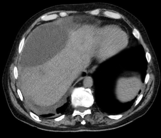
Hepatic Biloma on Computed Tomography. This image shows a well-defined, low-attenuation fluid collection adjacent to the liver, consistent with a hepatic biloma. The lesion appears hypodense compared to the surrounding hepatic parenchyma, with smooth margins and no internal enhancement, typical of a contained bile leak accumulating in the subcapsular region.
Contributed by R Kabir, MD
References
Gould L, Patel A. Ultrasound detection of extrahepatic encapsulated bile: "biloma". AJR. American journal of roentgenology. 1979 Jun:132(6):1014-5 [PubMed PMID: 108953]
Level 3 (low-level) evidenceCopelan A, Bahoura L, Tardy F, Kirsch M, Sokhandon F, Kapoor B. Etiology, Diagnosis, and Management of Bilomas: A Current Update. Techniques in vascular and interventional radiology. 2015 Dec:18(4):236-43. doi: 10.1053/j.tvir.2015.07.007. Epub 2015 Jul 15 [PubMed PMID: 26615164]
Vilar Tabanera A, Puerta Vicente A, López Buenadicha A, Peromingo R, Lopez Hervás P, Nuño Vasquez-Garza J. Luschka Duct Leak: An Unexpected Cause of Choleperitoneum After Liver Transplant. Experimental and clinical transplantation : official journal of the Middle East Society for Organ Transplantation. 2020 Aug:18(4):526-528. doi: 10.6002/ect.2018.0348. Epub 2019 Jun 25 [PubMed PMID: 31250744]
Lee CM, Stewart L, Way LW. Postcholecystectomy abdominal bile collections. Archives of surgery (Chicago, Ill. : 1960). 2000 May:135(5):538-42; discussion 542-4 [PubMed PMID: 10807277]
Shankar S, vanSonnenberg E, Silverman SG, Tuncali K, Morrison PR. Diagnosis and treatment of intrahepatic biloma complicating radiofrequency ablation of hepatic metastases. AJR. American journal of roentgenology. 2003 Aug:181(2):475-7 [PubMed PMID: 12876029]
Level 3 (low-level) evidenceKhalid TR, Casillas VJ, Montalvo BM, Centeno R, Levi JU. Using MR cholangiopancreatography to evaluate iatrogenic bile duct injury. AJR. American journal of roentgenology. 2001 Dec:177(6):1347-52 [PubMed PMID: 11717081]
Snyder E, Banks KP. Hepatobiliary Scintigraphy. StatPearls. 2025 Jan:(): [PubMed PMID: 30855831]
Southern Surgeons Club. A prospective analysis of 1518 laparoscopic cholecystectomies. The New England journal of medicine. 1991 Apr 18:324(16):1073-8 [PubMed PMID: 1826143]
Liu J, Wu Y, Xu E, Huang Q, Ye H, Tan L, Zheng R, Zeng Q, Li K. Risk factors of intrahepatic biloma and secondary infection after thermal ablation for malignant hepatic tumors. International journal of hyperthermia : the official journal of European Society for Hyperthermic Oncology, North American Hyperthermia Group. 2019:36(1):980-985. doi: 10.1080/02656736.2019.1660422. Epub [PubMed PMID: 31544547]
Sakamoto I, Iwanaga S, Nagaoki K, Matsuoka Y, Ashizawa K, Uetani M, Fukuda T, Okimoto T, Okudaira S, Omagari K, Hayashi K, Matsunaga N. Intrahepatic biloma formation (bile duct necrosis) after transcatheter arterial chemoembolization. AJR. American journal of roentgenology. 2003 Jul:181(1):79-87 [PubMed PMID: 12818833]
de Vries Y, von Meijenfeldt FA, Porte RJ. Post-transplant cholangiopathy: Classification, pathogenesis, and preventive strategies. Biochimica et biophysica acta. Molecular basis of disease. 2018 Apr:1864(4 Pt B):1507-1515. doi: 10.1016/j.bbadis.2017.06.013. Epub 2017 Jun 20 [PubMed PMID: 28645651]
Della Valle V, Eshja E, Bassi EM. Spontaneous biloma: a case report. Journal of ultrasound. 2015 Sep:18(3):293-6. doi: 10.1007/s40477-013-0053-6. Epub 2013 Nov 12 [PubMed PMID: 26261461]
Level 3 (low-level) evidenceGanipisetti VM, Bollimunta P, Dudiki N. Spontaneous Biloma With a Hepatic Mass. Cureus. 2023 Jun:15(6):e40249. doi: 10.7759/cureus.40249. Epub 2023 Jun 11 [PubMed PMID: 37440803]
Kośnik A, Stadnik A, Szczepankiewicz B, Patkowski W, Wójcicki M. Spontaneous rupture of a mucinous cystic neoplasm of the liver resulting in a huge biloma in a pregnant woman: A case report. World journal of clinical cases. 2021 Oct 26:9(30):9114-9121. doi: 10.12998/wjcc.v9.i30.9114. Epub [PubMed PMID: 34786394]
Level 3 (low-level) evidenceAlexander HC, Bartlett AS, Wells CI, Hannam JA, Moore MR, Poole GH, Merry AF. Reporting of complications after laparoscopic cholecystectomy: a systematic review. HPB : the official journal of the International Hepato Pancreato Biliary Association. 2018 Sep:20(9):786-794. doi: 10.1016/j.hpb.2018.03.004. Epub 2018 Apr 9 [PubMed PMID: 29650299]
Level 1 (high-level) evidencePeters JH, Ellison EC, Innes JT, Liss JL, Nichols KE, Lomano JM, Roby SR, Front ME, Carey LC. Safety and efficacy of laparoscopic cholecystectomy. A prospective analysis of 100 initial patients. Annals of surgery. 1991 Jan:213(1):3-12 [PubMed PMID: 1824674]
Handra-Luca A, Ben Romdhane HM, Hong SM. Luschka Ducts of the Gallbladder in Adults: Case Series Report and Review of the Medical Literature. International journal of surgical pathology. 2020 Aug:28(5):482-489. doi: 10.1177/1066896920901334. Epub 2020 Jan 26 [PubMed PMID: 31983263]
Level 2 (mid-level) evidenceGilsdorf JR, Phillips M, McLeod MK, Harness JK, Hoversten GH, Woodbury D, Daley K. Radionuclide evaluation of bile leakage and the use of subhepatic drains after cholecystectomy. American journal of surgery. 1986 Feb:151(2):259-62 [PubMed PMID: 3946761]
Level 1 (high-level) evidenceKang EH, Middleton WD, Balfe DM, Soper NJ. Laparoscopic cholecystectomy: evaluation with sonography. Radiology. 1991 Nov:181(2):439-42 [PubMed PMID: 1833786]
Alkhateeb HM, Aljanabi TJ, Al-Azzawi KH, Alkarboly TA. Huge biloma after endoscopic retrograde cholangiopancreatography and endoscopic biliary sphincterotomy. International journal of surgery case reports. 2015:16():7-11. doi: 10.1016/j.ijscr.2015.08.039. Epub 2015 Sep 1 [PubMed PMID: 26402876]
Level 3 (low-level) evidenceWilliams EJ, Taylor S, Fairclough P, Hamlyn A, Logan RF, Martin D, Riley SA, Veitch P, Wilkinson ML, Williamson PR, Lombard M. Risk factors for complication following ERCP; results of a large-scale, prospective multicenter study. Endoscopy. 2007 Sep:39(9):793-801 [PubMed PMID: 17703388]
Level 2 (mid-level) evidenceChang IS, Rhim H, Kim SH, Kim YS, Choi D, Park Y, Lim HK. Biloma formation after radiofrequency ablation of hepatocellular carcinoma: incidence, imaging features, and clinical significance. AJR. American journal of roentgenology. 2010 Nov:195(5):1131-6. doi: 10.2214/AJR.09.3946. Epub [PubMed PMID: 20966318]
Level 2 (mid-level) evidenceZhang B, Guo Y, Wu K, Shan H. Intrahepatic biloma following transcatheter arterial chemoembolization for hepatocellular carcinoma: Incidence, imaging features and management. Molecular and clinical oncology. 2017 Jun:6(6):937-943. doi: 10.3892/mco.2017.1235. Epub 2017 May 4 [PubMed PMID: 28588794]
Dell AJ, Krige JE, Jonas E, Thomson SR, Beningfield SJ, Kotze UK, Tromp SA, Burmeister S, Bernon MM, Bornman PC. Incidence and management of postoperative bile leaks: A prospective cohort analysis of 467 liver resections. South African journal of surgery. Suid-Afrikaanse tydskrif vir chirurgie. 2016 Sep:54(3):18-22 [PubMed PMID: 28240463]
Ragavan M, Duraiprabhu A, Madan R, Murali K, Francis G, Subramanian M. Posttraumatic Intrahepatic Bilioma. The Indian journal of surgery. 2015 Dec:77(Suppl 3):1399-400. doi: 10.1007/s12262-014-1071-0. Epub 2014 Apr 20 [PubMed PMID: 27011576]
Vazquez JL, Thorsen MK, Dodds WJ, Quiroz FA, Martinez ML, Lawson TL, Stewart ET, Foley WD. Evaluation and treatment of intraabdominal bilomas. AJR. American journal of roentgenology. 1985 May:144(5):933-8 [PubMed PMID: 3885693]
Level 3 (low-level) evidenceArramón M, Sciarretta M, Correa GJ, Yantorno M, Redondo A, Baldoni F, Tufare F. Spontaneous Biloma Secondary to Choledocholithiasis. ACG case reports journal. 2021 Jun:8(6):e00620. doi: 10.14309/crj.0000000000000620. Epub 2021 Jun 15 [PubMed PMID: 34150922]
Level 3 (low-level) evidenceYousaf MN, D'Souza RG, Chaudhary F, Ehsan H, Sittambalam C. Biloma: A Rare Manifestation of Spontaneous Bile Leak. Cureus. 2020 May 14:12(5):e8116. doi: 10.7759/cureus.8116. Epub 2020 May 14 [PubMed PMID: 32542169]
Lebensburger J, Esbenshade A, Blakely M, Hankins J, Wang W. Biloma and pneumobilia in sickle cell disease. Pediatric blood & cancer. 2008 Aug:51(2):288-90. doi: 10.1002/pbc.21575. Epub [PubMed PMID: 18421713]
Level 3 (low-level) evidenceTrivedi PJ, Gupta P, Phillips-Hughes J, Ellis A. Biloma: an unusual complication in a patient with pancreatic cancer. World journal of gastroenterology. 2009 Nov 7:15(41):5218-20 [PubMed PMID: 19891023]
Level 3 (low-level) evidenceAkhtar MA, Bandyopadhyay D, Montgomery HD, Mahomed A. Spontaneous idiopathic subcapsular biloma. Journal of hepato-biliary-pancreatic surgery. 2007:14(6):579-81 [PubMed PMID: 18040624]
Level 3 (low-level) evidenceKannan U, Parshad R, Regmi SK. An unusual presentation of biloma five years following cholecystectomy: a case report. Cases journal. 2009 Sep 10:2():8048. doi: 10.1186/1757-1626-0002-0000008048. Epub 2009 Sep 10 [PubMed PMID: 20181203]
Level 3 (low-level) evidenceWürstle S, Göß A, Spinner CD, Huber W, Algül H, Schlag C, Schmid RM, Weber A, Obermeier A, Schneider J. A retrospective clinical and microbial analysis of 32 patients with bilomas. BMC gastroenterology. 2019 Apr 4:19(1):50. doi: 10.1186/s12876-019-0968-2. Epub 2019 Apr 4 [PubMed PMID: 30947689]
Level 2 (mid-level) evidenceThomas S, Jahangir K. Noninvasive Imaging of the Biliary System Relevant to Percutaneous Interventions. Seminars in interventional radiology. 2016 Dec:33(4):277-282 [PubMed PMID: 27904246]
Akin K, Ozturk A, Guvenc Z, Isiklar I, Haberal M. Localized fluid collections after liver transplantation. Transplantation proceedings. 2006 Mar:38(2):627-30 [PubMed PMID: 16549192]
Level 2 (mid-level) evidenceWalker AT, Shapiro AW, Brooks DC, Braver JM, Tumeh SS. Bile duct disruption and biloma after laparoscopic cholecystectomy: imaging evaluation. AJR. American journal of roentgenology. 1992 Apr:158(4):785-9 [PubMed PMID: 1532111]
Chaudhary A, Negi SS, Puri SK, Narang P. Comparison of magnetic resonance cholangiography and percutaneous transhepatic cholangiography in the evaluation of bile duct strictures after cholecystectomy. The British journal of surgery. 2002 Apr:89(4):433-6 [PubMed PMID: 11952583]
Shami VM, Talreja JP, Mahajan A, Phillips MS, Yeaton P, Kahaleh M. EUS-guided drainage of bilomas: a new alternative? Gastrointestinal endoscopy. 2008 Jan:67(1):136-40 [PubMed PMID: 18155436]
Level 3 (low-level) evidenceTonozuka R, Itoi T, Tsuchiya T, Sofuni A, Ishii K, Ikeuchi N, Umeda J, Tanaka R, Mukai S, Gotoda T, Moriyasu F. EUS-guided drainage of hepatic abscess and infected biloma using short and long metal stents (with videos). Gastrointestinal endoscopy. 2015:81(6):1463-9. doi: 10.1016/j.gie.2015.01.023. Epub 2015 Apr 2 [PubMed PMID: 25843615]
Flum DR, Cheadle A, Prela C, Dellinger EP, Chan L. Bile duct injury during cholecystectomy and survival in medicare beneficiaries. JAMA. 2003 Oct 22:290(16):2168-73 [PubMed PMID: 14570952]
Level 2 (mid-level) evidence