 Tools
Tools
 Anesthesia Breathing Systems
Introduction
Anesthesia breathing systems are essential for delivering oxygen, eliminating carbon dioxide, and providing an appropriate depth of anesthesia with inhaled agents. Various designs of breathing systems have been created to serve this function. Anesthesia breathing systems can be classified based on the presence of a reservoir bag and the degree of rebreathing into open, semi-open, semi-closed, and closed.[1]
Function
Register For Free And Read The Full Article
 Search engine and full access to all medical articles
- 10 free questions in your specialty
- Free CME/CE Activities
- Free daily question in your email
- Save favorite articles to your dashboard
- Emails offering discounts
Learn more about a Subscription to StatPearls Point-of-Care
Function
Anesthesia breathing systems facilitate the delivery of oxygen, air, and volatile anesthetic agents—both halogenated (eg, sevoflurane and isoflurane) and nonhalogenated (eg, nitrous oxide)—while removing exhaled gases, including carbon dioxide, from patients. The following classification of these systems often depends on the presence or absence of specific physical and functional components, though alternative classification methods also exist:
- Open breathing systems: These systems, which include insufflation and open-drop anesthesia, do not have a reservoir breathing bag, functional rebreathing of exhaled gases, tubing, or valves.
- Semi-open breathing systems: This group includes Mapleson breathing systems, which have a reservoir breathing bag, lack functional rebreathing of exhaled gases, and require high fresh gas flows.
- Semi-closed breathing systems: These systems have a reservoir breathing bag, partial rebreathing of exhaled gases, unidirectional valves, carbon dioxide neutralization, and low fresh gas flows, including circle systems with an adjustable pressure-limiting (APL) valve at least partially open.
- Closed breathing systems: These systems have a reservoir breathing bag, total rebreathing of exhaled gases, unidirectional valves, and carbon dioxide neutralization, including circle systems with the APL valve closed.[2]
Open Breathing Systems
Open-drop anesthesia
The open-drop anesthesia breathing system is no longer used in modern medicine, but is historically significant and may occasionally be used in developing countries.[3][4] The Schimmelbusch mask, a wired mask invented by Curt Schimmelbusch in 1889 and used into the 1950s, is 1 of the simplest forms of an anesthesia breathing system. This system secures gauze to a wired mask, and liquid volatile anesthetics (ether, chloroform, or halothane) are dropped onto the gauze. As the patient inhales through the mask, air passes through the gauze, vaporizes the anesthetic liquid, and delivers high concentrations of anesthetic to the patient. In this system, exhaled gases are not rebreathed. The benefit of the system is its simple setup and limited equipment requirements.
Insufflation
The insufflation breathing system delivers oxygen, with or without anesthetic gases, through a mask across a patient’s face, without the mask being in direct contact with the patient. This breathing system is most commonly used with pediatric patients when placing a face mask directly on the child’s face is difficult or the patient resists.[5] High-flow insufflation has also been used in ophthalmology and otolaryngology procedures.[6] In this system, if the gas flow is high enough, virtually no exhaled gases are rebreathed.
Semi-Open Breathing Systems
Mapleson circuits
In 1954, William Mapleson designated various arrangements of breathing system components (eg, masks, breathing tubes, fresh gas flow inlets, adjustable pressure-limiting valves, and reservoir bags) as Mapleson A to E circuits.[7] An anesthesia mask is placed over a patient’s face with a firm seal to connect the patient to these systems. These systems can also be connected to laryngeal mask airways or endotracheal tubes. The system typically includes a fresh gas flow (FGF) source, reservoir bag, and adjustable-pressure limiting (APL) valve. Additionally, no corrugated tubing connects the mask or airway to the other system components.
An FGF inlet delivers mixed gases to the system, and as anesthetic and fresh gases enter the system, the pressure builds. To control pressure within the circuit, once the pressure exceeds a set value, the APL valve opens, allowing gases to exit the breathing system. An open APL valve allows all expired gases to exit the system. Partial closure of the APL valve allows continuous positive airway pressure (CPAP) to be delivered. Because semi-open systems lack components for carbon dioxide absorption, higher FGF rates are necessary to prevent rebreathing and carbon dioxide buildup.
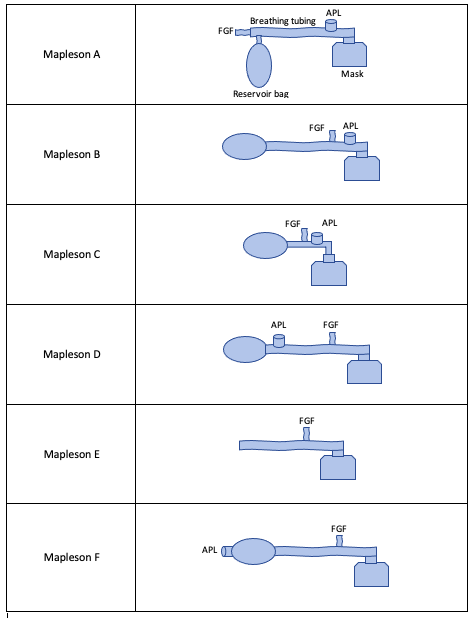
The varying arrangements of Mapleson circuits are discussed briefly (see Image. Mapleson Circuits).[7] Each circuit arrangement is described relative to the patient from most distal to most proximal.
Mapleson A
This circuit is arranged as an FGF inlet, a reservoir bag, an APL valve, and a mask. In this circuit, because the reservoir bag is between the FGF inlet valve and the APL valve, expired gas from the patient may re-enter the system and fill the reservoir bag during controlled ventilation (ie, when the APL valve is partially closed) if the fresh gas inflow is not adequate (defined as 2 to 3 times minute ventilation in this system). However, this is the most efficient system for spontaneous breathing, as the FGF rate must equal only a patient’s minute ventilation to prevent rebreathing.
Mapleson B
In this circuit, arranged as a reservoir bag, FGF inlet, APL valve, and mask, the FGF inlet is closer to the APL valve, which helps prevent the rebreathing concern in the Mapleson A circuit, as above, during controlled ventilation.
Mapleson C
The arrangement in this circuit, as a reservoir bag, FGF inlet, APL valve, and mask, is the same as the Mapleson B circuit. However, it is shorter as it does not contain elongated corrugated tubing. This circuit also has the FGF inlet close to the APL valve to prevent rebreathing.
Mapleson D
This circuit is arranged as a reservoir bag, an APL valve, an FGF inlet, and a mask. In this circuit, the arrangement interchanges the FGF inlet and APL valve of the Mapleson A circuit. This system prevents rebreathing by directing FGF toward the APL valve rather than toward the patient during exhalation. Therefore, in this circuit, as in circuits E and F, ventilation must be used 2 to 3 times a minute to prevent rebreathing during spontaneous breathing. In contrast, only 1 to 2 times the minute ventilation flow rate is required during controlled breathing. Clinicians should note that this is the opposite of the Mapleson A circuit.
Mapleson E
This circuit, arranged as corrugated tubing, an FGF inlet, and a mask, does not include a reservoir bag or an APL valve. Given the inability to alter the circuit's pressure, the Mapleson E is ideal for spontaneously ventilating neonates or pediatric patients who require low-pressure ventilation.[7] The system prevents rebreathing, as with the Mapleson D circuit.
Mapleson F
This circuit is arranged as an APL valve, directly connected to a reservoir bag, corrugated tubing, an FGF inlet, and a mask. The system prevents rebreathing, as with the Mapleson D, by directing FGF towards the APL valve.
Table. Summary of Mapleson Arrangements
|
Circuit Arrangement |
Fresh Gas Flow Inlet | Reservoir Bag | APL Valve |
Comments |
| A | Distal | Present | Patient Adjacent |
|
| B | By APL | Distal | Patient Adjacent | |
| C | By APL | Distal | Patient Adjacent | No corrugated tubing |
| D | Patient Adjacent | Present | Distal | Most efficient for controlled ventilation. |
| E | Patient Adjacent | Absent | Absent | Also known as Ayer's T-piece |
| F | Patient Adjacent | Present | Absent | Also known as Jackson Rees |
The Circle System
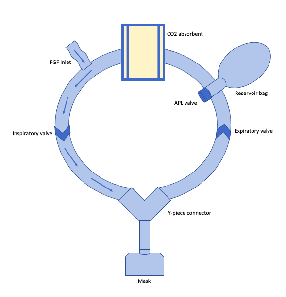
The circle system is the most clinically relevant breathing system (see Image. Circle System). Depending on the APL valve, the circle system can be semi-closed or closed. This system has more components than a Mapleson circuit and is arranged in a circular apparatus. Titration of flow rates will control the degree of rebreathing. In addition to a reservoir bag, tubing, an APL valve, and an FGF inlet, this system also has a carbon dioxide (CO2) absorber, unidirectional expiratory and inspiratory valves, and a Y-piece connector.
Unidirectional valves are located in the inspiratory and expiratory limbs, which are joined by a Y-piece that is connected to the patient’s anesthesia mask or airway. The FGF inlet delivers a gas mixture (oxygen, air, and volatile anesthetic) through the inspiratory tubing and Y-piece to the patient's mask or airway device. As the patient exhales, the gas exits through the Y-piece and into the expiratory breathing tubing. The reservoir bag and APL valve are located in the expiratory circuit to control pressurization.
The APL valve serves a critical role in preventing pressure buildup in the circle system. Imagine a closed system in which the APL valve is fully closed. Since no "pop-off" occurs, the additional volume from the FGF will distend the reservoir bag, continuously increasing the system pressure and the pressure the patient's lungs experience. If this goes unrecognized, the bag could potentially explode, and this could cause barotrauma. Thus, most circle systems are used in a semi-closed manner, with the APL valve partially closed/open to allow gas to escape above a set pressure.
The last component of a circle system is the CO2 absorber, located between the reservoir bag and the FGF inlet.[1] During exhalation, the patient expires alveolar gas, a heated and humidified mixture containing CO2. Expired CO2 must be removed from the system to prevent a buildup, which can cause hypercapnia and carbon dioxide narcosis. This is the function of the CO2 absorber. Exhaled CO2 from a patient combines with water to form carbonic acid. The CO2 absorber contains hydroxide salts (strong bases) that neutralize carbonic acid via a chemical reaction that produces additional heat and humidification, along with calcium carbonate. Soda-lime is 1 of the most common hydroxide salt absorbents and contains water, calcium hydroxide, sodium hydroxide, and potassium hydroxide.[8] Absorbents contain salt granules (sizes of approximately 4 to 8 mesh) that undergo a color change from the chemical reaction between these compounds and CO2. When approximately 50% to 70% of absorbent granules have changed color (a pH indicator dye that is typically a purple color known as ethyl violet, but may vary based on the manufacturer of the absorbent), it may indicate absorbent exhaustion requiring canister replacement to avoid rebreathing.[9]
The gas mixture purified by the CO2 absorbent is then combined with the fresh gas mixture from the FGF inlet and delivered to the patient via the inspiratory limb. Thus, a benefit of a circle system is that it conserves heat, humidity, and volatile anesthetics.
Reducing the Environmental Impact of Anesthetic Gases
Volatile anesthetics, eg, nitrous oxide and halogenated gases, contribute to environmental pollution.[10] Advocacy for the use of low-flow anesthesia (LFA) to limit the waste of volatile anesthetics has increased. Clinicians should note that variable bypass vaporizers for common volatile anesthetics, eg, sevoflurane, deliver a set partial pressure of gas; so the amount of anesthetic delivered is proportional to the FGF rate. Since anesthesia machines utilize a circle system, at high FGF rates, much of the exhaled gas, including carbon dioxide and volatile anesthetics, is wasted through the APL valve into the scavenging system and then out into the environment.
The goal of LFA is to titrate down the FGF rate to the minimum necessary to prevent loss of volatile anesthetics and humidity. A commonly cited reason against the use of LFA is the United States Food and Drug Administration (FDA) labeling on sevoflurane and recommendation against the use of FGF rates less than 2 L/min, citing evidence of harm, specifically nephrotoxicity from Compound A accumulation, in animal models. Systematic review of sevoflurane and reports of LFA in human patients have not found an association with nephrotoxicity.[11][12] While no specific target FGF rate has been identified, reports indicate FGF rates as low as 0.5 L/min are safe for use in adult patients. Finally, use of LFA requires additional vigilance of CO2 absorbent exhaustion and an understanding of equilibration times during induction, which is beyond the scope of this course.
Resuscitation Breathing Systems
Resuscitation breathing systems (including brand-name AMBU bags or bag-mask units) are simple, portable devices used in emergency situations or when transporting patients who need ventilation.[13] These systems can deliver nearly 100% oxygen to a patient. The system contains an inlet nipple (open to the air or connected to an oxygen source) connected through an intake valve to a ventilation bag. The ventilation bag, self-inflating based on inherent material memory, then delivers gas to the patient’s mask or invasive airway via a patient valve. An intake valve attached to the ventilation bag closes when compressed, allowing positive-pressure ventilation. Rebreathing is prevented in this system by venting exhaled gas through an exhalation port in the patient valve, which can be equipped with an additional positive end-expiratory pressure (PEEP) valve to provide positive end-expiratory pressure.[13][14]
Clinical Significance
Breathing systems, eg, Mapleson circuits and resuscitation bags, are commonly used across healthcare settings, from emergency medical services to the intensive care unit. Clinicians should understand the advantages and disadvantages to select the appropriate tool based on the patient's clinical needs.
The circle system is the most commonly used breathing system for anesthesia in the United States. Advantages of this breathing system include control of anesthetic depth, scavenging of exhaled gases, reduced requirements for fresh gas flow due to the CO2 absorber, conservation of heat and humidity, and minimal contribution to anatomic dead space, as only components distal to and including the Y-piece contribute to dead space. The CO2 absorber helps break down volatile anesthetics, generating carbon monoxide and thereby increasing clinically measurable carboxyhemoglobin concentrations. The highest risk of this reaction occurs when using desflurane and CO2 absorbents containing high concentrations of strong bases, eg, potassium hydroxide (KOH) or sodium hydroxide (NaOH).[8][15][16] In rat and non-human primate studies, sevoflurane degradation by strong base absorbents has produced a nephrotoxic byproduct called compound A. This nephrotoxicity has not been demonstrated in humans.[17][18]
Enhancing Healthcare Team Outcomes
Anesthesia breathing systems form the foundation of safe anesthetic delivery by enabling oxygen administration, carbon dioxide elimination, and regulation of anesthetic depth. These systems—classified as open, semi-open, semi-closed, and closed—differ in their degree of rebreathing and structural components such as reservoir bags, valves, and carbon dioxide absorbers. Mastery of their function, performance, and limitations ensures safe and efficient anesthesia management across surgical and critical care settings.
Effective team-based anesthesia care depends on every clinician’s understanding of these systems. Attending anesthesiologists, residents, certified registered nurse anesthetists, certified anesthesiologist assistants, and anesthesia technologists share responsibility for maintaining system integrity, troubleshooting malfunctions, and applying appropriate techniques. Interprofessional communication among physicians, advanced practitioners, nurses, and pharmacists fosters coordination in gas delivery management, medication administration, and monitoring. Collaboration and accountability within the anesthesia team enhance patient safety, optimize perioperative outcomes, and strengthen patient-centered care by ensuring continuous, well-coordinated vigilance during every phase of anesthesia delivery.
Media
(Click Image to Enlarge)
Mapleson Circuits
Contributed by A Garrett, MD
(Click Image to Enlarge)
Circle System
Contributed by A Garrett, MD
References
Conway CM. Anaesthetic breathing systems. British journal of anaesthesia. 1985 Jul:57(7):649-57 [PubMed PMID: 3925973]
Baum J. [Anesthesia systems]. Der Anaesthesist. 1987 Aug:36(8):393-9 [PubMed PMID: 3310726]
Lo R. The Schimmelbusch mask. Hong Kong medical journal = Xianggang yi xue za zhi. 2014 Dec:20(6):560-1 [PubMed PMID: 25632423]
Maltby JR, Malla DS, Dangol H. Open drop ether anaesthesia for caesarean section: a review of 420 cases in Nepal. Canadian Anaesthetists' Society journal. 1986 Sep:33(5):651-6 [PubMed PMID: 3768769]
Level 3 (low-level) evidenceTalyshinskiĭ AM, Baĭtiakov VV, Kosenko VF. [Use of insufflation method of inhalation anesthesia in tonsillectomy and adenoidectomy in children]. Vestnik otorinolaringologii. 1971 Jul-Aug:33(4):76-80 [PubMed PMID: 5112401]
Tautenhahn E. [Insufflation anesthesia in ophthalmic surgery]. Klinische Monatsblatter fur Augenheilkunde. 1972 Dec:161(6):703-5 [PubMed PMID: 4655902]
Kaul TK, Mittal G. Mapleson's Breathing Systems. Indian journal of anaesthesia. 2013 Sep:57(5):507-15. doi: 10.4103/0019-5049.120148. Epub [PubMed PMID: 24249884]
Keijzer C, Perez RS, de Lange JJ. Carbon monoxide production from desflurane and six types of carbon dioxide absorbents in a patient model. Acta anaesthesiologica Scandinavica. 2005 Jul:49(6):815-8 [PubMed PMID: 15954965]
Parthasarathy S. The closed circuit and the low flow systems. Indian journal of anaesthesia. 2013 Sep:57(5):516-24. doi: 10.4103/0019-5049.120149. Epub [PubMed PMID: 24249885]
Samad K, Yousuf MS, Ullah H, Ahmed SS, Siddiqui KM, Latif A. Anesthesia and its environmental impact: approaches to minimize exposure to anesthetic gases and reduce waste. Medical gas research. 2025 Mar 1:15(1):101-109. doi: 10.4103/mgr.MEDGASRES-D-23-00059. Epub 2024 Jun 26 [PubMed PMID: 39436173]
Kharasch ED, Frink EJ Jr, Zager R, Bowdle TA, Artru A, Nogami WM. Assessment of low-flow sevoflurane and isoflurane effects on renal function using sensitive markers of tubular toxicity. Anesthesiology. 1997 Jun:86(6):1238-53 [PubMed PMID: 9197292]
Sondekoppam RV, Narsingani KH, Schimmel TA, McConnell BM, Buro K, Özelsel TJ. The impact of sevoflurane anesthesia on postoperative renal function: a systematic review and meta-analysis of randomized-controlled trials. Canadian journal of anaesthesia = Journal canadien d'anesthesie. 2020 Nov:67(11):1595-1623. doi: 10.1007/s12630-020-01791-5. Epub 2020 Aug 18 [PubMed PMID: 32812189]
Level 1 (high-level) evidenceKhoury A, Hugonnot S, Cossus J, De Luca A, Desmettre T, Sall FS, Capellier G. From mouth-to-mouth to bag-valve-mask ventilation: evolution and characteristics of actual devices--a review of the literature. BioMed research international. 2014:2014():762053. doi: 10.1155/2014/762053. Epub 2014 May 27 [PubMed PMID: 24971346]
Bucher JT, Vashisht R, Cooper JS. Bag-Valve-Mask Ventilation. StatPearls. 2025 Jan:(): [PubMed PMID: 28722953]
Wissing H, Kuhn I, Warnken U, Dudziak R. Carbon monoxide production from desflurane, enflurane, halothane, isoflurane, and sevoflurane with dry soda lime. Anesthesiology. 2001 Nov:95(5):1205-12 [PubMed PMID: 11684991]
Berry PD, Sessler DI, Larson MD. Severe carbon monoxide poisoning during desflurane anesthesia. Anesthesiology. 1999 Feb:90(2):613-6 [PubMed PMID: 9952168]
Level 3 (low-level) evidenceKharasch ED. [Compound A: toxicology and clinical relevance]. Der Anaesthesist. 1998 Nov:47 Suppl 1():S7-10 [PubMed PMID: 9893874]
Level 3 (low-level) evidenceFrink EJ Jr, Green WB Jr, Brown EA, Malcomson M, Hammond LC, Valencia FG, Brown BR Jr. Compound A concentrations during sevoflurane anesthesia in children. Anesthesiology. 1996 Mar:84(3):566-71 [PubMed PMID: 8659785]