 Tools
Tools
 Cleft Palate Repair
Introduction
Management of palatal clefts requires an understanding of the anatomy, embryology, and physiology of the upper aerodigestive tract, as well as their relationship to speech, swallowing, breathing, and hearing. The incidence of palatal clefts ranges from 1 to 25 per 10,000 live births, with the rate primarily dependent on ethnicity; White individuals are most likely to be affected, while Black individuals are least likely.[1] Cleft palates come in many forms, including submucous clefting, secondary palate clefting, primary and secondary palate clefting, and complete clefting, which includes the alveolar ridge and lip (see Image. Oral Cleft).
Cleft width, extent, and degree of velopharyngeal dysfunction vary among individuals, as do comorbidities. Roughly half of all palatal clefting cases are nonsyndromic, but myriad syndromes may also be associated with cleft palates. Every patient is different, and every cleft palate is potentially unique.[2] For this reason, a thorough understanding of treatment options and the potential sequelae of both palatal clefting and cleft palate repair is essential for the interprofessional healthcare team to treat these patients safely, optimize outcomes, and minimize complications.
Anatomy and Physiology
Register For Free And Read The Full Article
 Search engine and full access to all medical articles
- 10 free questions in your specialty
- Free CME/CE Activities
- Free daily question in your email
- Save favorite articles to your dashboard
- Emails offering discounts
Learn more about a Subscription to StatPearls Point-of-Care
Anatomy and Physiology
Anatomically, the palate is divided into primary and secondary palates by the incisive foramen (also called the anterior palatine or nasopalatine foramen), with the primary palate located anterior to this foramen and the secondary palate located posterior to it. The primary palate, along with the central segment of the alveolar ridge and the maxillary incisors, is known as the premaxilla. The secondary palate consists of the soft palate and the remainder of the hard palate posterior to the premaxilla. The incisive foramen's location is in the midline, osseous part of the oral hard palate, immediately posterior to the central incisor teeth, and corresponds to the medial palatine and incisive sutures' junction.
In addition to bony anomalies, patients with palatal clefting often exhibit abnormal velopharyngeal musculature, as initially described by Fergusson and Veau. In cases of cleft palate, rather than inserting into the midline soft palate aponeurosis, the tensor veli palatini muscle is partially attached to the hard palate's posterior border laterally and partially to the margin of the soft palate cleft. The muscle is not aligned correctly to glide around the pterygoid hamuli and function as a sling. The levator veli palatini muscle also demonstrates an abnormal insertion at the cleft margins in the anterior half to two-thirds of the velum rather than meeting the palatine aponeurosis.[3]
As described below, careful dissection, release, and reorientation of the palatal muscles will determine, to a great extent, the mobility of the soft palate postoperatively. The blood supply to the soft and hard palates comes from the descending palatine arteries, which emerge from the greater palatine foramina medial to the third molars (see Image. Anatomy of Upper Palate). When reconstructing the palate, it is crucial to preserve these blood vessels, as inadvertent injury may lead to palatal necrosis, particularly when the palatal flaps are monopedicled.
Some patients may present with submucous palatal clefting, characterized by abnormal speech and velopharyngeal insufficiency in the absence of an overt palatal cleft. The physical examination may reveal a bifid uvula, a zona pellucida, and/or a palpable notch on the posterior margin of the hard palate (see Image. Submucous Cleft Palate). Depending on the patient's age and the severity of the cleft, management may include or be limited to speech therapy. However, most patients with submucous palatal clefting require a velopharyngoplasty, such as the Furlow or Sommerlad procedures, followed by speech therapy.
Ideally, the soft palate moves cephalically and posteriorly to separate the nasopharynx from the oropharynx when pronouncing syllables that contain "b," "p," "k," and "s" due to the action of the levator veli palatini muscle. For this reason, it is crucial to carefully reorient the fibers of this muscle during cleft palate repair. Clinically, an unrepaired cleft of the secondary palate will limit the ability of a child to generate adequate levels of oral air pressure needed for the production of stop consonants, also known as plosives, which in English include /b/, /d/, /g/, /k/, /t/, and /p/. Moreover, the production of /b/ is deemed a significant predictor of the success of palatal surgery, given that this is an early-developing phoneme that is dependent on an adequate velopharyngeal closure.[4]
Embryologically, most craniofacial structures form between 4 and 8 weeks of gestation, and developmental abnormalities during this period may result in orofacial clefting. A cleft of the primary palate occurs due to a failure of fusion between the lateral palatine processes and the median palatine process, and this is typically accompanied by a cleft of the secondary palate and the upper lip (complete cleft palate). Primary palate clefts may be unilateral or bilateral, as the cleft runs in a paramedian position between the incisive foramen and the gap between the lateral maxillary incisive and the canine tooth.
Clefting of the secondary palate may affect the soft palate only or a combination of the soft and hard palates, manifesting as anything from a bifid uvula with or without a submucosal muscular dehiscence, up to a cleft that follows the midline from the uvula all the way to the incisive foramen (see Image. Secondary Cleft Palate of Soft Palate Only and Image. Preoperative View of Isolated Cleft Palate). If the secondary hard palate cleft is narrow, the nasal septum may be attached to one of the cleft's edges; however, if the cleft is wide, the inferior border of the septum may hang above the center of the gap. Secondary palate clefts result from a failure of the lateral palatine processes and the nasal septum to fuse. Histologically, when the palatal epithelium forms in utero, the nasal cavity epithelium will differentiate into columnar ciliated epithelium between the sixth and twelfth weeks of gestation. Conversely, the epithelium covering the palate's oral cavity side will differentiate into the stratified squamous epithelium.[5]
Indications
In general, the presence of an overt palatal cleft constitutes a sufficient indication for repair, primarily due to the speech and swallowing sequelae of the anomaly. Patients are typically between 4 and 18 months old at the time of repair. Similarly, patients with submucous palatal clefting may be candidates for surgery if they experience those same symptoms, especially if they have unsuccessfully completed a course of speech therapy previously. Additionally, the persistence of speech and swallowing dysfunction after cleft palate repair and speech therapy may necessitate a revision operation.
Contraindications
There are no absolute contraindications for cleft palate repair. Relative contraindications include medical conditions that preclude the administration of general anesthesia (eg, cardiac comorbidities, severe illness). Some patients with airway obstruction or craniofacial syndromes may require preoperative assessment before palatoplasty, including those with retrognathia or Pierre-Robin sequence.
Equipment
Cleft palate repair sets should include the following:
- A fine suction cannula (eg, 7 French Frazier tip)
- Mouth retractor (eg, Dingman, Fisher) with tongue blades of different sizes
- Periosteal elevators (eg, Freer, Cronin, Molt #9, Warwick-James, Mitchel trimmer, Barsky cleft palate rasp)
- Retractors (eg, single hook, Guthrie hook)
- Long forceps (eg, Gerald with or without teeth)
- Long scissors (eg, Metzenbaum, Reynolds tenotomy, Gorney swan neck)
- Long needle holder (eg, Rider)
- Monopolar cautery pencil with long Colorado tip
- Long bipolar electrocautery forceps
Surgical loupes with magnification of 2.5x to 4x are recommended for cleft palate repair; however, some surgeons prefer to use an operating microscope.
Personnel
The following personnel are required for a cleft palate repair:
- A reconstructive surgeon with expertise in cleft palate repair
- Surgical assistant
- Anesthesiologist, preferably a pediatric anesthesiologist
- Nurse circulator
- A surgical technician or scrub nurse to aid in passing the instruments
Preparation
During prenatal consultations, a common parental concern is the risk of having a child with a cleft lip and/or cleft palate. For parents with no history of cleft lip or palate and who have had a child with a cleft lip and palate, the chances of having a second child with a cleft are approximately 4%. When parents have had 2 children with cleft lip and palate, the likelihood of having a third child with cleft lip and palate increases to approximately 9%. If 1 parent has had a cleft lip and palate and 1 sibling has a cleft lip or palate, the risk increases further to approximately 17%. Recurrence risk increases with more severe clefts.
Approximately 50% of patients with cleft palate present with nonsyndromic clefting, and the remaining 50% have clefting in the context of a genetic syndrome. Some syndromes are linked to single-gene alterations (monogenic syndromes), whereas others result from multiple-gene abnormalities. Among monogenic syndromes, velocardiofacial syndrome (also known as Shprintzen syndrome) is an autosomal-dominant condition associated with a 22q11.2 deletion, characterized by a prominent nose, notched nostrils, and a small chin. In patients with suspected velocardiofacial syndrome, preoperative imaging is critical to verify the location of the internal carotid arteries before pharyngeal or oral surgery, as these arteries may be displaced medially. Should a posterior pharyngeal flap be performed, injury to the internal carotid artery could occur and have potentially fatal consequences.
Preoperative counseling should include a comprehensive discussion of surgical and anesthetic risks. These include bleeding (with the potential need for blood product administration), infection, wound dehiscence, need for reoperation, and the possible need to keep the patient intubated after surgery (with postoperative monitoring in the pediatric intensive care unit [ICU]), and death.[6] The patient must be healthy enough to undergo palate repair under general anesthesia, free of active infections, and have a hemoglobin level ≥10 mg/dL. Confirmation of a nil per os status before surgery is mandatory.
Cleft palate repair is performed with the patient under general endotracheal anesthesia in the supine position. If the patient has no contraindication to neck extension (eg, atlantoaxial instability in trisomy 21), placing a shoulder roll can assist in extending the neck, thereby improving visualization of the oral cavity and oropharynx. A mouth retractor (eg, Dingman, Fisher) will be employed to maintain exposure of the oral cavity throughout the procedure (see Image. Secondary Cleft Palate Involving Hard and Soft Palate). The lips and tongue must be appropriately protected and lubricated during surgery to prevent inadvertent injury or substantial tongue swelling.
Individuals with a cleft palate commonly develop otitis media with effusion in the first 2 years of life (with incidence rates of ≥90%).[7][8] Recurrent otitis media can have a significant adverse impact on speech, language, and emotional and intellectual development.[9] Therefore, infants with cleft palate should undergo newborn hearing screening after birth, as well as tympanometry and otoscopy, before undergoing cleft palate repair. Tympanometry will help identify middle ear dysfunction, prompting the otorhinolaryngologist to place pressure-equalizing tubes during the same anesthetic event as the cleft palate repair. Performing both procedures under one anesthetic event minimizes exposure to anesthesia and its associated risks.
Technique or Treatment
Numerous surgical techniques for cleft palate repair have been developed; this article focuses on the most frequently used procedures. Regardless of the method selected, it is essential to irrigate the nose, mouth, and pharynx thoroughly with chlorhexidine or another antiseptic solution before commencing the operation. Additionally, infiltration of the palate with a local anesthetic containing epinephrine reduces blood loss and improves intraoperative visualization. The palate is highly vascular.
Von Langenbeck Palatoplasty
This is a commonly used surgical technique for repairing incomplete palatal clefts. The von Langenbeck procedure advances bilateral flaps medially to cover the cleft, and each flap is bipedicled to improve vascularity (see Image. Von Langenbeck Cleft Palate Repair).[10][11][12] The steps are as follows:
- Incisions are made along the cleft soft palate's medial margins, from the cleft's midportion proceeding posteriorly towards the uvula.
- Releasing incisions are made on the soft palate posterior to the maxillary tuberosity, followed by careful blunt dissection in the plane between the superior constrictor muscle and the velar musculature.
- Dissection of the nasal mucosa from the palatal musculature is performed, followed by approximating the nasal mucosa with simple interrupted sutures.
- Approximation of both hemiuvulae using interrupted horizontal mattress sutures is then performed, taking care to coapt muscle to muscle and mucosa to mucosa.
- Incisions are made along the medial margins of the cleft hard palate, followed by releasing incisions along the hard palate's lateral margins, just medial to the alveolar ridge. To preserve perfusion to the palatal flaps, the releasing incisions should stop several millimeters short of the incisive foramen anteriorly; posteriorly, they stop before the greater palatine foramina. Bipedicled mucoperiosteal flaps are then dissected off the underlying hard palate bone (periosteal attachments provide the anterior blood supply; the posterior blood supply comes from the descending palatine arteries).
- The mucoperiosteal flaps are then approximated in the midline with either a horizontal mattress or simple interrupted sutures (see Image. Postoperative View of Isolated Cleft Palate). If the flaps are insufficiently mobile to meet in the midline, fracture of the pterygoid hamuli may provide additional movement. Lateral defects may be closed to cover the bare bone if this can be done without excess tension on the sutures. While it may be counterintuitive that this closure should be possible, elevating flaps from a highly arched palate and then closing the cleft in a lower position may provide enough soft tissue coverage to suture the lateral incisions primarily. When lateral closure is not practical, there may be a benefit in placing hemostatic agents or fibrin glue on the site of exposed bare bone.
- Intravelar veloplasty may also be performed (see below). Although not described in the original technique, most cleft surgeons perform this procedure during the von Langenbeck palatoplasty.
Bardach Palatoplasty
This technique represents a modification of the von Langenbeck palatoplasty in which bilateral monopedicled mucoperiosteal flaps are elevated and advanced to the midline. This technique is more applicable to complete palatal clefts and wider clefts of the secondary palate (see Image. Bardach Cleft Palate Repair).[11] The steps are as follows:
- Incisions are made along the medial margins of the cleft soft palate at the junction between the nasal and oral mucosal surfaces. The incisions extend to the tip of the uvula, dividing it into nasal and oral portions. Incisions are then made along both sides of the hard palate cleft to create mucoperiosteal flaps.
- Releasing incisions similar to von Langenbeck are then made. Still, these proceed all the way to the margin of the cleft, creating monopedicled flaps based posteriorly on the greater palatine arteries.
- Dissection of the palatal flaps proceeds first on the oral side, in the subperiosteal plane. The greater palatine foramen is identified, preserving the neurovascular bundle. Releasing the neurovascular bundle from the foramen via osteotomy may increase flap mobility, if necessary, but this maneuver must be performed with great care. In the event of bleeding, pressure should be applied, and cautery should be avoided. The nasal mucoperiosteum is then dissected in a similar manner.
- Dissection of the palatal muscles off the hard palate's posterior edge and from the periosteum on the nasal side is performed. Then, the muscles are repositioned medially and distally—a pivotal step in recreating the palatal muscle sling and lengthening the soft palate.
- Muscle repair is performed, starting distally at the uvula and proceeding anteriorly toward the hard palate's posterior edge. While the original technique advocates the use of vertical mattress sutures (including the oral mucosa and muscle), placing horizontal mattress or simple interrupted sutures in the muscle is an acceptable alternative.
- Approximation of the flaps along the hard palate is then performed.
- The areas with exposed bare bone lateral to the mucoperiosteal flaps are assessed. Bardach advocated placing loose sutures to reduce the area of bare bone exposure. Hemostatic agents or fibrin glue may be used if closure is not feasible.
Veau-Wardill-Kilner (V-to-Y pushback) Palatoplasty
This technique is a modification of the Bardach palatoplasty, in which the bilateral flaps are advanced posteromedially to close the palatal defect and lengthen the soft palate (see Image. Veau-Wardill-Kilner “V-to-Y Pushback” Cleft Palate Repair).[11][13] Because of the high incidence of postoperative fistula formation due to single-layer closure and midfacial growth disturbance, however, most surgeons have abandoned this technique.[14] The steps are as follows:
- Incisions are made as in the von Langenbeck technique, except that the hard palate-releasing incisions do not end behind the medial alveolar ridge but instead angulate posteromedially to meet the anterior aspect of the cleft, avoiding the incisive foramen. This design produces monopedicled mucoperiosteal flaps that can then be advanced posteromedially during closure.
- While a true V-to-Y advancement is not performed during this procedure because the secondary defects remain open rather than being closed to push tissue into the primary defect, the overall effect is the advancement and elongation of the palate via V-shaped incisions. Like the Bardach repair, the Veau-Wardill-Kilner technique produces bilateral monopedicled flaps dependent solely on perfusion from the descending palatine arteries.
- Closure proceeds with simple interrupted or horizontal mattress sutures. The anterior palate repair consists of nasal mucosal closure only, which relies on a 1-layer, unilateral or bilateral, superiorly-based mucosal advancement from the vomer.
Furlow Double Opposing Z-Palatoplasty
This technique is used to lengthen and close soft palate clefts, including submucous clefts. Still, it may be combined with any of the techniques mentioned above when the hard palate is involved as well (see Image. Furlow “Double-Opposing Z-Plasty").[11][15][16][17] The steps are as follows:
- A Z-plasty is marked such that its central segment corresponds to the palatal cleft, one limb runs along the posterior border of the hard palate, and the other limb runs posterior and parallel to the posterior border of the soft palate. If the cleft is submucous rather than overt, an incision may be made in the mucosal midline to expose the muscular cleft; this will then serve as the central segment of the Z-plasty (see Image. Secondary Cleft Palate of Soft Palate).
- The incisions are made, and flaps elevated such that the flap with the incision at the posterior border of the hard palate contains muscle and oral mucosa. In contrast, the contralateral flap contains only oral mucosa and submucosa.
- A second, deep Z-plasty is then marked such that its central segment corresponds to the palatal cleft, and its other limbs run opposite to those of the first Z-plasty.
- The incisions are made and flaps elevated, again ensuring that the flap based on the posterior border of the hard palate contains mucosa and muscle. In contrast, the other flap consists solely of nasal mucosa and submucosa.
- The deep Z-plasty flaps are then transposed, and the deep incisions are closed. Blunt dissection into the space of Ernst between the constrictor and pterygoid muscles or fracture of the pterygoid hamuli will help to mobilize the flaps, if necessary. The superficial Z-plasty flaps are then transposed and closed, with the overall effect being to reorient the tensor veli palatini sling into a more normal anatomical position and increase the length of the soft palate, ideally improving the efficacy of velopharyngeal closure (see Image. Repaired Secondary Soft Palate).
Sommerlad Technique
This technique aims to reconstruct the palatal muscle sling, as does the Furlow palatoplasty, but avoids Z-plasty incisions, thereby expediting recovery at the expense of soft-palate lengthening (see Image. Sommerlad Repair of Soft Palate Clefts and Muscular Sling Reconstruction). Sommerlad advocates the use of the operating microscope for this procedure because it provides greater magnification and superior visualization. However, using a microscope in patients with limited mouth opening (eg, patients with Pierre Robin sequence) and operating in environments without surgical microscopes (eg, on medical missions) has made surgeons comfortable employing this technique under loupe magnification (3.5x or greater).[3][18] The steps are as follows:
- Incisions are made along the soft palate's cleft margin, usually placing the incision slightly more on the oral side of the junction between the nasal and oral mucosal surfaces. The incision is extended to the posterior hard palate. Releasing incisions on the hard palate may also be performed if the Sommerlad technique is combined with hard palate reconstruction.
- If performing a hard palate repair, a periosteal elevator is used to elevate posteriorly-based mucoperiosteal flaps, passing it carefully behind the descending palatine vessels, and then back to the posterior edge of the hard palate.
- Elevation of the oral mucosa investing the musculature of the velum follows.
- Then, the dissection of the palatal shelves' nasal mucosa and careful approximation in the midline is performed. Once the nasal mucosa has been sutured, additional dissection proceeds laterally, following the plane between the nasal mucosa and the soft palate musculature.
- Dissection of the palatal muscles from the posterior edge of the palatal shelf and from the nasal mucosa is carried out, followed by division of the tensor veli palatini muscle medial to the hamulus.
- After adequate muscle mobilization, the tensor veli palatini sling is reconstructed with loop-mattress sutures or interrupted sutures.
- Suture approximation of the oral layer completes the repair.
Timing of Cleft Palate Repair
There is controversy among experts regarding the ideal timing of cleft palate repair in nonsyndromic individuals. Following Dorf and Curtin's 1982 report of improved speech outcomes in children who underwent repair before 12 months of age, the target age was advanced from 18 to 24 months to 9 to 12 months.[19] However, other groups reported improved feeding and a lower incidence of velopharyngeal insufficiency among patients who underwent surgery before 6 to 7 months of age.[20][21] Proponents of early repair argue that the palate should be repaired before the crucial phase of speech development begins. Whether an early repair is associated with midface or palatal growth disturbance has not yet been elucidated.
There is also some debate regarding whether cleft palate repair should be performed in a single surgical intervention, after cleft lip repair and when the infant is close to 12 months of age, or if the patient should undergo hard and soft and palate repair in stages beginning at an earlier age (eg, hard palate closure at the time of cleft lip repair around 3 months of age). Based on favorable long-term growth outcomes reported by the Oslo cleft lip and palate team, which performs primary hard palate repair concurrently with lip reconstruction, many groups have adopted a similar protocol.[22] Orr et al reported favorable outcomes in a series of 71 patients who underwent hard palate closure with a vomer flap during cleft lip repair at a median age of 3.5 months, followed by soft palate repair at a later stage.[23]
Advocates of a hard palate repair with a primary vomer flap in the same intervention as the cleft lip repair postulate that the soft palate can be repaired during a subsequent operation with minimal dissection of palatal mucoperiosteum, minimal or no releasing incisions in the hard palate, and consequently more favorable outcomes in dentofacial anatomy, including a higher success rate of later bone grafting.[24] Some surgeons advocate a single-stage cleft repair that includes palate, lip, alveolus, and nasal repair and is often performed between 6 and 9 months of age. This approach is considered safe in patients without significant comorbidities who are healthy enough for longer anesthetic durations.[25] Additionally, this protocol entails lower financial costs than a cleft lip repair at 3 months of age, followed by palatoplasty at 12 months of age.[26]
Ancillary Procedures with Cleft Palate Repair
Intravelar veloplasty
This term refers to the reapproximation of the disrupted palatal sling to improve velopharyngeal function. This procedure is typically performed via incisions along the soft-palate cleft margins, as described in the section on the Sommerlad technique above. After the cleft margin incisions are made, careful dissection of the muscle from the nasal and oral mucosal layers proceeds. The muscles are then detached from the palatal shelf, realigned, and sutured transversely to form a sling.[27][28]
Vomer flap
This technique aims to obtain additional mucoperiosteal tissue from the vomer to reconstruct the nasal mucosal layer in patients with wide palatal clefts. The procedure involves a paramedian incision of the mucosa overlying the vomer, followed by careful dissection in the subperiosteal plane. Once free, the flap is mobilized and sutured to the nasal mucosa layer on the cleft side or "double-breasted" against the raw surface of a ledge of mucoperiosteum raised from the oral layer of the lateral palatal element. A double-breasted vomer flap can also be sutured to the nasal floor mucosa on the lateral side of the cleft, but opposing the raw surface of the vomer flap to the raw surface of the oral palatal mucosal flap seems to have a substantial impact on improving vomer flap survival.[23] While the initial descriptions of the vomer flap were based on a caudal or inferior blood supply, most currently used vomer flaps rely on a cephalic or superior blood supply.[11]
Posterior Pharyngeal Flap
A broad mucosal flap (approximately 3 cm in width, or the distance between the tonsils) can be dissected from the posterior pharyngeal wall down to the level of the middle of the tonsils, leaving the flap pedicled superiorly at the level of the Passavant ridge. Dissection is performed such that the prevertebral fascia is left exposed and intact in the posterior oropharynx. After hemostasis is achieved, the flap is inset into the soft palate, thereby improving anteroposterior closure and maintaining lateral "ports" patent for breathing. While this technique is highly effective for reducing velopharyngeal insufficiency due to a short or hypomobile soft palate, it can significantly worsen obstructive sleep apnea.[11][28] Patients with poor soft palate mobility and adenoid hypertrophy may still have acceptable velopharyngeal closure due to the contribution of the adenoid; these patients may not require a posterior pharyngeal flap unless adenoidectomy is indicated and hypernasality subsequently develops.
Utilization of a Composite Membrane or Scaffold
Placement of a scaffold, made from human amniotic membrane, acellular dermal/collagen matrix, or a synthetic composite, has been adopted by several groups and used on the raw surfaces created during cleft palate repair (ie, on the palatal bone exposed by von Langenbeck releasing incisions), or to help obliterate the space created between the oral and nasal surfaces of flaps. Fujiwara et al report the use of an amniotic membrane in patients who underwent primary cleft palate repair, resulting in good healing of the donor site within a short period and decreased scarring.[29] Additionally, several authors have reported the use of an acellular dermal matrix to reduce the risk of oronasal fistula formation in patients presenting with wide clefts or with large residual defects after failed palatoplasty.[30][31]
Alveolar Bone Grafting
If the cleft includes the primary palate, the alveolar ridge will often require bone grafting to improve the continuity and stability of the alveolar arch, permit orthodontia and dental implant placement, separate the nasal and oral cavities to rehabilitate speech and swallowing, and restore the symmetry of the premaxilla, which will in turn improve the appearance of the central midface. Bone grafting is typically performed between the ages of 7 and 11, before the eruption of the permanent lateral incisor and canine teeth. Cancellous bone is preferred due to its ability to integrate rapidly with the bone on either side of the cleft.
The graft is most often harvested from the iliac crest, or occasionally the tibia, and covered with a local gingivoperiosteal flap. Despite the graft being avascular, survival rates are nearly 100%. Subsequently, orthodontic treatment may be necessary to realign tooth position; in some cases, preoperative orthodontic treatment may be required to improve access to the alveolar cleft.[32]
Tympanostomy Tube Placement
While not a palate reconstruction technique in its own right, myringotomy with tympanostomy tube placement is nearly universally performed for patients with palate clefts due to Eustachian tube dysfunction caused by aberrant orientation of the levator and tensor veli palatini muscles. Reconstruction of the palatal muscular sling typically improves velopharyngeal function with respect to speech and swallowing; however, many patients with repaired palate clefts continue to require tympanostomy tubes in the long term to mitigate the sequelae of Eustachian tube dysfunction, including recurrent acute otitis media, middle ear effusion, tympanic membrane retraction, middle ear barotrauma, and development of cholesteatoma.[33]
Complications
Immediate
Immediate complications occur within 2 weeks of surgical repair. They include:
- Postoperative airway obstruction due to tongue edema
- When an improperly sized mouth retractor is used, or when the mouth gag exerts sufficient pressure to compromise perfusion, the tongue may swell substantially after surgery and occlude the oral airway.[34][35] Given that infants are typically laid supine to rest, an edematous tongue may fall posteriorly and contact the posterior pharyngeal wall, obstructing airflow.
- Postoperative palatal edema may have a similar effect on airway patency, occluding the posterior oro- and nasopharynx. Two commonly employed maneuvers to combat airway compromise after cleft palate repair are the placement of a suture that permits anterior retraction of the tongue to open the oropharyngeal airway and the placement of a nasopharyngeal airway (nasal trumpet) via one or both nares to displace the edematous soft palate out of the airway and permit nasal breathing.[36]
- Prolonged intubation
- Patients with multiple comorbidities, syndromic patients, or patients with intraoperative cardiopulmonary instability may not tolerate immediate postoperative extubation and instead require transfer to the pediatric intensive care unit for monitoring.
- Laryngospasm
- As with many other procedures involving the upper aerodigestive tract, a persistent spasm of the larynx may occur upon extubation. While this condition is often controlled with positive-pressure mask ventilation, reintubation may be required.
- Postoperative hemorrhage
- In some situations, when the patient emerges from general anesthesia or begins to cry vigorously, a substantial increase in blood pressure may dislodge clots from the raw, hard palate or along the edges of the palatal flaps. In most of these scenarios, gentle pressure for five minutes with a small piece of gauze will stop the bleeding. Still, pressure should not be applied so forcefully that the suture lines are disrupted unless the bleeding becomes life-threatening.
- Wound dehiscence
- A partial or complete separation of previously approximated tissues may occur if excessive tension is present across the repair or if the repair is traumatized postoperatively.
- Infection
- This complication typically manifests as a fistula that forms during palate repair. However, it may present as an overt infection, with redness, warmth, purulent exudate, and pain.
Long-Term
Long-term complications occur more than 2 weeks after surgical repair. They include:
- Fistula
- Communication between the oral and nasal cavities may result from localized repair failure, infection, or trauma (see Image. Anterior Fistula of the Hard Palate After Repair). Fistulas may run between the soft palate and the nasal cavity (palatal fistula), between the dental alveolus and the nasal cavity (nasoalveolar), or between the oral cavity and the nasal cavity via the hard palate (oronasal fistula).
- Oronasal fistulas usually present with the passage of fluids and solid foods from the oral cavity into the nasal cavity. The diagnosis of a fistula, regardless of its location, is clinical. The condition is confirmed by visualizing the communication or by gently introducing a cotton-tipped applicator through the nostril and observing its passage into the oral cavity.
- Treatment of fistulas requires additional surgery to separate the oral and nasal cavities; these surgeries, while appearing comparatively simple, are often complicated by recurrence of the fistula, even for tiny ones.
- Repetition or revision of the initially employed technique may correct the defect; however, there is often a reason the procedure failed in the first place, and vascularized tissue from a different location may be required. Commonly employed options include the facial artery musculomucosal flap and the anteriorly based tongue flap, both of which are well-vascularized, interpolated flaps that typically require pedicle division 3 weeks after inset and may cause significant oral functional deficits during the interim between procedures.[37][38]
- Partial dehiscence and bifid uvula
- This complication may present a few weeks after surgical repair and is often associated with improper technique during uvula closure. Because the uvula has both muscular and mucosal layers, the raw surfaces of both layers must be carefully approximated to prevent wound dehiscence or recurrent uvular bifidity.[39]
- Inadequate palatal movement
- The palate should ideally extend cephalically and posteriorly to separate the oropharynx from the nasopharynx during speech and swallowing. Inadequate movement may be due to poor surgical technique, excess scarring, suboptimal healing, or neurological conditions. If velopharyngeal function does not improve sufficiently following surgery, the patient should undergo flexible, fiberoptic nasopharyngoscopy and may be referred to a speech-language pathologist.
- Palatal necrosis
- This is a devastating complication resulting from injury to the descending palatine artery during palatal flap elevation; it may occur immediately or in a delayed fashion.
Clinical Significance
The objectives of cleft palate repair are:
- Reorientation of the palatal muscles to provide the foundation for adequate palatal movement and intelligible speech.
- Isolate the oral cavity from the nasal cavity.
- Repair the palatal defect.
One of the surgical principles of cleft palate reconstruction is "borrowing from Peter to pay Paul." Dissection and mobilization of the hard palate mucosa to close a midline defect will leave demucosalized donor areas on the lateral aspects of the roof of the mouth, which typically remucosalize in 48 to 72 hours. From a technical standpoint, it is essential to dissect, reposition, and suture the tensor and levator veli palatini into a position as close as possible to the normal palatal muscle configuration, thereby improving speech and swallowing function.[3] Lastly, the most important technical consideration is to avoid tension across the repair. Tension is the main contributor to partial or total dehiscence and fistula formation.
Enhancing Healthcare Team Outcomes
Effective management of cleft palate repair requires a highly coordinated, multidisciplinary approach in which each professional contributes specialized skills to optimize patient-centered care and outcomes. Surgeons and clinicians lead the diagnostic evaluation, timing, and technical aspects of repair, while advanced practitioners support perioperative assessment, parental counseling, and continuity of care. Nurses play a central role in airway monitoring, feeding support, wound care, and early identification of complications, ensuring patient safety throughout the perioperative period. Pharmacists contribute by optimizing analgesia, antibiotic stewardship, and medication safety, particularly for infants, who have unique dosing considerations. At the same time, speech-language pathologists, audiologists, and nutritionists provide essential evaluations and interventions that guide both preoperative readiness and postoperative recovery.
Interprofessional communication and structured care coordination underpin the success of cleft palate management. Regular team conferences, shared electronic care plans, and unified counseling for families ensure that all professionals deliver consistent, evidence-based guidance and avoid fragmented care. By integrating surgical planning with speech therapy goals, audiologic monitoring, feeding strategies, and psychosocial support, the team enhances functional outcomes. Collaborative surgical planning also reduces long-term morbidity, including velopharyngeal insufficiency, chronic otitis media, and feeding difficulties. This combined, anticipatory strategy strengthens team performance, minimizes safety risks, and delivers a cohesive, family-centered care experience that supports the child’s overall growth and development.
Nursing, Allied Health, and Interprofessional Team Interventions
Airway monitoring in the early postoperative period is essential, and nurses should be prepared to identify and manage any potential obstructions. Two common interventions are the placement of a nasal trumpet and the retraction of an intraoperatively placed tongue suture, both of which help displace edematous tissue from the airway. Additionally, putting the patient in the lateral decubitus or prone position typically improves airway patency.
As the patient becomes more alert after emergence from general anesthesia, they will open their eyes and become more active. Sudden awakening or crying due to uncontrolled pain may cause intraoral bleeding due to a dramatic increase in blood pressure. If this occurs, applying gentle, direct pressure with a small piece of gauze for 5 minutes should usually reduce or stop bleeding.
However, it is imperative to ensure that the airway is not compromised during finger or gauze insertion. Patients with persistent or recurrent bleeding may require assessment in the operating suite under general anesthesia. Postoperatively, it takes approximately two weeks for the palate to heal; therefore, ensuring the palate remains free of trauma during this time period is critical.
Patients' diets should consist of milk or puréed foods, and carbonated beverages; drinking through a straw should be avoided. Oral hygiene is crucial, and for some patients, it may require rinsing with a syringe and normal saline after meals. Children older than 18 months, or those who tend to insert fingers or objects into their mouths, should wear arm splints to protect the palate repair during the first 2 postoperative weeks.
Media
(Click Image to Enlarge)
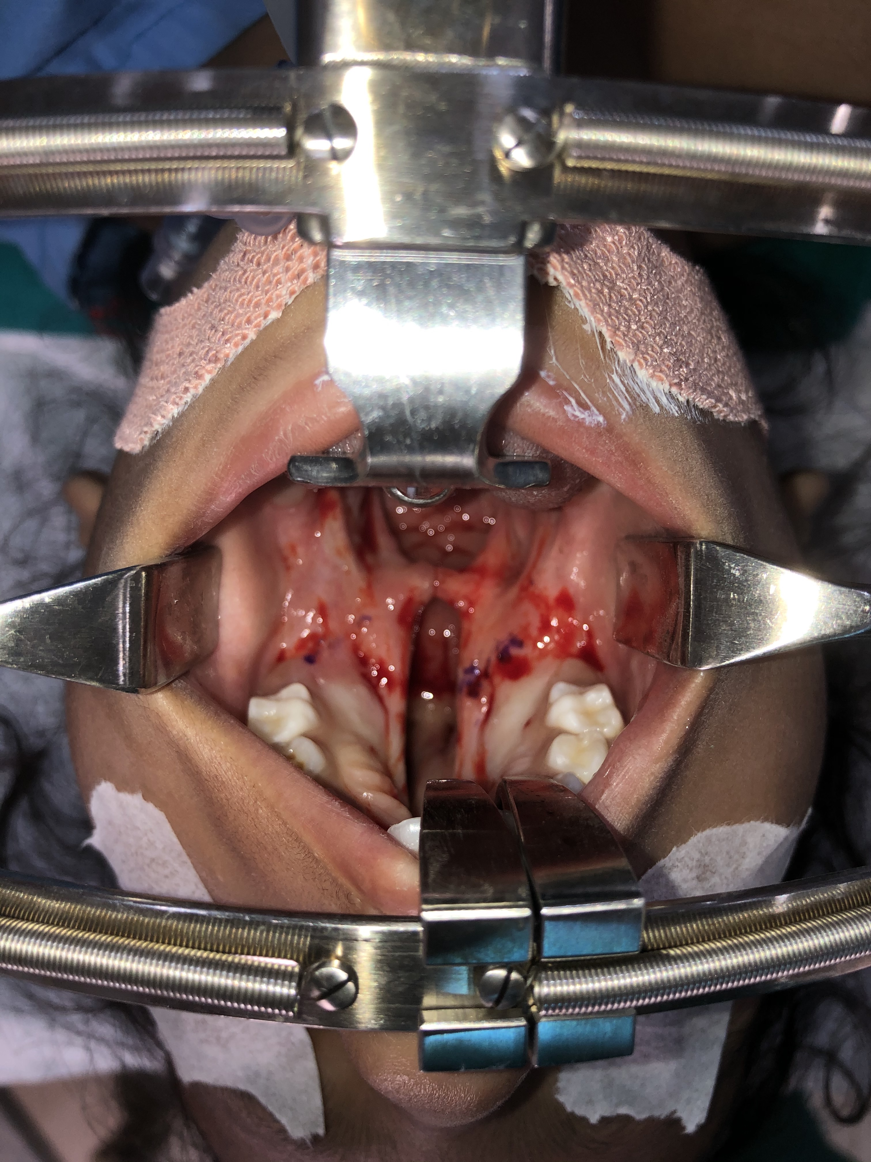
Secondary Cleft Palate Involving Hard and Soft Palate. This is an operative view of a secondary cleft palate involving the hard and soft palate.
Copyright and contributed by R Winters, MD
(Click Image to Enlarge)
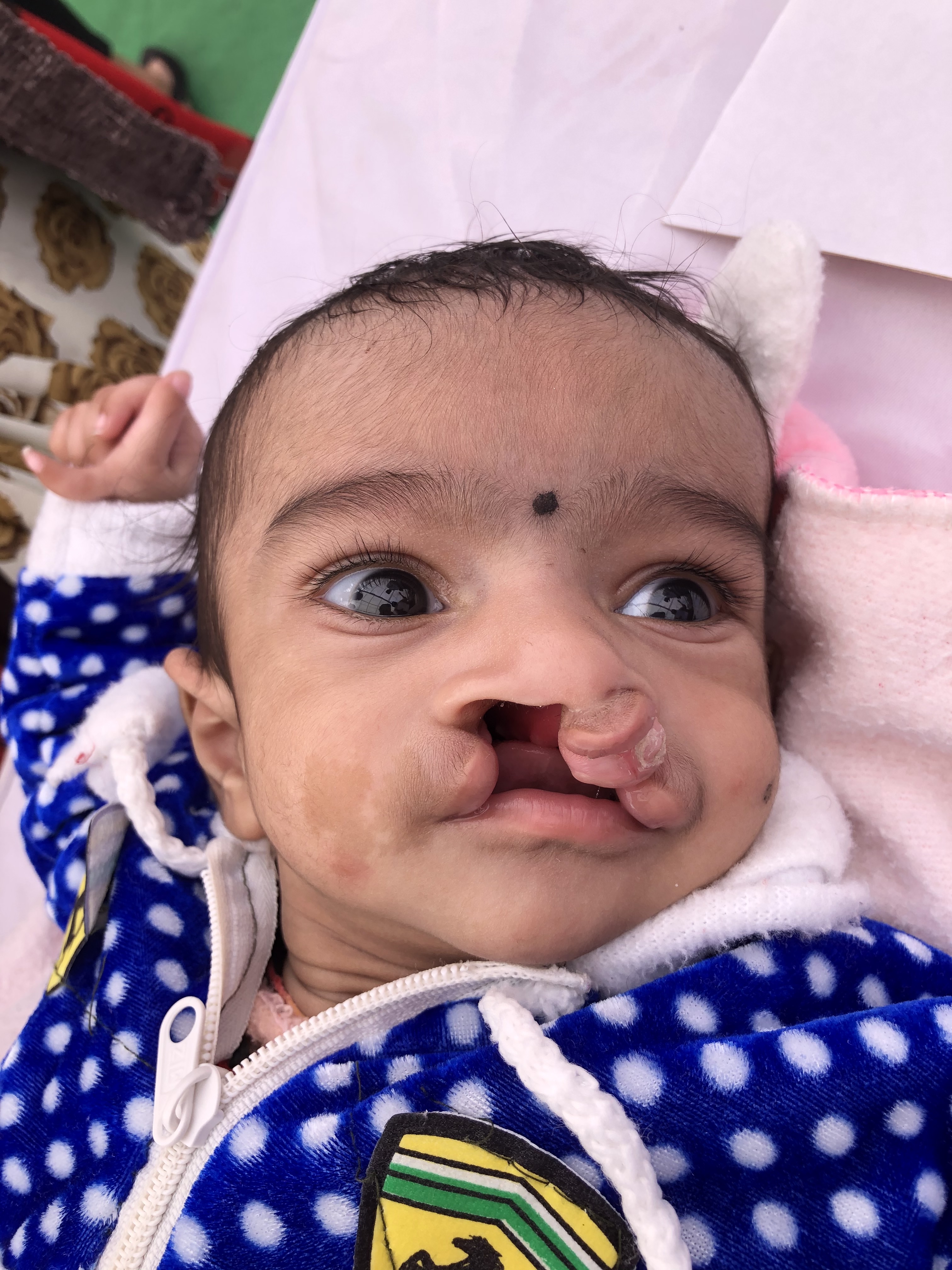
Oral Cleft. This is an image of a patient with a bilateral, complete cleft lip and palate. The condition involves the lip and primary and secondary palates. Note the protrusion of the premaxillary segment.
Copyright and contributed by R Winters, MD
(Click Image to Enlarge)
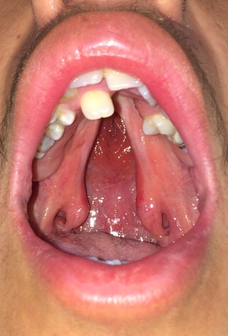
Preoperative View of Isolated Cleft Palate. This is an adolescent patient with an isolated cleft palate involving the hard and soft palate.
Copyright and contributed by J Espel, MD
(Click Image to Enlarge)
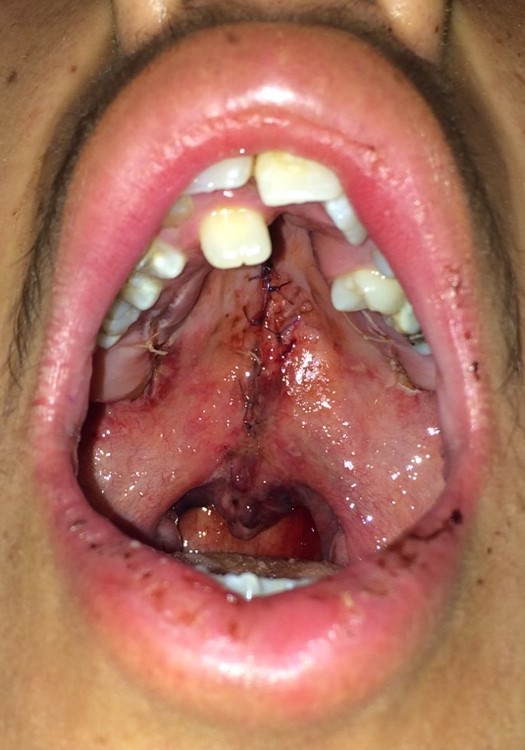
Postoperative View of Isolated Cleft Palate. This is an adolescent patient with an isolated cleft palate involving the hard and soft palate.
Copyright and contributed by J Espel, MD
(Click Image to Enlarge)
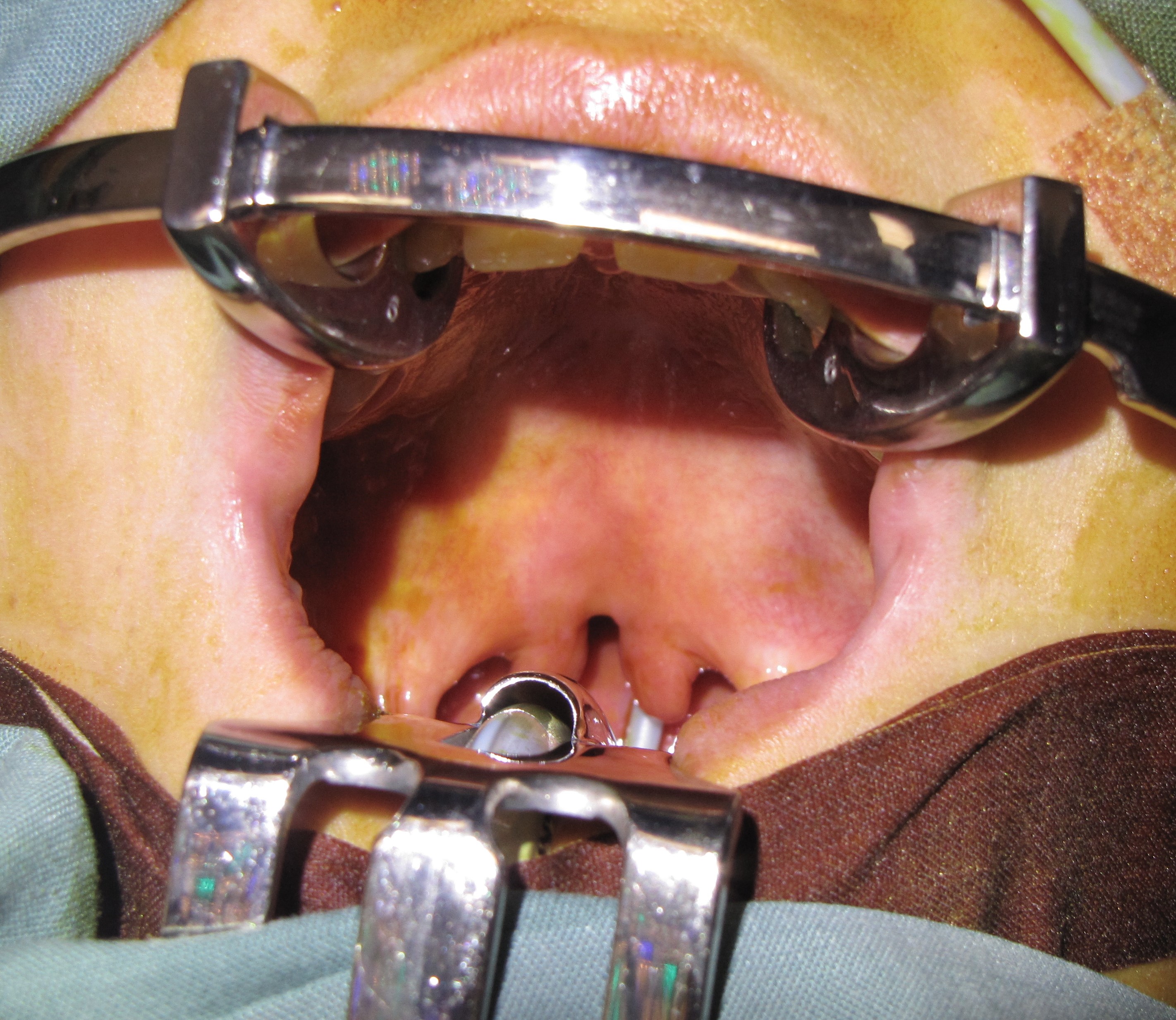
Secondary Cleft Palate of Soft Palate Only. This is an operative view of a secondary cleft palate involving the soft palate exclusively.
Copyright and contributed by J Espel, MD
(Click Image to Enlarge)
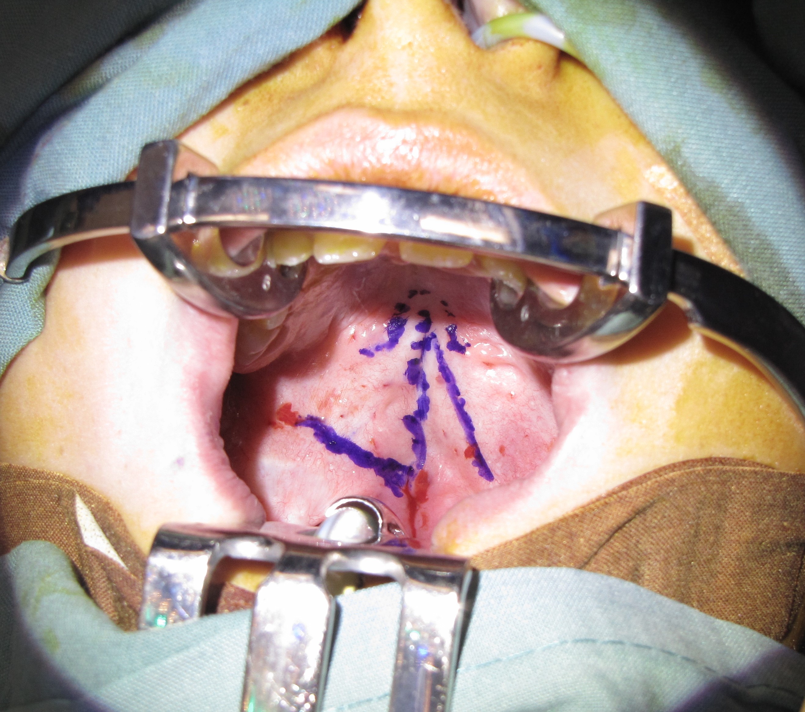
Secondary Cleft Palate of Soft Palate. Operative view of a secondary cleft palate involving the soft palate exclusively. The markings are for a Furlow double opposing Z-plasty.
Copyright and contributed by J Espel, MD
(Click Image to Enlarge)
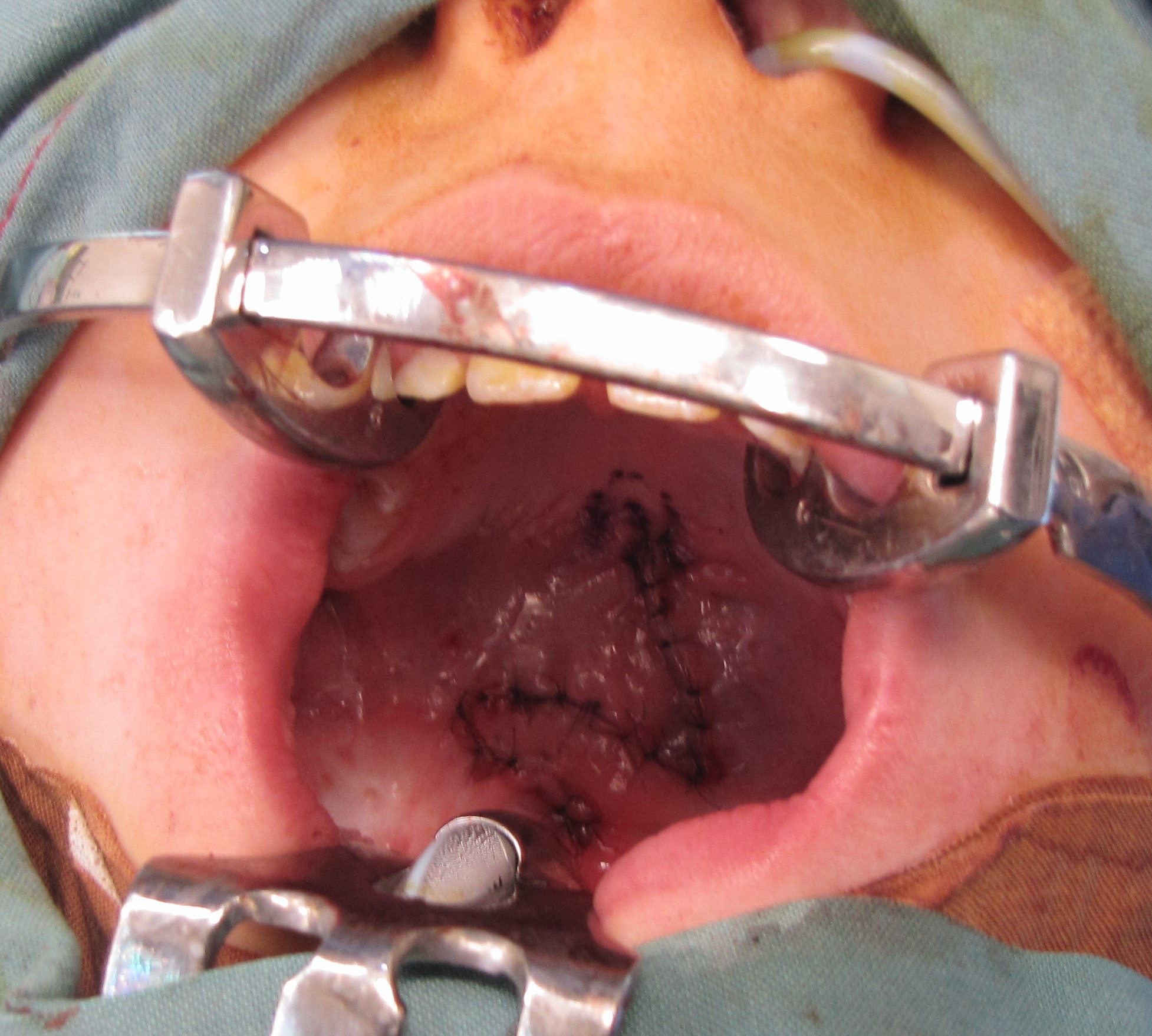
Repaired Secondary Soft Palate. This is an operative view of a secondary cleft palate involving the soft palate, after repair following Furlow double opposing Z-plasty.
Copyright and contributed by J Espel, MD
(Click Image to Enlarge)
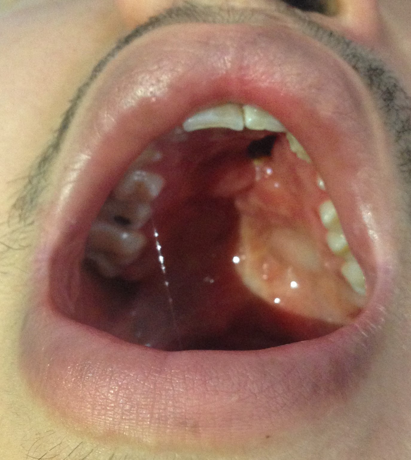
Anterior Fistula of the Hard Palate After Repair. This is an adolescent patient with an anterior fistula involving the hard palate, several years after cleft palate repair.
Copyright and contributed by J Espel, MD
(Click Image to Enlarge)
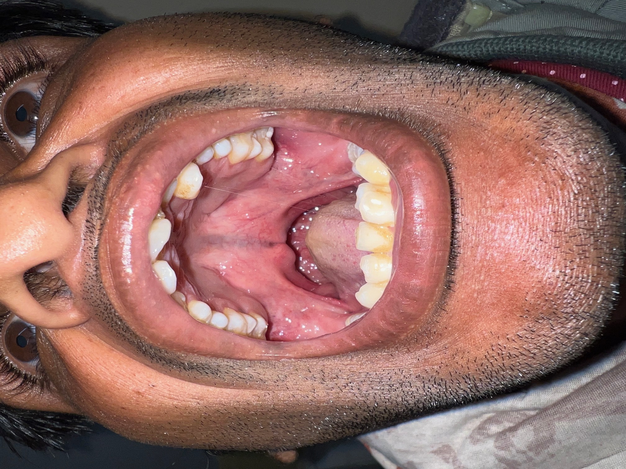
Submucous Cleft Palate. This is an image of a zona pellucida (and bifid uvula) in a submucous cleft palate.
Contributed by R Winters, MD
(Click Image to Enlarge)
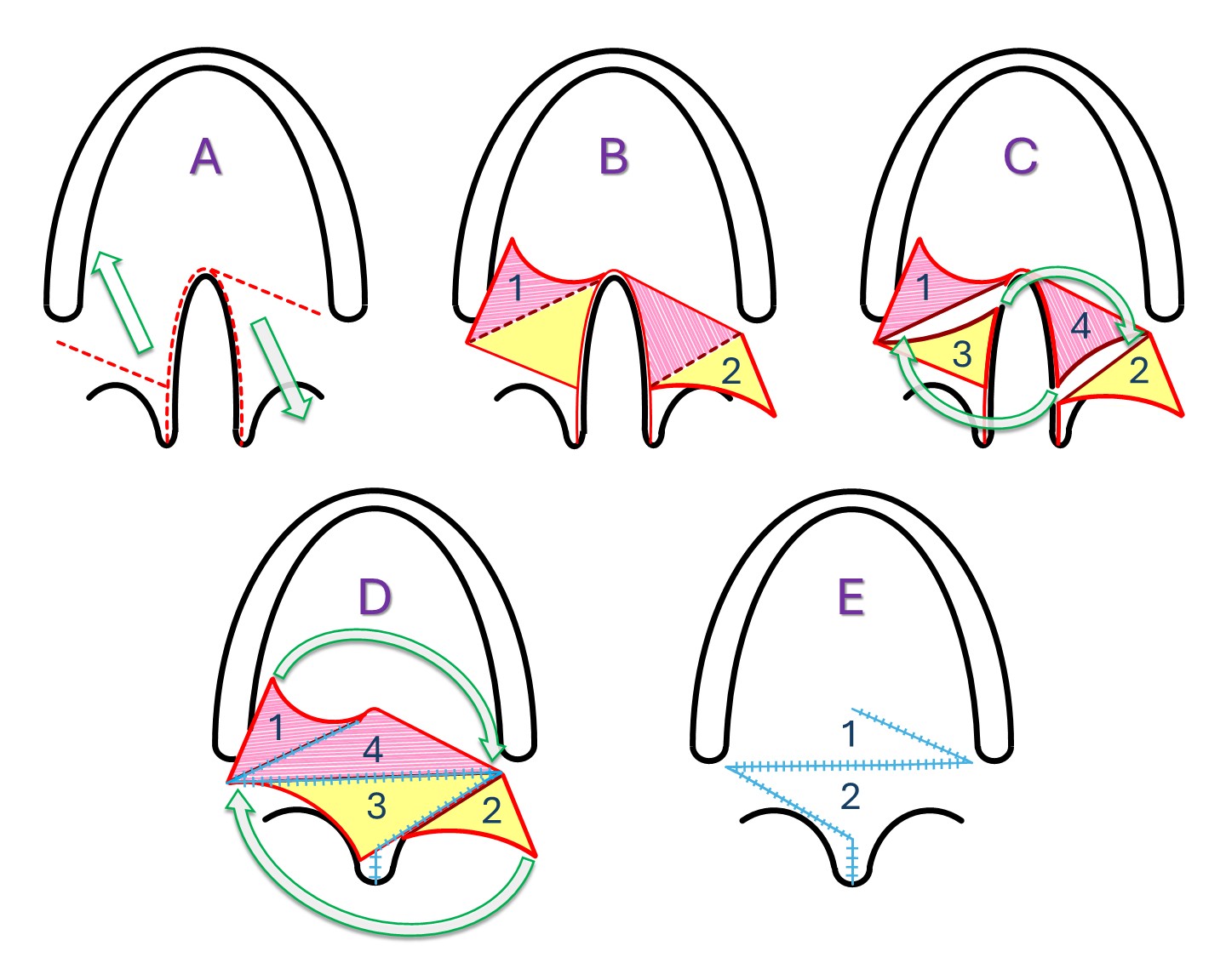
Furlow “Double-Opposing Z-Plasty." A) Incisions and vector of closure. B) The superficial flaps are raised, with the flap containing the muscle fibers (flap 1) based along the posterior border of the hard palate and the other containing only mucosa and submucosa (flap 2). C) Incisions are made for deep flaps, keeping the muscle contained within the flap based on the posterior border of the hard palate (flap 4). D) The deep flaps (flaps 3 and 4) are transposed as a Z-plasty, reconstructing the nasal surface of the soft palate and reorienting the muscle fibers in flap 4 to course transversely, parallel to the posterior border of the hard palate, thus recreating part of the muscular sling. The soft palate is also lengthened in an anteroposterior direction, and the uvula is reapproximated. E) Palatoplasty finished with flap 1 overlying flap 4 and completing the muscular sling.
Contributed by MH Hohman, MD, FACS
(Click Image to Enlarge)
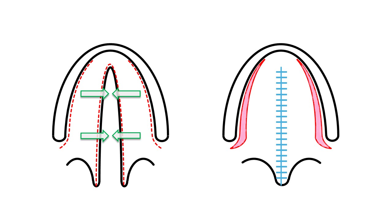
Von Langenbeck Cleft Palate Repair. Left: incisions and vector of closure. Note that the flaps remain pedicled anteriorly and posteriorly. Right: palatoplasty finished. The pink areas represent exposed bone; they may be sutured closed, or they may be left to heal by secondary intention if the cleft is wide and closure of the lateral defects will require excessive tension.
Contributed by MH Hohman, MD, FACS
(Click Image to Enlarge)
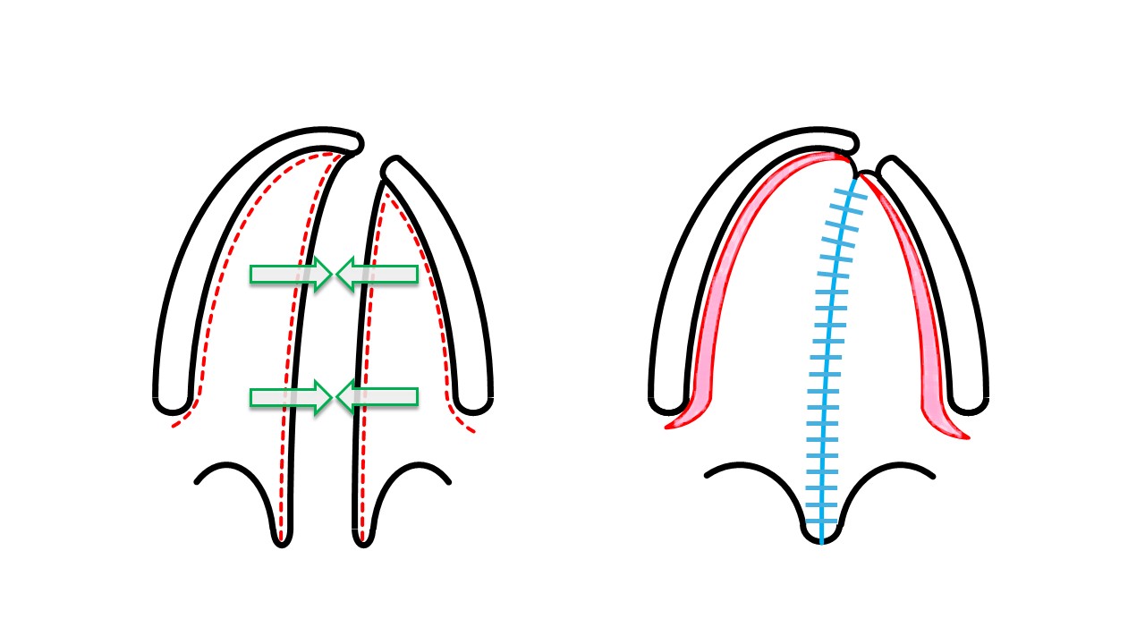
Bardach Cleft Palate Repair. This technique may be employed for wider clefts than are appropriate for the von Langenbeck approach or for complete clefts. Left: incisions and vector of closure. Note that the flaps remain pedicled only posteriorly, dependent upon perfusion from the greater palatine vessels. Right: palatoplasty complete. The pink areas represent exposed bone; they may be sutured closed, or they may be left to heal by secondary intention, if the cleft is wide and closure of the lateral defects will require excessive tension.
Contributed by MH Hohman, MD, FACS
(Click Image to Enlarge)
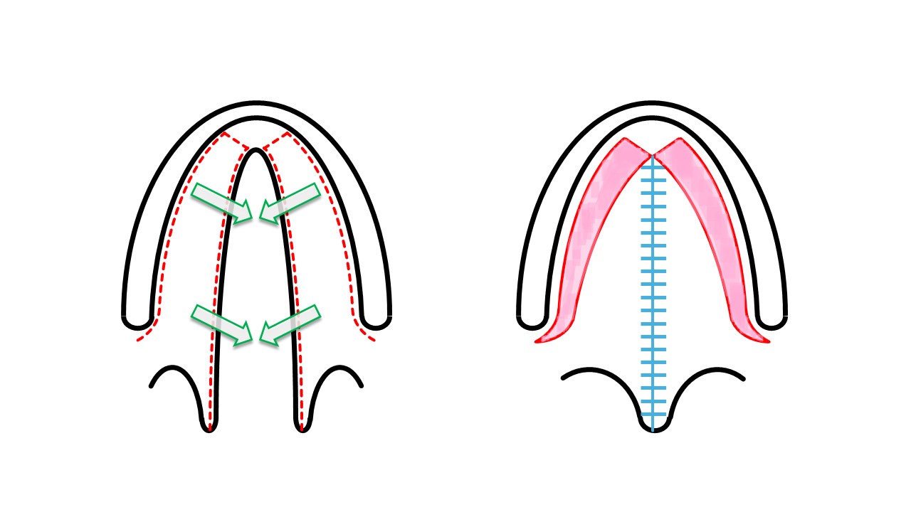
Veau-Wardill-Kilner “V-to-Y Pushback” Cleft Palate Repair. The V-to-Y advancement permits lengthening of the palate at the expense of a higher rate of fistula formation near the incisive foramen. Left: incisions and vector of closure. Note that the flaps remain pedicled only posteriorly, dependent upon perfusion from the greater palatine vessels. Right: palatoplasty complete. The pink areas represent exposed bone; they may be sutured closed, or they may be left to heal by secondary intention, if the cleft is wide and closure of the lateral defects will require excessive tension.
Contributed by MH Hohman, MD, FACS
(Click Image to Enlarge)
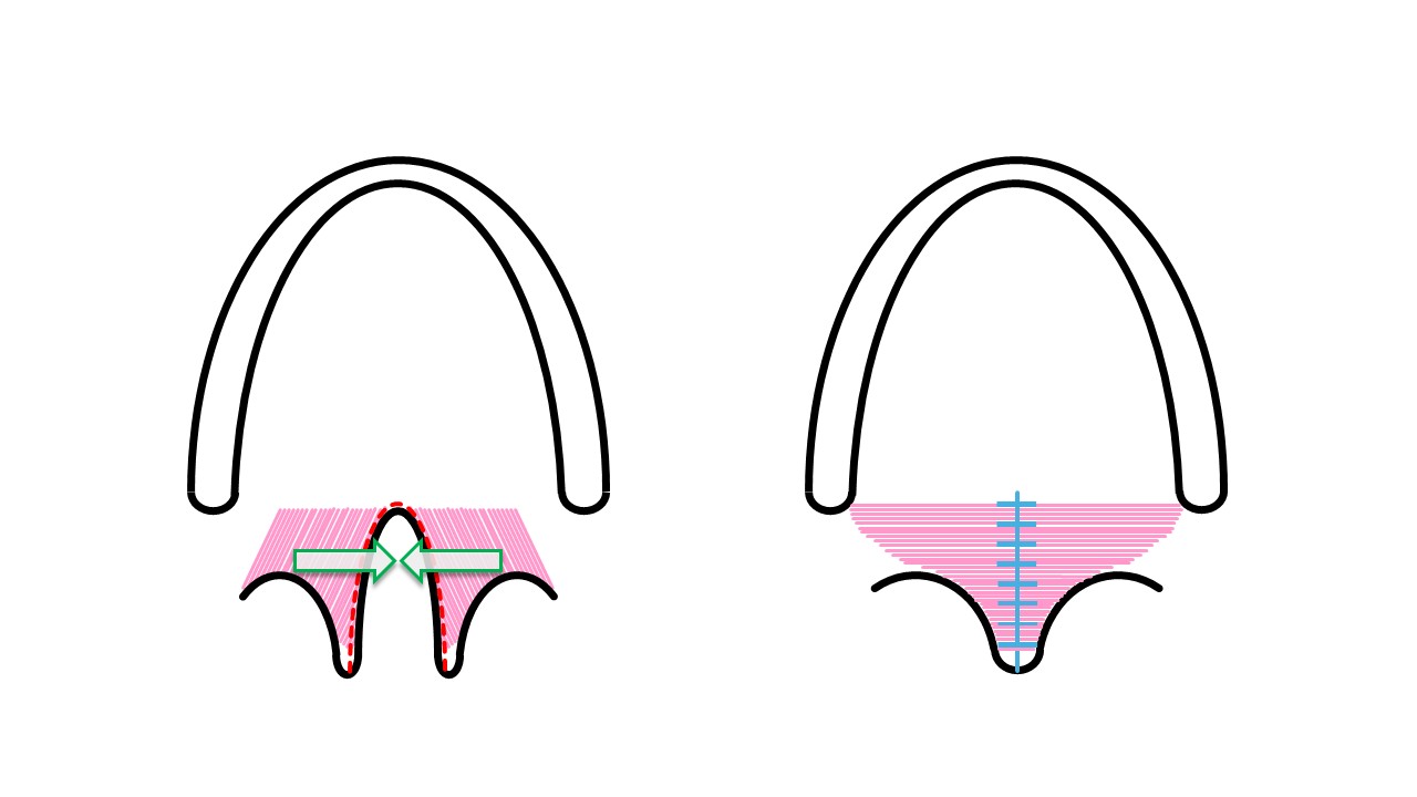
Sommerlad Repair of Soft Palate Clefts and Muscular Sling Reconstruction. Note the change in muscle fiber orientation at the conclusion of the operation. This technique may be combined with other techniques to repair longer clefts while also reconstructing the muscular soft palate sling. Left: incisions and vector of closure. The levator veli palatini muscle is dissected free from the overlying mucosa and closed as a deep layer in its new position. Right: palatoplasty complete.
Contributed by MH Hohman, MD, FACS
(Click Image to Enlarge)
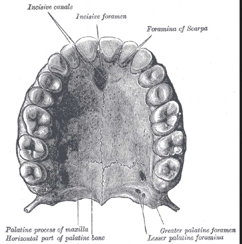
Anatomy of Upper Palate. Palatine process, palatine foramen, incisive canals, incisive foramen, and foramina of scarpa are shown.
Henry Vandyke Carter, Public Domain, via Wikimedia Commons
References
Burg ML, Chai Y, Yao CA, Magee W 3rd, Figueiredo JC. Epidemiology, Etiology, and Treatment of Isolated Cleft Palate. Frontiers in physiology. 2016:7():67. doi: 10.3389/fphys.2016.00067. Epub 2016 Mar 1 [PubMed PMID: 26973535]
Acharya P, Sahana BA, Desai A, Jalisatgi RR, Kumar N. Pre-operative and post-operative audiological assessment in cleft lip and palate patients-a prospective study. Journal of oral biology and craniofacial research. 2021 Apr-Jun:11(2):321-329. doi: 10.1016/j.jobcr.2021.03.003. Epub 2021 Mar 17 [PubMed PMID: 33786295]
Sommerlad BC. A technique for cleft palate repair. Plastic and reconstructive surgery. 2003 Nov:112(6):1542-8 [PubMed PMID: 14578783]
Zajac DJ, Vallino LD, Baylis AL, Adatorwovor R, Preisser JS, Vivaldi D. Emergence of Prevocalic Stop Consonants in Children With Repaired Cleft Palate. Journal of speech, language, and hearing research : JSLHR. 2021 Jan 14:64(1):30-39. doi: 10.1044/2020_JSLHR-20-00282. Epub 2020 Dec 2 [PubMed PMID: 33444099]
Narayanraopeta S, Vemisetty HK, Marri T, Konda P. Rehabilitation of a Unilateral Cleft Palate with Endosseous Implants in an Edentulous Elderly Patient. Contemporary clinical dentistry. 2020 Jul-Sep:11(3):285-289. doi: 10.4103/ccd.ccd_193_19. Epub 2020 Nov 26 [PubMed PMID: 33776358]
MUSGRAVE RH, BREMNER JC. Complications of cleft palate surgery. Plastic and reconstructive surgery and the transplantation bulletin. 1960 Aug:26():180-9 [PubMed PMID: 14425494]
Dhillon RS. The middle ear in cleft palate children pre and post palatal closure. Journal of the Royal Society of Medicine. 1988 Dec:81(12):710-3 [PubMed PMID: 3065499]
Level 1 (high-level) evidenceKaranth TK, Whittemore KR. Middle-ear disease in children with cleft palate. Auris, nasus, larynx. 2018 Dec:45(6):1143-1151. doi: 10.1016/j.anl.2018.04.012. Epub [PubMed PMID: 29764722]
Grant HR, Quiney RE, Mercer DM, Lodge S. Cleft palate and glue ear. Archives of disease in childhood. 1988 Feb:63(2):176-9 [PubMed PMID: 3348665]
Level 1 (high-level) evidenceGoldwyn RM. Bernhard Von Langenbeck. His life and legacy. Plastic and reconstructive surgery. 1969 Sep:44(3):248-54 [PubMed PMID: 4895616]
Leow AM, Lo LJ. Palatoplasty: evolution and controversies. Chang Gung medical journal. 2008 Jul-Aug:31(4):335-45 [PubMed PMID: 18935791]
Stewart TL, Fisher DM, Olson JL. Modified Von Langenbeck cleft palate repair using an anterior triangular flap: decreased incidence of anterior oronasal fistulas. The Cleft palate-craniofacial journal : official publication of the American Cleft Palate-Craniofacial Association. 2009 May:46(3):299-304. doi: 10.1597/07-185.1. Epub 2008 Oct 22 [PubMed PMID: 19642748]
Level 2 (mid-level) evidenceWallace AF. A history of the repair of cleft lip and palate in Britain before World War II. Annals of plastic surgery. 1987 Sep:19(3):266-75 [PubMed PMID: 3310812]
Semb G, Shaw WC. Facial growth after different methods of surgical intervention in patients with cleft lip and palate. Acta odontologica Scandinavica. 1998 Dec:56(6):352-5 [PubMed PMID: 10066115]
Furlow LT Jr. Cleft palate repair by double opposing Z-plasty. Plastic and reconstructive surgery. 1986 Dec:78(6):724-38 [PubMed PMID: 3786527]
Zito PM, Jawad BA, Hohman MH, Mazzoni T. Z-Plasty. StatPearls. 2026 Jan:(): [PubMed PMID: 29939552]
Moores C, Shah A, Steinbacher DM. Cleft Palate Repair Using a Double Opposing Z-Plasty. The Journal of craniofacial surgery. 2016 Jul:27(5):e444-5. doi: 10.1097/SCS.0000000000002751. Epub [PubMed PMID: 27315321]
Sakran KA, Yin J, Yang R, Elayah SA, Alkebsi K, Zhang S, Wang Y, Shi B, Huang H. Early Cleft Palate Repair by a Modified Technique Without Relaxing Incisions. The Cleft palate-craniofacial journal : official publication of the American Cleft Palate-Craniofacial Association. 2024 Apr:61(4):646-653. doi: 10.1177/10556656221135288. Epub 2022 Oct 26 [PubMed PMID: 36300250]
Dorf DS, Curtin JW. Early cleft palate repair and speech outcome. Plastic and reconstructive surgery. 1982 Jul:70(1):74-81 [PubMed PMID: 7089110]
Ysunza A, Pamplona MC, Mendoza M, García-Velasco M, Aguilar MP, Guerrero ME. Speech outcome and maxillary growth in patients with unilateral complete cleft lip/palate operated on at 6 versus 12 months of age. Plastic and reconstructive surgery. 1998 Sep:102(3):675-9 [PubMed PMID: 9727430]
Stanton EW, Rochlin D, Lorenz HP, Sheckter CC. Early Cleft Palate Repair is Associated With Lower Incidence of Velopharyngeal Insufficiency Surgery. The Journal of craniofacial surgery. 2025 May 1:36(3):781-785. doi: 10.1097/SCS.0000000000010540. Epub 2024 Aug 26 [PubMed PMID: 39178397]
Semb G. A study of facial growth in patients with unilateral cleft lip and palate treated by the Oslo CLP Team. The Cleft palate-craniofacial journal : official publication of the American Cleft Palate-Craniofacial Association. 1991 Jan:28(1):1-21; discussion 46-8 [PubMed PMID: 2004087]
Martin-Smith JD, Fitzgerald L, Orr DJA. How reliable is the vomer flap in early hard palate repair? Journal of plastic, reconstructive & aesthetic surgery : JPRAS. 2017 Jun:70(6):828-832. doi: 10.1016/j.bjps.2017.02.026. Epub 2017 Mar 10 [PubMed PMID: 28343783]
Bergland O, Semb G, Abyholm FE. Elimination of the residual alveolar cleft by secondary bone grafting and subsequent orthodontic treatment. The Cleft palate journal. 1986 Jul:23(3):175-205 [PubMed PMID: 3524905]
Kantar RS, Rifkin WJ, Cammarata MJ, Maliha SG, Diaz-Siso JR, Farber SJ, Flores RL. Combined Primary Cleft Lip and Palate Repair: Is It Safe? The Journal of craniofacial surgery. 2019 Mar/Apr:30(2):384-389. doi: 10.1097/SCS.0000000000005039. Epub [PubMed PMID: 30531281]
Holzmer S, Davila A, Martin MC. Cost Utility Analysis of Staged Versus Single-Stage Cleft Lip and Palate Repair. Annals of plastic surgery. 2020 May:84(5S Suppl 4):S300-S306. doi: 10.1097/SAP.0000000000002255. Epub [PubMed PMID: 32049761]
Kriens OB. An anatomical approach to veloplasty. Plastic and reconstructive surgery. 1969 Jan:43(1):29-41 [PubMed PMID: 5765081]
Holmström H, Stenborg R, Blomqvist G. Elongated pharyngeal flap in extensive clefts of the hard and soft palate. The Cleft palate journal. 1986 Jan:23(1):41-7 [PubMed PMID: 3455902]
Fujiwara K, Tsuno H, Okabe M, Yoshida T, Imaue S, Tomihara K, Arai N, Noguchi M. Clinical Application of Hyperdry Amniotic Membrane in Cleft Palate Repair. The Cleft palate-craniofacial journal : official publication of the American Cleft Palate-Craniofacial Association. 2023 Jun:60(6):701-705. doi: 10.1177/10556656221075937. Epub 2022 Feb 28 [PubMed PMID: 35225019]
Aldekhayel SA, Sinno H, Gilardino MS. Acellular dermal matrix in cleft palate repair: an evidence-based review. Plastic and reconstructive surgery. 2012 Jul:130(1):177-182. doi: 10.1097/PRS.0b013e318254b2dc. Epub [PubMed PMID: 22743882]
Boot M, Winters R. Managing massive palatial defect secondary to palatoplasty failures: an in-depth analysis. Current opinion in otolaryngology & head and neck surgery. 2024 Aug 1:32(4):269-277. doi: 10.1097/MOO.0000000000000968. Epub 2024 Feb 23 [PubMed PMID: 38393699]
Level 3 (low-level) evidenceLilja J. Alveolar bone grafting. Indian journal of plastic surgery : official publication of the Association of Plastic Surgeons of India. 2009 Oct:42 Suppl(Suppl):S110-5 [PubMed PMID: 19884665]
Wijnants N, Widdershoven JCC, van Boxel SCJ, van Kuijk SMJ, Brunings JW, Booi DI, van der Hulst RRWJ, Schols RM. Impact of Tympanostomy Tube Placement on Hearing Outcomes in Children With Cleft Palate-Related Otitis Media: Retrospective Analysis of a Primary Single-Stage Cleft Palate Repair Cohort. Ear, nose, & throat journal. 2025 Sep 25:():1455613251376792. doi: 10.1177/01455613251376792. Epub 2025 Sep 25 [PubMed PMID: 40995937]
Level 2 (mid-level) evidenceBell C, Oh TH, Loeffler JR. Massive macroglossia and airway obstruction after cleft palate repair. Anesthesia and analgesia. 1988 Jan:67(1):71-4 [PubMed PMID: 3337349]
Level 3 (low-level) evidenceChan MT, Chan MS, Mui KS, Ho BP. Massive lingual swelling following palatoplasty. An unusual cause of upper airway obstruction. Anaesthesia. 1995 Jan:50(1):30-4 [PubMed PMID: 7702141]
Level 3 (low-level) evidenceGallagher S, Ferrera A, Spera L, Eppley BL, Soleimani T, Tahiri Y, Sood R, Flores RL, Wooden WA, Tholpady SS. Utility of Tongue Stitch and Nasal Trumpet in the Immediate Postoperative Outcome of Cleft Palatoplasty. Plastic and reconstructive surgery. 2016 Feb:137(2):569-573. doi: 10.1097/01.prs.0000475786.60069.a3. Epub [PubMed PMID: 26818292]
Ali AAA, Hasan MHO. Outcomes of Facial Artery Musculomucosal Flap in Repair of Post-palatoplasty Fistula - A Retrospective Study. Annals of maxillofacial surgery. 2024 Jul-Dec:14(2):180-186. doi: 10.4103/ams.ams_123_24. Epub 2024 Dec 12 [PubMed PMID: 39957871]
Level 2 (mid-level) evidenceMeneses Argalle JD, Espinosa Orozco AM, Prada Madrid JR. Tongue Flap for Closure of Complex Oronasal Fistula. The Journal of craniofacial surgery. 2023 Sep 1:34(6):1872-1875. doi: 10.1097/SCS.0000000000009468. Epub 2023 Jun 22 [PubMed PMID: 37344931]
Smith DM, Vecchione L, Jiang S, Ford M, Deleyiannis FW, Haralam MA, Naran S, Worrall CI, Dudas JR, Afifi AM, Marazita ML, Losee JE. The Pittsburgh Fistula Classification System: a standardized scheme for the description of palatal fistulas. The Cleft palate-craniofacial journal : official publication of the American Cleft Palate-Craniofacial Association. 2007 Nov:44(6):590-4. doi: 10.1597/06-204.1. Epub [PubMed PMID: 18177198]
Level 2 (mid-level) evidence