 Tools
Tools
 Intestinal Ultrasonography: Technique, Indications, and Clinical Applications
Introduction
Intestinal or bowel ultrasonography is a noninvasive sonographic examination of the gastrointestinal tract. This modality serves as a complementary examination to standard abdominal ultrasonography, providing clinical data that can be beneficial for patients who are unable to undergo CT or MRI examinations for various reasons.[1] Intestinal ultrasonography also provides a portable option for patients who are immobile.[2]
As a hollow organ, the gastrointestinal tract contains air and fluid. While fluid is considered a good acoustic medium, air is a poor acoustic medium, typically impairing ultrasonographic imaging. Thus, patient preparation often improves the diagnostic quality of ultrasonographic evaluation of the small and large bowel. Bowel ultrasonography can be used in the detection, diagnosis, and follow-up of various gastrointestinal conditions. Ultrasonography does not involve radiation and is, therefore, safe for pregnant women and children and for patients who require repeated radiological examinations. Operator technique and time allocation may pose limitations to this examination.[3][4]
Anatomy and Physiology
Register For Free And Read The Full Article
 Search engine and full access to all medical articles
- 10 free questions in your specialty
- Free CME/CE Activities
- Free daily question in your email
- Save favorite articles to your dashboard
- Emails offering discounts
Learn more about a Subscription to StatPearls Point-of-Care
Anatomy and Physiology
Normal bowel anatomy determined by ultrasonography differs from that seen with other radiological techniques and from histological appearance under light microscopy. Multiple diagnostic scoring systems are available to detect gastrointestinal diseases.[5]
- Normal bowel wall appearance and thickness: The normal bowel wall consists of 5 layers, each differing in echogenicity, allowing some degree of distinction among these layers. Normal bowel diameter varies by intestinal segment. Bowel wall thickness is the most important feature in intestinal ultrasonographic assessment and is an important parameter for detecting intestinal disease. Thickness is determined by measuring all wall layers from the lumen interface to the serosa. Typically, a wall thickness of 3 to 4 mm is considered normal, except for the gastric wall, which may measure up to 5 to 6 mm.[6]
- Normal bowel diameter: Pathology of the small and large bowel can manifest as an obstructive or paralytic process, causing bowel dilation. Consequently, bowel loop diameter can help assess pathology. As a rule of thumb, the small intestine diameter should not exceed 2.5 to 3 cm. Similarly, the large intestine typically does not exceed 5 cm in diameter. Please see StatPearls' companion reference, "Small Bowel Obstruction," for further information.
- Echo pattern: Although the arrangement of wall layers does not correlate exactly with histological layers, this stratification remains important for identifying diseases by detecting loss of stratification.
- The lumen of the gut interface: hyperechoic
- The mucosa: hypoechoic
- The submucosa: hyperechoic
- The muscularis propria: hypoechoic
- The serosa: hyperechoic
- Vascularity: Determining abnormal vascularity, such as hyperemia or neovascularization, is important for diagnosing intestinal diseases, including edema, inflammation, and malignant neoplasms. Clinicians usually assess vascularity with color Doppler in addition to grayscale ultrasonographic evaluation.[7] Another critical parameter in sonographic bowel evaluation is assessing the appearance of large vessels supplying the gastrointestinal tract, including the superior mesenteric artery, inferior mesenteric artery, and celiac trunk. Evaluating the superior and inferior mesenteric veins, as well as the portal and splenic veins, can also provide important diagnostic information.
- Motility: Motility is a subjective measure of bowel health and is operator-dependent. Peristalsis is limited in cases of bowel inflammation or fibrous strictures. Increased peristalsis is present in diarrhea, celiac disease, and intestinal obstruction, among other causes.[8]
- Compressibility: Noncompressible bowel could indicate an inflammatory or malignant change in the bowel wall, because infiltrative neoplasms result in the loss of normal bowel wall pliability.[9]
Other important signs that could be found on intestinal ultrasound and are an indication of the underlying disease:
- Mesenteric fat: Mainly assessed from the epigastrium to the right iliac fossa. Increased thickness >6 mm on occasion may be associated with an abnormality, such as an inflammatory process, such as diverticulitis.
- Extraluminal gas: This usually indicates luminal perforation at the site of pathology.
Different Scoring Systems in Intestinal Ultrasonography
The use of regular ultrasonography or bubble contrast (contrast-enhanced ultrasonography) is common in Europe and North America.[10] Different scoring systems have been evaluated, but no universal scoring system has been proven effective to date. Most scoring systems include parameters such as bowel wall thickness, wall stratification, detection of fistula formation, ascites, mesenteric fat, lymph nodes, compressibility, peristalsis, and inflammatory signs in the bowel wall on color Doppler.[2][11] A simple ultrasonographic score for monitoring Crohn disease was validated in clinical studies. Results from the studies indicated that the score could be used during follow-up instead of endoscopy for patients with Crohn disease.[12]
The Difference in Wall Thickness in Children with Crohn Disease
The normal bowel thickness in children is typically less than 2 mm, similar to that of adults. Intestinal ultrasonography may be helpful in the detection and follow-up of Crohn disease in children.[13]
Indications
Intestinal ultrasonography is a noninvasive procedure that could be used as an adjunct to more frequently performed abdominal ultrasonography. Some clinicians can use intestinal ultrasonography as the initial examination of choice, depending on operator comfort with ultrasonography technique and image interpretation.[14][15][16]
Intestinal Ultrasonography Could Assist in Evaluating the Following Pathologies
Acute abdomen:
- Acute appendicitis: Different compression techniques are used to assess for it. Diagnostic signs on ultrasonography are a noncompressible, enlarged, blind-ending tubular structure representing an inflamed appendix in the right iliac fossa (see Image. Appendix in Longitudinal Plane with Fecolith).[17]
- Acute diverticulitis: Ultrasonography can demonstrate bowel wall thickening, diverticulosis, and foci of varying echogenicity, as well as hyperechoic pericolic inflammatory fat.[18] Perforation can manifest as foci of dirty shadowing representing gas.
- Ischemic colitis: An increase in the colon wall thickness greater than 5 mm, typically involving the left colon, with loss of bowel wall stratification may be seen. Absent or markedly increased vascular flow on color Doppler can also be demonstrated.[19]
- Intestinal obstruction: Intestinal ultrasonography is not the most conclusive method for diagnosing intestinal obstruction because gaseous distention associated with obstruction can be misleading. However, if small bowel loops are distended with fluid, sonography can assess this finding to some degree. Rarely, the underlying cause of obstruction, such as a large mass, can be visualized. The small bowel diameter is typically greater than 2.5 cm in the setting of obstruction, whereas the length of the obstructed segment typically exceeds 10 cm, with affected bowel loops distended with fluid or debris (see Images. Small Bowel Obstruction on Ultrasound and Abdomen Ultrasound, Intussuception Appendicolith).[20]
- Enlargement of mesenteric lymph nodes: While paraaortic lymph nodes are typically too deep for adequate visualization, mesenteric lymph nodes can often be seen.[21] Examples include enlarged mesenteric lymph nodes adjacent to a thickened ileum in Crohn disease-related inflammation, or generalized adenopathy, such as sclerosing mesenteritis.
Chronic Diseases or Nonurgent Conditions
Crohn disease: Ultrasonography has a limited role in evaluating Crohn disease, but can be used as a screening or follow-up modality to assess complications. Sonographic findings can often be nonspecific. Findings include loss of peristalsis, mural hyperemia of the affected bowel, a hyperechoic layer surrounding the bowel wall representing fibrofatty proliferation, and small bowel wall thickening. Additional findings include a loss of compressibility and bowel wall fibrosis, which can mimic normal bowel wall submucosa. Other nonspecific findings include mesenteric lymphadenopathy and intraperitoneal fluid.[22] Complications of Crohn disease can also be seen, including abscess formation and fistula formation.[23]
Ulcerative colitis: Ultrasonography can demonstrate increased bowel wall thickness of the rectosigmoid colon, typically greater than 4 mm in patients with ulcerative colitis. Loss of wall haustrations, wall stratification, hyperemia on power color Doppler, and enlarged mesenteric fat can also be seen.[24]
Infectious diseases: Infectious diseases include bacterial enteritis, tuberculous enteritis, pseudomembranous colitis, amebic or parasitic enteritis, and ascariasis. Depending on the bacterial or parasitic organism, findings can range from nonspecific wall thickening secondary to inflammation to localized fluid collections, such as in a hydatid cyst. In rare cases, the actual parasite can be visualized, such as in the setting of ascariasis.[25]
Colorectal and gastric cancers: Malignant neoplasms can present in various shapes, from a focal endophytic or occasionally exophytic mass to infiltrative wall thickening, either circumferential or involving limited portions of the wall circumference.[26]
Peritoneal metastasis: Peritoneal metastasis can often manifest as multifocal masses adherent to the bowel, with increased vascularity. An associated complex ascitic fluid can also be seen on occasion.[27]
Appendiceal mucoceles: Appendiceal mucoceles are often well visualized on ultrasonography, especially when large, and typically appear as a unilocular mass. Appendiceal mucoceles have been described as having a whipped cream appearance.[28]
Contraindications
No contraindications exist to this noninvasive procedure. When appropriate, such as in the setting of acute pain, CT should be prioritized over sonographic bowel imaging when available and clinically appropriate.
Equipment
During bowel ultrasonography, the sonographer uses the same equipment as standard abdominal ultrasonography but requires more experience to detect abnormalities in the intestinal wall. Bowel screening can start with the common convex low-frequency probe (3.5 to 5 MHz) used in a standard abdominal ultrasonography examination. Detailed visualization of the bowel can be performed using a linear high-frequency (4 to 13 MHz) probe, which has high resolution.[8] When possible, tissue harmonic imaging should be used because it can provide additional diagnostic information regarding the bowel wall, lumen, and fluid content.
Personnel
Intestinal ultrasonography is typically performed by:
- Sonographers
- Radiologists
- Gastroenterologists
- Emergency medicine clinicians with specialized ultrasonography training
Findings from a recent case report suggested that, on an individual basis, a patient could perform self-monitoring using a portable ultrasonography device to monitor severe ulcerative colitis.[29]
Preparation
The patient should be fasting for at least 8 to 10 hours. Food can cause gaseous distention, making bowel visualization difficult. Ingestion of a large amount of fluid could fill the bowel with water and mimic intestinal obstruction. Therefore, patients should ideally fast for better intestinal visualization. Although fasting is preferable, fasting is not mandatory when evaluating an acute abdomen.
Technique or Treatment
The Sequence of Intestinal Ultrasonography Examination
In chronic conditions, such as ulcerative colitis, Crohn disease, and celiac disease, the examiner should preferably start from the epigastric region or the left iliac or sigmoid quadrant and then proceed to examine the rest of the intestine and colon, including the terminal ileum and appendix in the right iliac quadrant. Although sequential search patterns can vary with operator training, a consistent pattern helps prevent missed diagnoses. In acute conditions, such as an acute abdomen, the examiner should start with the abdominal quadrant the patient identifies as most tender.
Limitations
If intestinal visualization is not possible due to gaseous distention, graded compression of the bowel will help shift gaseous shadows and improve image quality. Another maneuver is to turn the patient to the right or left side, depending on the bowel segment examined, to help displace visually obstructing gas.
Complications
No complications are associated with this noninvasive procedure.
Clinical Significance
The Impact of Intestinal Ultrasonography on the Medical Care of Inflammatory Bowel Disease
Intestinal ultrasonography could be used to diagnose multiple gastrointestinal diseases (acute and chronic).[30][31][24] Inflammatory bowel diseases can be diagnosed and monitored using intestinal ultrasonography, as these patients require close monitoring and follow-up. Using a cost-effective, noninvasive, radiological-hazard-free method, such as intestinal ultrasonography, is greatly beneficial to patients.
Intestinal ultrasonography scores: Many scores are available for ulcerative colitis and Crohn disease. Most scores depend on bowel wall thickness and the Doppler signal. When these scores, such as Milan Ultrasound Criteria and Ulcerative Colitis–Intestinal Ultrasound, are applied to the pediatric population, they correlate significantly with endoscopic scores.[32][33]
Results from a recent systematic review included 51 studies. Bowel wall thickness, with a cutoff of 3 mm, and Limberg score, with 0 or 1 considered normal, are considered the standard in 53% of studies, with only 16% using intestinal ultrasonography scores. These scores included 2 Crohn disease studies using the Bowel Ultrasound Score, 1 Crohn disease study using the International Bowel Ultrasound Segmental Activity Score, 1 ulcerative colitis study using the Milan Ultrasound Criteria score, and 4 studies using unvalidated scores.[34]
Inflammatory bowel disease intestinal ultrasonography guidelines: Recent European Crohn’s and Colitis Organisation–European Society of Gastrointestinal and Abdominal Radiology–European Society of Pathology–International Bowel Ultrasound Group guidelines recommend the use of intestinal ultrasonography in the initial diagnosis of inflammatory bowel disease, treatment initiation, and follow-up, along with endoscopy, inflammatory markers, and radiological examination in the form of CT and magnetic resonance enterography (MRE). Some of these scores are validated. For Crohn disease, validated scores include the International Bowel Ultrasound Segmental Activity Score, Simple Ultrasound Score for Crohn Disease, and Bowel Ultrasound Score. For ulcerative colitis, validated scores include Milan Ultrasound Criteria and Ulcerative Colitis–Intestinal Ultrasound, all of which correlate significantly with endoscopic findings.[35][36]
Using artificial intelligence in intestinal ultrasonography: Intestinal ultrasonography is a very efficient point-of-care ultrasonography tool in Crohn disease and ulcerative colitis. This modality can optimize the clinical and therapeutic treatment of patients on-site without the need for more costly cross-sectional radiography, such as CT or magnetic resonance enterography. While convolutional neural networks and support vector machines are widely used in CT and MRI, they are still in their early stages in intestinal ultrasonography. These artificial intelligence models improve image classification and data extraction, reducing the tool's subjectivity. Results from 3 studies using CNNs to detect bowel wall thickness from images showed high accuracy, with an area under the curve reaching 98%.[37]
Enhancing Healthcare Team Outcomes
Intestinal ultrasonography as part of the follow-up for patients with inflammatory bowel disease, including Crohn disease and ulcerative colitis, has an important role, especially when performed by a trained operator. Intestinal ultrasonography can decrease the need for repeated CT or MRI of the abdomen. An interprofessional team approach in which family clinicians, specialists, nurses, advanced practice clinicians, and ultrasonography technologists communicate openly about examination performance and resulting findings can help the team guide patient care, leading to better outcomes.
Media
(Click Video to Play)
Small Bowel Obstruction on Ultrasound. Small bowel obstruction with dilated bowel, thick bowel wall, adjacent intraperitoneal fluid, and back-and-forth peristalsis.
Contributed by M Schick, DO, MA
(Click Image to Enlarge)
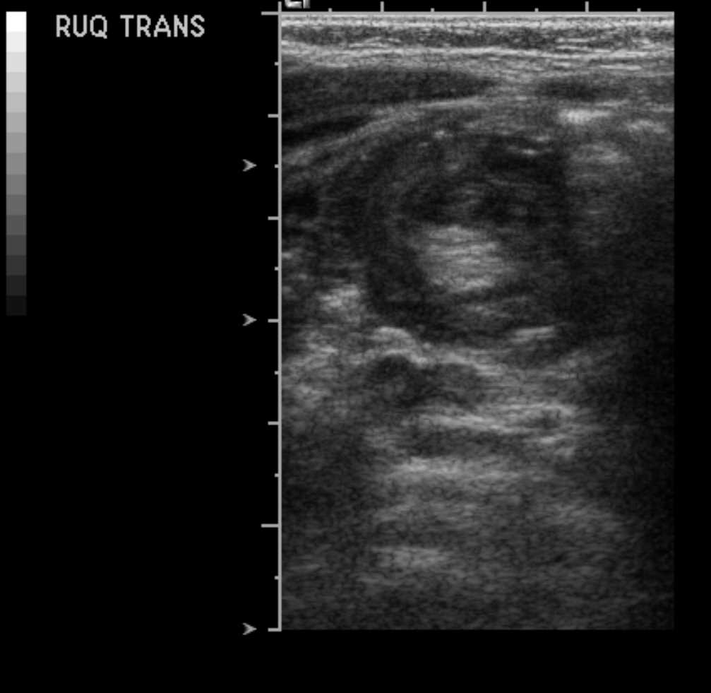
Abdomen Ultrasonography, Intussuception Appendicolith. A classic target or doughnut sign pathognomonic for intussusception, likely secondary to an appendicolith acting as a lead point.
Contributed by S Dulebohn, MD
(Click Video to Play)
Appendix in Longitudinal Plane with Fecolith. A highly echogenic fecolith (appendicolith) with characteristic posterior acoustic shadowing is a common finding in cases of acute obstructive appendicitis.
Contributed by M Lambert, MD
References
Maconi G, Terracciano F, de Sio I, Rigazio C, Roselli P, Radice E, Castellano L, Farci F, Francica G, Giannetti A, Marcucci F, Dalaiti A, Badini M, Fraquelli M, Massironi S. Referrals for bowel ultrasound in clinical practice: a survey in 12 nationwide centres in Italy. Digestive and liver disease : official journal of the Italian Society of Gastroenterology and the Italian Association for the Study of the Liver. 2011 Feb:43(2):165-8. doi: 10.1016/j.dld.2010.05.017. Epub 2010 Jul 7 [PubMed PMID: 20615765]
Level 3 (low-level) evidenceDeepak P, Kolbe AB, Fidler JL, Fletcher JG, Knudsen JM, Bruining DH. Update on Magnetic Resonance Imaging and Ultrasound Evaluation of Crohn's Disease. Gastroenterology & hepatology. 2016 Apr:12(4):226-36 [PubMed PMID: 27231453]
Parente F, Greco S, Molteni M, Cucino C, Maconi G, Sampietro GM, Danelli PG, Cristaldi M, Bianco R, Gallus S, Bianchi Porro G. Role of early ultrasound in detecting inflammatory intestinal disorders and identifying their anatomical location within the bowel. Alimentary pharmacology & therapeutics. 2003 Nov 15:18(10):1009-16 [PubMed PMID: 14616167]
Pinto J, Azevedo R, Pereira E, Caldeira A. Ultrasonography in Gastroenterology: The Need for Training. GE Portuguese journal of gastroenterology. 2018 Nov:25(6):308-316. doi: 10.1159/000487156. Epub 2018 Feb 27 [PubMed PMID: 30480048]
Atkinson NSS, Bryant RV, Dong Y, Maaser C, Kucharzik T, Maconi G, Asthana AK, Blaivas M, Goudie A, Gilja OH, Nuernberg D, Schreiber-Dietrich D, Dietrich CF. How to perform gastrointestinal ultrasound: Anatomy and normal findings. World journal of gastroenterology. 2017 Oct 14:23(38):6931-6941. doi: 10.3748/wjg.v23.i38.6931. Epub [PubMed PMID: 29097866]
Fernandes T, Oliveira MI, Castro R, Araújo B, Viamonte B, Cunha R. Bowel wall thickening at CT: simplifying the diagnosis. Insights into imaging. 2014 Apr:5(2):195-208. doi: 10.1007/s13244-013-0308-y. Epub 2014 Jan 10 [PubMed PMID: 24407923]
Drews BH, Barth TF, Hänle MM, Akinli AS, Mason RA, Muche R, Thiel R, Pauls S, Klaus J, von Boyen G, Kratzer W. Comparison of sonographically measured bowel wall vascularity, histology, and disease activity in Crohn's disease. European radiology. 2009 Jun:19(6):1379-86. doi: 10.1007/s00330-008-1290-5. Epub 2009 Jan 30 [PubMed PMID: 19184036]
Andrzejewska M, Grzymisławski M. The role of intestinal ultrasound in diagnostics of bowel diseases. Przeglad gastroenterologiczny. 2018:13(1):1-5. doi: 10.5114/pg.2018.74554. Epub 2018 Mar 26 [PubMed PMID: 29657604]
Roccarina D, Garcovich M, Ainora ME, Caracciolo G, Ponziani F, Gasbarrini A, Zocco MA. Diagnosis of bowel diseases: the role of imaging and ultrasonography. World journal of gastroenterology. 2013:19(14):2144-53. doi: 10.3748/wjg.v19.i14.2144. Epub [PubMed PMID: 23599640]
Quaia E. Contrast-enhanced ultrasound of the small bowel in Crohn's disease. Abdominal imaging. 2013 Oct:38(5):1005-13. doi: 10.1007/s00261-013-0014-8. Epub [PubMed PMID: 23728306]
Goodsall TM, Jairath V, Feagan BG, Parker CE, Nguyen TM, Guizzetti L, Asthana AK, Begun J, Christensen B, Friedman AB, Kucharzik T, Lee A, Lewindon PJ, Maaser C, Novak KL, Rimola J, Taylor KM, Taylor SA, White LS, Wilkens R, Wilson SR, Wright EK, Bryant RV, Ma C. Standardisation of intestinal ultrasound scoring in clinical trials for luminal Crohn's disease. Alimentary pharmacology & therapeutics. 2021 Apr:53(8):873-886. doi: 10.1111/apt.16288. Epub 2021 Feb 28 [PubMed PMID: 33641221]
Novak KL, Kaplan GG, Panaccione R, Afshar EE, Tanyingoh D, Swain M, Kellar A, Wilson S. A Simple Ultrasound Score for the Accurate Detection of Inflammatory Activity in Crohn's Disease. Inflammatory bowel diseases. 2017 Nov:23(11):2001-2010. doi: 10.1097/MIB.0000000000001174. Epub [PubMed PMID: 28644185]
Chiorean L, Schreiber-Dietrich D, Braden B, Cui X, Dietrich CF. Transabdominal ultrasound for standardized measurement of bowel wall thickness in normal children and those with Crohn's disease. Medical ultrasonography. 2014 Dec:16(4):319-24 [PubMed PMID: 25463885]
Cavalcoli F, Zilli A, Fraquelli M, Conte D, Massironi S. Small Bowel Ultrasound beyond Inflammatory Bowel Disease: An Updated Review of the Recent Literature. Ultrasound in medicine & biology. 2017 Sep:43(9):1741-1752. doi: 10.1016/j.ultrasmedbio.2017.04.028. Epub 2017 Jul 11 [PubMed PMID: 28625560]
Dietrich CF, Lembcke B, Jenssen C, Hocke M, Ignee A, Hollerweger A. Intestinal Ultrasound in Rare Gastrointestinal Diseases, Update, Part 2. Ultraschall in der Medizin (Stuttgart, Germany : 1980). 2015 Oct:36(5):428-56. doi: 10.1055/s-0034-1399730. Epub 2015 Jun 19 [PubMed PMID: 26091002]
Mazzei MA, Guerrini S, Cioffi Squitieri N, Cagini L, Macarini L, Coppolino F, Giganti M, Volterrani L. The role of US examination in the management of acute abdomen. Critical ultrasound journal. 2013 Jul 15:5 Suppl 1(Suppl 1):S6. doi: 10.1186/2036-7902-5-S1-S6. Epub 2013 Jul 15 [PubMed PMID: 23902801]
Chan I, Bicknell SG, Graham M. Utility and diagnostic accuracy of sonography in detecting appendicitis in a community hospital. AJR. American journal of roentgenology. 2005 Jun:184(6):1809-12 [PubMed PMID: 15908535]
Level 2 (mid-level) evidenceWilson SR, Toi A. The value of sonography in the diagnosis of acute diverticulitis of the colon. AJR. American journal of roentgenology. 1990 Jun:154(6):1199-202 [PubMed PMID: 2110728]
Danse EM, Jamart J, Hoang P, Laterre PF, Kartheuser A, Van Beers BE. Focal bowel wall changes detected with colour Doppler ultrasound: diagnostic value in acute non-diverticular diseases of the colon. The British journal of radiology. 2004 Nov:77(923):917-21 [PubMed PMID: 15507414]
Ko YT, Lim JH, Lee DH, Lee HW, Lim JW. Small bowel obstruction: sonographic evaluation. Radiology. 1993 Sep:188(3):649-53 [PubMed PMID: 8351327]
Level 2 (mid-level) evidenceSivit CJ, Newman KD, Chandra RS. Visualization of enlarged mesenteric lymph nodes at US examination. Clinical significance. Pediatric radiology. 1993:23(6):471-5 [PubMed PMID: 8255656]
Level 2 (mid-level) evidenceSarrazin J, Wilson SR. Manifestations of Crohn disease at US. Radiographics : a review publication of the Radiological Society of North America, Inc. 1996 May:16(3):499-520; discussion 520-1 [PubMed PMID: 8897619]
Khalid A, Faisal MF. Endoscopic Ultrasound-Guided Transrectal Drainage of Perirectal Abscess in a Patient with Crohn Disease. The American journal of case reports. 2021 Jun 8:22():e930698. doi: 10.12659/AJCR.930698. Epub 2021 Jun 8 [PubMed PMID: 34099613]
Level 3 (low-level) evidenceMaaser C, Petersen F, Helwig U, Fischer I, Roessler A, Rath S, Lang D, Kucharzik T, German IBD Study Group and the TRUST&UC study group, German IBD Study Group and TRUST&UC study group. Intestinal ultrasound for monitoring therapeutic response in patients with ulcerative colitis: results from the TRUST&UC study. Gut. 2020 Sep:69(9):1629-1636. doi: 10.1136/gutjnl-2019-319451. Epub 2019 Dec 20 [PubMed PMID: 31862811]
Mahmood T, Mansoor N, Quraishy S, Ilyas M, Hussain S. Ultrasonographic appearance of Ascaris lumbricoides in the small bowel. Journal of ultrasound in medicine : official journal of the American Institute of Ultrasound in Medicine. 2001 Mar:20(3):269-74 [PubMed PMID: 11270532]
Level 3 (low-level) evidenceTafti D, Krause K, Dillon R, Flores R, Cecava ND. Sigmoid cancer mimicking ovarian echotexture on transvaginal ultrasound: Case report with literature review. Radiology case reports. 2020 Nov:15(11):2482-2492. doi: 10.1016/j.radcr.2020.09.041. Epub 2020 Sep 28 [PubMed PMID: 33014234]
Level 3 (low-level) evidenceSavelli L, De Iaco P, Ceccaroni M, Ghi T, Ceccarini M, Seracchioli R, Cacciatore B. Transvaginal sonographic features of peritoneal carcinomatosis. Ultrasound in obstetrics & gynecology : the official journal of the International Society of Ultrasound in Obstetrics and Gynecology. 2005 Oct:26(5):552-7 [PubMed PMID: 16184510]
Jansen E, Fransis S, Ahmad S, Timmerman D, Van Holsbeke C. Imaging in gynaecological disease: clinical and ultrasound characteristics of mucocele of the appendix. A pictorial essay. Facts, views & vision in ObGyn. 2013:5(3):209-12 [PubMed PMID: 24753946]
Level 3 (low-level) evidenceKrugliak Cleveland N, Miyatani Y, Picker EA, Rubin DT. At-home Disease Monitoring by Patient-performed Intestinal Ultrasound in Severe Ulcerative Colitis. Inflammatory bowel diseases. 2023 Dec 5:29(12):1997-1998. doi: 10.1093/ibd/izad237. Epub [PubMed PMID: 37843049]
Bryant RV, Friedman AB, Wright EK, Taylor KM, Begun J, Maconi G, Maaser C, Novak KL, Kucharzik T, Atkinson NSS, Asthana A, Gibson PR. Gastrointestinal ultrasound in inflammatory bowel disease: an underused resource with potential paradigm-changing application. Gut. 2018 May:67(5):973-985. doi: 10.1136/gutjnl-2017-315655. Epub 2018 Feb 3 [PubMed PMID: 29437914]
Fraquelli M, Castiglione F, Calabrese E, Maconi G. Impact of intestinal ultrasound on the management of patients with inflammatory bowel disease: how to apply scientific evidence to clinical practice. Digestive and liver disease : official journal of the Italian Society of Gastroenterology and the Italian Association for the Study of the Liver. 2020 Jan:52(1):9-18. doi: 10.1016/j.dld.2019.10.004. Epub 2019 Nov 13 [PubMed PMID: 31732443]
Hudson AS, Isaac DM, Ma H, Lo C, Kim J, Kuc A, Novak KL, Carroll MW, Wine E, Huynh HQ. Four intestinal ultrasound scores and bowel wall thickness alone correlated well with pediatric ulcerative colitis disease activity. Journal of pediatric gastroenterology and nutrition. 2024 Nov:79(5):1000-1008. doi: 10.1002/jpn3.12358. Epub 2024 Aug 28 [PubMed PMID: 39193928]
Innocenti T, Rocco C, Balena E, Petrucci G, Lynch EN, Bagnoli S, Macrì G, Rogai F, Orlandini B, Bonanomi AG, Milani S, Galli A, Biagini MR, Milla M, Dragoni G. The use of International Bowel Ultrasound Segmental Activity Score (IBUS-SAS) in patients with ulcerative colitis: applicability and comparison with other ultrasound scores. Journal of Crohn's & colitis. 2025 May 8:19(5):. pii: jjaf050. doi: 10.1093/ecco-jcc/jjaf050. Epub [PubMed PMID: 40127042]
Allocca M, D'Amico F, Fiorino G, Jairath V, Kucharzik T, Peyrin-Biroulet L, Danese S. Systematic review on definitions of intestinal ultrasound treatment response and remission in inflammatory bowel disease. Journal of Crohn's & colitis. 2025 Feb 4:19(2):. pii: jjaf011. doi: 10.1093/ecco-jcc/jjaf011. Epub [PubMed PMID: 39825748]
Yanai H, Feakins R, Allocca M, Burisch J, Ellul P, Iacucci M, Maaser C, Zilli A, Zidar N, Wilkens R, Verstockt B, Vavricka S, Uzzan M, Stoker J, Scharitzer M, Rubin D, Rosini F, Rimola J, van Rheenen P, Borralho Nunes P, Novak K, Noor NM, Nardone OM, Lu C, Kopylov U, Katsanos K, de Kock I, Kellar A, Karmiris K, Maconi G, Furfaro F, Flanagan E, Driessen A, Chavannes M, Bettenworth D, Ben-Horin S, Bhatnagar G, Baldin P, Kucharzik T, Taylor S. ECCO-ESGAR-ESP-IBUS Guideline on Diagnostics and Monitoring of Patients with Inflammatory Bowel Disease: Part 2. Journal of Crohn's & colitis. 2025 Jul 3:19(7):. pii: jjaf107. doi: 10.1093/ecco-jcc/jjaf107. Epub [PubMed PMID: 40742985]
Kucharzik T, Taylor S, Allocca M, Burisch J, Ellul P, Iacucci M, Maaser C, Baldin P, Bhatnagar G, Ben-Horin S, Bettenworth D, Chavannes M, Driessen A, Flanagan E, Furfaro F, Maconi G, Karmiris K, Kellar A, De Kock I, Katsanos K, Kopylov U, Lu C, Nardone OM, Noor NM, Novak K, Borralho Nunes P, van Rheenen P, Rimola J, Rosini F, Rubin D, Scharitzer M, Stoker J, Uzzan M, Vavricka S, Verstockt B, Wilkens R, Zidar N, Zilli A, Yanai H, Feakins R. ECCO-ESGAR-ESP-IBUS Guideline on Diagnostics and Monitoring of Patients with Inflammatory Bowel Disease: Part 1. Journal of Crohn's & colitis. 2025 Jul 3:19(7):. pii: jjaf106. doi: 10.1093/ecco-jcc/jjaf106. Epub [PubMed PMID: 40741688]
Zammarchi I, Nardone OM, Cannatelli R, Ghosh S, Ricci C, Iacucci M. AI-powered intestinal ultrasound in inflammatory bowel disease: Advancing toward automated disease assessment. Current opinion in pharmacology. 2026 Feb:86():102598. doi: 10.1016/j.coph.2025.102598. Epub 2025 Dec 8 [PubMed PMID: 41468711]
Level 3 (low-level) evidence