 Tools
Tools
 Gender-Affirming Surgery of the Head and Neck
Introduction
Globally, between 0.02% and 8.4% of the population identifies as transgender, depending on the study population and methodology.[1][2] Although not every transgender individual seeks care, the prevalence of transgender people requires healthcare professionals to understand transgender health needs, particularly gender dysphoria. Gender dysphoria is distress or impairment in social, occupational, or other important areas of functioning that results from marked incongruence between an individual’s experienced or expressed gender and assigned gender, and serves as the primary indication for gender-affirming surgical procedures.
Many factors contribute to gender dysphoria, including how individuals see themselves and how others treat them. Misgendering, which occurs when another person refers to an individual in a manner that implicitly or explicitly conveys a perception of the individual’s gender that differs from the individual’s gender identity, is a common source of distress. Furthermore, according to the 2022 United States Transgender Survey (USTS), which compiled responses from 92,329 individuals who identify as transgender or gender nonconforming, 47% of respondents reported at least 1 adverse interaction with a healthcare professional related to their transgender identity.[USTS. 2022 United States Transgender Survey]
For this reason, clinicians caring for transgender individuals should be prepared to identify and address their needs in a safe, supportive environment. A welcoming, accepting, and nonjudgmental approach is an important first step toward building therapeutic rapport. An inclusive clinical setting, where staff members are trained to ask about patients’ pronouns, share their own pronouns, and provide gender-neutral restrooms, creates a substantially more welcoming healthcare environment for transgender patients.
Gender dysphoria often responds well to behavioral health interventions, endocrine therapy (estrogen and antiandrogen therapy for transgender women and testosterone therapy for transgender men), and gender-affirming surgical care. Facial gender-affirming procedures are performed primarily in transgender women, who were assigned male at birth. Results from the USTS indicated that 35% of respondents identified as transgender women, 25% as transgender men, 38% as nonbinary, and 2% as cross-dressers.
More transgender women seek facial surgical procedures than transgender men, primarily because testosterone therapy often produces sufficient changes in facial secondary sex characteristics, including facial hair growth, increased frontal bossing, and voice deepening, that surgical procedures may not be necessary (see Image. CT Scans Demonstrating Different Thicknesses of the Anterior Tables of the Frontal Sinuses).[3][4] Implant placement or fat transfer can increase volume in specific areas of the face and provide additional masculinization for patients assigned female at birth. For facial masculinization, a broad, lantern-jawed appearance is most likely to lead others to perceive the patient as male in the absence of other cues, whereas research indicates that reducing brow prominence is the most critical change for facial feminization.[5][6]
Feminization of the upper third of the face often requires a combination of interventions, including hairline advancement, hair transplant, brow lifting, and frontal bossing reduction, or frontal cranioplasty.[7][8][9] Scalp flap advancement, hair transplant, and pretrichial brow lifting are commonly used cosmetic surgical procedures, whereas frontal cranioplasty requires special consideration. The 3 most common methods of reducing brow prominence are type 1, type 2, and type 3 frontal cranioplasties.[10]
Type 1 cranioplasty reduces the supraorbital ridge’s protrusion, usually with a drill. This technique is the simplest, but it is only effective in patients with either a very thick anterior frontal sinus table or absent frontal sinus pneumatization. Type 2 cranioplasty involves augmentation of the forehead’s convexity using bone cement or methyl methacrylate along with reduction of the supraorbital ridge. Type 3 cranioplasty, which is most common, consists of removing the anterior table of the frontal sinus, thinning the bone flap, and replacing the bone over the frontal sinus in a more recessed position, along with reducing the remainder of the supraorbital ridge.[5][11]
Other common surgical interventions requested by transgender women include feminization of the eyes with lateral canthoplasty, reduction rhinoplasty, malar implant placement or fat transfer, upper lip lift, mandibular angle reduction, genioplasty, rhytidectomy, laser hair removal, and laryngeal chondroplasty, commonly called a tracheal shave. Although not all of these procedures can be discussed in depth, this article focuses on facial feminization surgical procedures. The breadth of procedures performed during gender affirmation of the head and neck requires an interdisciplinary model of care in which plastic, facial plastic, oculoplastic, oral and maxillofacial, and head and neck surgeons collaborate with dermatologists, endocrinologists, primary care clinicians, psychologists, speech therapists, and nurses.
Most surgeons who perform gender-affirming surgical procedures consider them medically necessary. However, despite evidence supporting their efficacy, insurance coverage for these procedures and adjunctive interventions remains inconsistent. Facelift procedures, for example, can substantially feminize the faces of older patients who were assigned male at birth. After aggressive mandibular reduction, facelift procedures may also reduce jowling, particularly in individuals with higher body weight who were assigned male at birth. Regardless of coverage decisions by healthcare institutions or insurers, results from studies have demonstrated the need for and efficacy of gender-affirming surgical procedures.
Results from the 2022 USTS showed that 44% of respondents reported serious psychological distress during the month before the survey, and 40% reported having attempted suicide at some point. Additionally, results from the survey demonstrated that, during the 12 months before completing the survey, respondents experienced verbal assault (30%), online assault (39%), or physical assault (3%) because of their gender expression. Moreover, 30% of transgender individuals had experienced homelessness (compared with 6%–14% of the overall United States population), 34% were experiencing poverty when they completed the survey (compared with 10.6% of the general population), 18% were unemployed (compared with 4.3% of the general population), and 11% reported losing a job because of their gender expression. Results from the USTS indicated that gender-affirming surgical procedures improved quality of life, with 88% of respondents reporting substantially greater life satisfaction after treatment. Furthermore, results from numerous studies have corroborated the importance of gender-affirming surgical procedures for individuals with gender dysphoria by demonstrating improved mental health and quality of life following procedures intended to improve gender congruence.[12][13][14][15][16]
Anatomy and Physiology
Register For Free And Read The Full Article
 Search engine and full access to all medical articles
- 10 free questions in your specialty
- Free CME/CE Activities
- Free daily question in your email
- Save favorite articles to your dashboard
- Emails offering discounts
Learn more about a Subscription to StatPearls Point-of-Care
Anatomy and Physiology
Male and female head and neck morphology differ in numerous ways, including the features listed below (see Image. Differences Between Male and Female Skulls):
- Hairline shape: Men typically have an M-shaped hairline, whereas women typically have a rounded hairline.
- Hairline position: Men often have a higher hairline than women, particularly with aging.
- Eyebrow position: Men’s eyebrows are typically located at the supraorbital rim, whereas women’s eyebrows are often positioned above the rim laterally.
- Eyebrow shape: Men’s eyebrows are frequently described as flatter than women’s eyebrows, although shape depends largely on grooming preference and fashion.
- Frontal bossing: Men almost always have a more prominent supraorbital ridge than women, although ridge prominence also varies by racial background.
- Eyelid creases: Men generally have lower supratarsal creases than women, typically 7 to 8 mm above the lash line in White men compared with 10 to 12 mm above the lash line in women, although the creases may be lower or absent in patients from racial and ethnic minority groups.
- Zygomatic arch width: Men frequently have relatively narrower midfaces than women because male skulls tend to be larger overall.
- Facial fat pads: Men commonly have fuller buccal fat pads, whereas women have greater malar fat volume.
- Nasofrontal angle: Men have a more acute nasofrontal angle and a deeper radix than women because of greater frontal bossing.
- Nasal dorsum height: Men often have a higher, straighter, or more convex nasal dorsum than women, whose ideal nasal dorsum is lower and may be slightly concave.
- Nasal tip rotation: Leptorrhine nasal tips in men are typically less rotated than those in women, with a nasolabial angle of 90° to 95° compared with 100° to 115° in women, with more obtuse angles possible in shorter women.
- Lip thickness: Men usually have thinner lips than women, although age may be a greater determinant of lip volume than gender.
- Dental show: Men tend to display less maxillary dental show at rest than women, and younger women are particularly likely to display the maxillary teeth at rest.
- Dental profile: Men’s teeth, particularly the incisors, tend to have a more square shape with sharper corners, whereas women’s teeth tend to have rounded contours and may be smaller overall.
- Facial hair: Facial and cervical hair generally occur only in men, and cervical hair may blend with thoracic hair, whereas women typically have very limited facial hair unless advanced age or an endocrine abnormality is present.
- Mandibular angle: Men often have more acute mandibular angles than women, making the posterior jaw more defined.
- Mandibular height: Men generally have taller mandibular rami than women, which also contributes to greater prominence of the mandibular angle.
- Mandibular width: Men tend to have a wider mandible than women when measured between the angles, with greater outward flare of the angles.
- Masseter volume: Men’s masseter muscles are often larger than women’s, making the mandible and lower face appear fuller.
- Chin width: Men’s chins are usually wider than women’s, paralleling the greater mandibular width at the angles.
- Chin protrusion: Men’s chins tend to project farther anteriorly than women’s.
- Laryngeal prominence: The thyroid cartilage is generally more visible in men than in women, partly because the 2 halves of the thyroid cartilage meet at a more acute angle in the midline.
Over time, soft tissues descend, and facial sexual dimorphism becomes less distinct, which likely accounts for the adage that old married couples start to look alike. For this reason, surgical procedures addressing facial aging are often combined with gender-affirming surgical procedures in older individuals to reestablish sexual dimorphism.[17][18][19][20][21]
Indications
The World Professional Association for Transgender Health (WPATH) suggests the following criteria should be met in order to consider gender affirming surgery:
- Gender incongruence is marked and sustained.
- The patient meets diagnostic criteria for gender incongruence prior to gender-affirming surgical intervention in regions where a diagnosis is necessary to access health care.
- The patient demonstrates capacity to consent for the specific gender-affirming surgical intervention.
- The patient understands the effects of gender-affirming surgical intervention on reproduction, and they have explored reproductive options.
- Other possible causes of apparent gender incongruence have been identified and excluded.
- Mental and physical health conditions that may negatively affect the outcome of gender-affirming surgical intervention have been assessed, and the associated risks and benefits have been discussed.
- The patient is receiving a stable gender-affirming hormone therapy regimen, which may include at least 6 months of hormone therapy or longer if needed to achieve the desired surgical result, unless hormone therapy is not desired or medically contraindicated.[22][WPATH. Standards of Care for the Health of Transgender and Gender Diverse People, Version 8]
The WPATH does not impose strict requirements regarding the duration of hormone therapy or behavioral health approval before a surgical procedure. However, both can help establish candidacy for facial gender-affirming procedures. Many surgeons prefer that patients, especially transgender women, undergo at least 6 to 12 months of hormone therapy before a surgical procedure to permit changes in skin thickness and hair growth to become apparent preoperatively. Psychological evaluation may likewise help reduce the risk of postoperative depression or suicide because recovery from a surgical procedure can be highly stressful.[23]
For patients who have received hormone therapy for an appropriate duration and are emotionally prepared for a surgical procedure, procedure selection depends on personal preference and a thorough discussion of goals and expectations with the surgeon. Individual needs vary, but frontal cranioplasty with brow ridge reduction and hairline advancement and mandibular angle reduction and genioplasty are common interventions for facial feminization. For transgender women with greater lower facial fullness, reduction of buccal fat and masseter volume can further feminize the face. Lip lifting, rhinoplasty, and laryngeal chondroplasty are frequently selected procedures, and many patients, particularly older transgender women, may also benefit from concomitant fat transfer or cheek implants and rhytidectomy. Regarding timing, rhytidectomy should be delayed until after mandibular reduction if the latter is planned, because reducing mandibular bone decreases facial soft-tissue support and may increase jowling.
Transgender men are less likely to require facial surgical procedures because of the effects of testosterone therapy but may still benefit from augmentation of the brow ridge, buccal fat pads, and mandible and hair transplant to enhance beard and mustache growth. Vocal surgical procedures may occasionally benefit transgender women or men. However, speech therapy may be sufficient to develop a more gender-congruent cadence and intonation.
Contraindications
Contraindications to facial gender-affirming surgical procedures are similar to those that would prevent any other type of major facial surgical procedure. Cardiopulmonary disease, bleeding diatheses, malnutrition, a history of poor wound healing or anesthetic complications, and other major comorbidities should be evaluated and optimized whenever possible. Additionally, psychological instability should be addressed before a surgical procedure.
Behavioral health diagnoses are common among transgender individuals undergoing preoperative evaluation and may include a history of repeated dissatisfaction with surgical outcomes, body dysmorphic disorder, or depression with or without suicidal ideation. Patients with a history of dissatisfaction after multiple plastic surgical procedures or those with body dysmorphic disorder or a personality disorder, primarily narcissistic or borderline personality disorder, should be evaluated carefully and may not be ideal candidates because they may have a low likelihood of satisfaction after gender-affirming surgical procedures.[24][25][26][27][28] Similarly, patients with anxiety or depression and those pursuing a surgical procedure to improve a romantic relationship may be more likely to be dissatisfied postoperatively.[29][30][31][32] Results from studies have also identified younger age as a risk factor for surgical dissatisfaction.[33][34]
Clinicians should also consider the risk of venous thromboembolism. Numerous factors contribute to the risk of clotting, including a body mass index greater than 35 kg/m2, inability to ambulate, smoking, and estrogen therapy.[35][36][37] Results from studies showed that smoking and estrogen therapy interact synergistically, increasing clotting risk by up to 9-fold.[38][39]
Because smoking is a risk factor for poor outcomes after plastic surgical procedures, clinicians generally avoid performing surgical procedures on patients who use nicotine-containing products until at least 2 weeks after cessation.[40][41] Regarding venous thromboembolism risk associated with hormone therapy, some surgeons elect to discontinue estrogen treatment for 1 to 2 weeks preoperatively. Results from studies suggested that temporary estrogen discontinuation does not significantly reduce clotting risk but may cause estrogen withdrawal symptoms, including hot flashes, and exacerbate gender dysphoria. Results from studies also suggested that venous thromboembolism risk depends on hormone type and route of administration, with ethinyl estradiol conferring a greater risk than other formulations, and oral formulations conferring a greater risk than transdermal patches.[42][43]
Equipment
Required equipment varies according to the procedures performed:
Frontal Cranioplasty, Brow Lift, and Hairline Adjustment
- Skin marker
- No 15 blade scalpel
- Adson-Brown forceps
- Raney clips and applier
- Bipolar electrocautery
- Freer elevator
- Cottle elevator
- 1/4-curved Daniel endoscopic forehead elevator
- Stevens tenotomy scissors
- Double-pronged Joseph skin hooks
- High-speed drill with 6-mm and 2-mm cutting burs, 5-mm and 4-mm diamond burs, and Endotine drill bit
- Through-cutting Janssen-Middleton forceps
- Endotine implants or similar, if desired
- Midface plating set
- 2- and 3-mm straight osteotomes
- Mallet
- Goodhill suction
- Halsey needle driver
- Suture scissors
- 3-0 polyglactin suture
- 4-0 poliglecaprone suture
- 5-0 plain gut suture
- Bacitracin ointment
Lateral Canthopexy
- Hartmann clamp
- No 15 blade scalpel
- Curved iris scissors
- Westcott scissors
- Adson-Brown forceps
- 0.5 mm Castroviejo forceps
- Castroviejo needle driver
- Small malleable retractor
- 5-0 polyglactin suture
- 6-0 plain gut suture
- Ophthalmic antibiotic ointment
Lip Lift
- Skin marker
- No 6700 Beaver blade
- Westcott scissors
- Kaye blepharoplasty scissors
- 0.5 mm Castroviejo forceps
- Adson-Brown forceps
- Suture scissors
- Castroviejo needle driver
- Bipolar electrocautery
- 5-0 poliglecaprone suture
- 6-0 polypropylene suture
- Bacitracin ointment
Mandibular Angle Reduction
- Headlight
- Monopolar electrocautery
- Molt elevator
- Minnesota retractor
- Army-Navy retractor
- Cheek retractor
- Goodhill suction
- Reciprocating or oscillating saw
- Kocher clamp
- High-speed drill with pineapple bur
- Halsey needle driver
- Gerald forceps
- 3-0 polyglactin suture
- Suture scissors
Buccal Fat Pad Reduction
- Bite block or mouth gag
- Minnesota retractor
- Needle-point monopolar electrocautery
- Crile clamp
- DeBakey forceps
- 3-0 chromic gut or polyglactin suture
- Suture scissors
Genioplasty
- Headlight
- Monopolar electrocautery
- No 15 blade scalpel
- Molt No 9 elevator
- Freer elevator
- Woodson elevator
- Minnesota retractor
- Army-Navy retractor
- Cheek retractor
- Goodhill suction
- Reciprocating saw
- High-speed drill
- Mandible plating set
- Halsey needle driver
- Gerald forceps
- 3-0 polyglactin suture
- Suture scissors
Laryngeal Chondroplasty
- No. 15 blade scalpel
- Army-Navy retractor
- Senn retractors
- Goodhill suction
- Monopolar electrocautery
- DeBakey forceps
- Freer elevator
- Petit-point Crile forceps
- High-speed drill with pineapple bur
- Halsey needle driver
- Adson-Brown forceps
- Suture scissors
- 3-0 silk suture
- 3-0 polyglactin suture
- 4-0 poliglecaprone suture
- 5-0 polypropylene suture
- Bacitracin ointment
For the equipment requirements for additional procedures, please see StatPearls' companion references "Rhinoplasty," "Autologous Fat Grafting for Facial Rejuvenation," and "Cervicofacial Rhytidectomy" for further information.
Personnel
To maximize surgical outcomes, the healthcare team should include the following personnel:
- Surgeon (plastic surgeon, facial plastic surgeon, oculoplastic surgeon, or oral and maxillofacial surgeon)
- Surgical first assistant (second attending surgeon, surgical trainee, physician assistant, or registered nurse first assistant)
- Anesthesia clinician
- Circulating nurse
- Surgical technologist
- Endocrinologist
- Behavioral health professional (psychiatrist, psychologist, or counselor)
Preparation
Preparation for a surgical procedure centers on determining the patient's goals, identifying the changes supported by the patient’s anatomy, and having a candid discussion about what to expect during the perioperative period. Thorough counseling during several visits to establish realistic expectations is critical for all patients, and these discussions, along with the active involvement of an interdisciplinary team, may help prevent postoperative regret related to gender transition.[44][45] A complete history helps clinicians determine candidacy for a surgical procedure and minimize the risks associated with general anesthesia, with particular attention given to the patient’s risk of thrombosis.
Most patients requiring facial gender-affirming surgical procedures undergo facial feminization while receiving estrogen therapy, which increases the risk of deep vein thrombosis and pulmonary embolism.[46] In many regions, gender-affirming surgical procedures are unavailable outside urban academic medical centers. Patients may need to travel relatively long distances to receive care, which further increases the risk of thromboembolic events during the perioperative period. Consequently, many surgeons recommend that patients discontinue hormone therapy for 2 weeks before the surgical procedure.
Photographic and radiographic imaging is frequently obtained to support preoperative planning. Standard photography similar to that used for patients undergoing facial rejuvenation surgical procedures and rhinoplasty is recommended, ideally using a digital single-lens reflex camera in a dedicated photography laboratory, with the patient positioned in the Frankfort horizontal plane. Multiple views at various angles and magnifications are needed to inform preoperative planning, provide the surgeon with intraoperative reference images, and permit postoperative comparison.
Computed tomography scanning may also support osteotomy planning by defining frontal bone thickness, informing the selection of cranioplasty technique, and identifying the borders of the frontal sinus, potentially reducing time under anesthesia (see Image. Anterior Table Thickness of the Frontal Sinuses). Additionally, CT scans may be used to plan mandibular reduction and chin reshaping while minimizing the risk of injury to the inferior alveolar nerve. Reformatted CT images can be used for preoperative surgical simulation, which can be combined with 3D printing to produce cutting guides that may improve surgical symmetry and consistency of outcomes (see Image. Surgical Simulation).[47] However, high-volume centers often forgo radiographic imaging and surgical simulation to reduce procedural costs, as cutting guides are unlikely to substantially reduce intraoperative time.
Technique or Treatment
Frontal Cranioplasty
The most commonly described approach to frontal cranioplasty uses a pretrichial incision. The incision provides excellent exposure of the frontal calvarium and supraorbital region and facilitates frontal hairline adjustment without the additional incision required with a traditional coronal approach.[8][11][48][49] A pretrichial incision starts at the root of the helix and extends superiorly within the temporal hair tuft until reaching the hairless temporal skin characteristic of the male hairline.
From this point, the incision extends just anterior to the hairline, behind the vellus hairs, and across the forehead to the contralateral side, where it reenters the temporal hair tuft and terminates at the contralateral helical root (see Image. Preoperative Marking of the Pretrichial Brow Lift Incision). For patients whose hairline does not require adjustment, a traditional coronal incision may be considered, as it avoids a scar at the frontal hairline but may leave a visible scar if the patient wears their hair short. However, many transgender women prefer to wear wigs or keep their hair long, making a coronal incision a viable option for these patients.
After the incision is created, Raney clips may be applied to prevent bleeding from the skin edges. A flap is then developed in the central forehead, following a subperiosteal plane down to the supraorbital rim (see Image. Periosteum Elevation Using a Freer Elevator). Laterally, the flap must be elevated deep to the temporoparietal fascia (superficial temporal fascia) to avoid injury to the frontal branch of the facial nerve and subsequent brow paralysis (see Image. Fascial Planes of the Face).
The authors prefer to dissect between the temporoparietal fascia and the deep fascia of the temporalis muscle (see Image. Temporoparietal Fascia Elevation). However, some surgeons dissect deep to the deep temporal fascia, which can be associated with greater bleeding but leaves an additional fascial layer between the dissection and the nerve. The nerve runs deep within the temporoparietal fascia or on its deep surface, and its course is approximated by the Pitanguy line, which extends between a point 0.5 cm inferior to the tragus and a point 1.5 cm superior to the lateral brow (see Image. Pitanguy Line).[50] The frontal branch of the facial nerve is one of the most commonly injured nerves during brow lifting and rhytidectomy.[51]
The central and lateral dissection compartments are separated by the conjoint tendons, which must be divided to release the forehead flap and permit exposure of the brow ridge. The conjoint tendons are fascial condensations that occur along the temporalis muscle border, where the temporoparietal fascia, deep temporal fascia, galea aponeurotica, and pericranium converge and adhere to the skull. After releasing the conjoint tendons, the surgeon should elevate the periosteum between the lateral canthi and divide and spread the arcus marginalis of both orbits superiorly to release the brows sufficiently for an effective brow lift (see Image. Sharp Division of the Periosteum Along the Arcus Marginalis).
The arcus marginalis is a fascial condensation of pericranium, periorbita, and orbital septum that occurs around the orbital rim. Exposure of the supraorbital rims should include identification and preservation of the supraorbital neurovascular bundles; however, the supratrochlear bundles need not be identified in every patient. In approximately 25% of patients, the supraorbital nerve exits the skull through a foramen. Removing bone from the inferior aspect of the foramen with a 3-mm straight osteotome releases the bundle and permits reflection of the flap inferiorly enough to expose the bone of the brow.
After complete exposure of the supraorbital ridge, frontal cranioplasty may begin (see Image. Supraorbital Rim and Frontal Bone Exposure). Some surgeons start by burring down bone from the region of the zygomaticofrontal suture and moving medially toward the lateral border of the frontal sinus; others prefer to begin by identifying the frontal sinus borders when preoperative CT findings confirm sinus pneumatization. Several methods can be used to determine the extent of the frontal sinus.
The traditional method involves printing a Caldwell-view radiograph obtained from a distance of 6 feet from the patient and bringing it to the operating room for sterilization and use as a template. Printing radiographs in the United States has become increasingly difficult from a logistical standpoint because most radiographs are now viewed on computer monitors. Transillumination of the sinus is another option, using a sinus endoscope applied to the superomedial orbit.
If the intersinus septa are not thick and the sinus is not large, the light may travel through the whole sinus and define its boundaries. Transillumination does not provide acceptable visualization when the intersinus septa are thick or numerous or when the frontal sinus is large. If the patient has had a prior frontal sinusotomy, placement of an endoscope in the middle meatus of the nasal cavity may provide sufficient illumination to transilluminate the entire frontal sinus, but the endoscope will usually not pass into this location in the absence of a prior surgical procedure. Nevertheless, a lighted fiberoptic wand can be passed intranasally through the frontal sinus outflow tract in patients without previous frontal sinus procedures and used to transilluminate the frontal sinus effectively and reliably, although positioning the fiberoptic wand can be challenging in some patients.
A 3D printed cutting guide based on the preoperative CT scan can efficiently outline the frontal sinus and reduce operating room time, although this method is correspondingly expensive (see Image. Frontal Sinus Borders Identified Using a 3D Printed Cutting Guide).[47] Alternatively, experienced surgeons may identify the sinus borders directly by burring down bone from lateral to medial until reaching the point where the bone becomes thin and translucent enough to visualize the bluish tint of the frontal sinus submucosa, a technique known as blue-lining the sinus. Once the frontal sinus borders are defined, the anterior table can be removed using a combination of a 2-mm cutting bur on a high-speed drill and 2- and 3-mm osteotomes (see Image. Anterior Table of the Frontal Sinus Outlined for Removal).
The bone flap is then thinned on the back table in preparation for replacement in a recessed position. Recessing the anterior table requires removing the intersinus septum, which can be accomplished with a drill or a rongeur, such as Janssen-Middleton forceps. The bone flap is then replaced in its new position and fixed with low-profile titanium miniplates measuring 0.3 to 0.6 mm and 1.5 × 4 mm screws, or with a resorbable plating system (see Image. Replacement and Fixation of the Anterior Table).
When the frontal sinus is absent, or the bone of the anterior table is exceptionally thick, simple burring of the bone may produce the desired contour (type 1 frontal cranioplasty) and obviate the need for removal and recession of the anterior table (type 3 frontal cranioplasty). An alternative to removal and recession of the frontal sinus anterior table involves thinning the bone with a drill and then infracturing it in a controlled fashion to produce the desired contour. Controlled infracture may be more effective when the intersinus septum is absent or very thin because the septa limit the extent to which the anterior table bone can be recessed.[52]
Once the frontal sinus has been addressed, the remainder of the supraorbital ridge can be reduced with a high-speed drill and pineapple bur or a 6-mm otologic cutting bur, with a 5-mm diamond bur used to smooth the bone after the reduction (see Image. Supraorbital Ridge Reduction). Bone dust is frequently collected during drilling because it can be used as bone pâté to fill gaps around the anterior table of the frontal sinus after recession and fixation. Additionally, some surgeons elect to open the orbits slightly by burring the undersurface of the supraorbital rims, which can feminize the eyes but should be performed judiciously to avoid injuring the orbital contents.
Excessive burring of the supraorbital rims can increase intraorbital volume and contribute to orbital hollowing. Conversely, aggressive reduction of the bony brow ridge facilitates a more effective soft-tissue brow lift. Furthermore, overfeminization of the surgically addressed features is desirable because it helps compensate for the masculine appearance of features that cannot be changed surgically, such as shoulder width and hand shape, and may create an overall appearance consistent with the patient’s female gender identity despite features that remain characteristically masculine.
Brow Lift
After frontal cranioplasty, the brow lift is performed during closure of the incision. Because of the reduction of the supraorbital bone and the release of the arcus marginalis, the brow will often elevate without much additional effort. Nevertheless, some surgeons prefer to place resorbable forehead implants or sutures to aid fixation of the forehead flap in an elevated position during the healing process. If used, the implants or sutures are placed superior to the peak of the brow, which is usually located between the medial limbus of the iris and the lateral canthus. Inferior implant placement produces a more effective lift but also makes the implants more visible beneath the skin until they dissolve several months later.
Hairline Adjustment
Hairline advancement and reshaping occur immediately before closure. Some surgeons employ curvilinear temporal scalp-releasing incisions roughly parallel to the temporal lines, depending on the exact shape of the hairline. The incisions permit advancement and rotation of hair-bearing skin to reduce temporal recession associated with Norwood 2 and 3 hairlines.
Although effective, this technique creates scars that can be readily visible unless the hair is worn long. An alternative is to advance the entire scalp posterior to the pretrichial incision as a single flap and then tailor the hairline to create a continuous feminine curve rather than a male double arch with a widow's peak. When elevated in the standard subgaleal plane, the scalp is notoriously inelastic and difficult to advance; however, elevation in the supragaleal, subdermal plane substantially increases scalp mobility.
Subdermal elevation is substantially more hemorrhagic and time-consuming than subgaleal elevation; therefore, surgeons should consider these factors during procedural planning. The authors prefer to perform the subdermal scalp elevation before forehead elevation. Once the scalp has been elevated back to the nuchal line using a scalpel, Mayo scissors, and blunt digital dissection, hemostasis is achieved with conservative bipolar electrocautery to minimize thermal alopecia, and the wound is packed with enough gauze sponges to fill the dissected space.
The sponges are then soaked in saline to expand them, improving hemostasis and distending the scalp while attention is directed to cranioplasty and brow lifting. The resulting scalp laxity permits a substantial reduction of temporal hairline recession (see Image. Hairline Adjustment After Frontal Cranioplasty and Brow Lift). Reducing the widow's peak helps smooth and feminize the hairline. Judicious skin removal near the temporal hair tuft can provide additional lateral brow elevation and increase the distance between the tail of the brow and the temporal hair tuft. The surgeon should avoid closing the pretrichial incision under tension, even if it is sutured in 3 layers (galea aponeurotica, subdermis, and skin surface), as excessive tension can produce a wide, unsightly scar that may require future revision.
A closed-suction drain is unlikely to be helpful because it will usually draw air through the nose and frontal sinus into the potential space under the flap rather than evacuate much fluid. Conversely, a pressure dressing, such as a Barton dressing, may be applied for 24 hours to prevent accumulation of a hematoma or seroma. If scalp advancement is insufficient to adequately feminize the hairline, hair transplant may be considered at a later date. For further information, see StatPearls' companion resource, "Hair Transplantation.
Lateral Canthopexy
Feminization of the lateral canthi is often desired by transgender women, who may request a cat-eye appearance in which the lateral canthus is elevated relative to the medial canthus (see Image. Lateral Tarsal Strip Canthoplasty). Typically, the lateral canthus should be positioned approximately 2 mm above the medial canthus, although some patients prefer an even higher position. When the lateral canthus is lower than the medial canthus, the eyes may appear more masculine or fatigued. Several methods can adjust the position of the lateral canthus, but the lateral tarsal strip procedure is easily performed concurrently with other surgical procedures or under local anesthesia in the clinical setting.
The lateral tarsal strip procedure begins with a lateral canthotomy in a crow's-foot wrinkle, using a No 15 scalpel blade or iris scissors, followed by an inferior cantholysis with scissors to release the lateral aspect of the inferior eyelid from the orbital rim. A full-thickness incision is then made just inferior to the lateral aspect of the inferior tarsal plate, after which the tarsus is isolated by denuding the gray line along the lid margin, the conjunctiva posteriorly, and the skin and orbicularis oculi muscle anteriorly. Before suspension of the tarsal strip, a 5-0 polyglactin suture should be placed laterally between the margins of the upper and lower lids, with the needle passed along the gray lines rather than perpendicular to them, to recreate a sharp lateral canthus. The tarsal strip is then suspended to the periorbita in the desired position using the same suture, which tightens and elevates the lower eyelid as necessary (see Image. Tarsal Strip Lateral Canthopexy). The procedure requires different ergonomics on the left and right sides when performed by a surgeon who is not ambidextrous; however, careful technique is necessary to produce the greatest possible symmetry.
Rhinoplasty
Rhinoplasty can be an essential part of facial feminization for many patients because of the numerous differences between male and female noses.[53] Although an in-depth description of rhinoplasty techniques is beyond the scope of this article, many techniques used in gender-affirming surgical procedures are also used in cosmetic rhinoplasty. Specific examples include dorsal hump reduction, upward rotation and deprojection of the tip, cephalic trimming of the lateral crura of the lower lateral cartilages, and dome divisions.[54]
When only dorsal hump reduction and cephalic trimming are planned, the rhinoplasty can be performed through a closed approach using intracartilaginous and intercartilaginous incisions. If more extensive tip modification is required, such as dome division or grafting, an open approach may be preferred. When a lip lift is also planned, the rhinoplasty may be approached through the central portion of the bullhorn incision (see Image. Central Bullhorn Incision for Open Rhinoplasty).[55]
For additional information regarding rhinoplasty, please see StatPearls' companion resources, "Rhinoplasty" and "Tip-Shaping Rhinoplasty." When performing aggressive reduction rhinoplasty, surgeons should consider the potential effects on the nasal airway and incorporate techniques such as inferior turbinoplasty, septoplasty, and internal nasal valve support to prevent the nasal skeleton's reduction from causing nasal airway obstruction.[56] Placement of thin spreader grafts or upper lateral cartilage turn-in flaps, also called autospreader flaps, can significantly improve nasal airflow without noticeably widening the middle third of the nose.[57][58][59]
Lip Lift
Lip lifting is a procedure frequently performed as a cosmetic intervention in cisgender women to shorten the cutaneous upper lip, which often elongates with age, and to evert and enlarge the upper lip vermilion while increasing maxillary dental show at rest. Each effect enhances the femininity of the upper perioral region, making lip lifting a beneficial adjunct to facial feminization. A common approach to the lip lift employs a bullhorn subnasal incision, which extends around the lateral aspect of the nasal ala, proceeds under the columella, and continues to the contralateral ala (see Image. Bullhorn Lip Lift Incision).[60]
The incision is designed to hide the final scar at the junction between the nasal and upper lip facial subunits. The marked excision height depends on the degree of cutaneous lip shortening and upper lip eversion desired but usually includes one-fourth to one-third of the height of the cutaneous upper lip. If possible, the lip lift should be performed before any mandibular procedures because these procedures produce substantial edema that can obscure precise placement of the bullhorn incision.
The incision should be made carefully using a fine scalpel, such as a No 15C blade or a No 6700 Beaver blade. Once the incision is complete, the skin is excised completely using sharp scissors, and no dermis should remain. However, a thin layer of fat should remain over the barely visible orbicularis oris muscle.
Hemostasis is achieved with conservative application of bipolar electrocautery. Undermining of the cutaneous lip and tacking the skin flap to the underlying muscle may help reduce tension on the wound and minimize scarring. Skin closure is performed in layers with several carefully placed deep dermal sutures of 5-0 poliglecaprone or polydioxanone, ensuring proper alignment of the philtrum columns and avoiding step-offs.
The skin surface is closed with interrupted 6-0 polypropylene or nylon sutures, and antibiotic ointment is applied. Clinicians should advise patients to avoid sun exposure to the area for 1 year after the surgical procedure to prevent scar hyperpigmentation. Postoperative laser treatment may help minimize the appearance of the scar if necessary.
Alternatively, the incision may be carried up and over the nasal sill, advancing portions of the cutaneous upper lip onto the anterior nasal floor to provide additional scar camouflage.[61] Additional options include scarless suture techniques and incisions placed at the vermilion border itself; however, these approaches do not typically provide the combination of scar concealment and an effective, natural-appearing lift routinely achieved with a bullhorn incision.[62][63] For patients who prefer nonsurgical treatments, hyaluronic acid dermal filler or botulinum toxin lip flip injections are additional options.
The filler can be injected along the vermilion border to reshape the lip, or within the body of the lip to volumize the wet-dry mucosal junction, depending on the patient's preference.[64] Lower-viscosity products are used for reshaping, whereas higher-viscosity products are more appropriate for increasing volume. Conversely, the lip flip technique uses a small volume of neurotoxin injected into 4 to 6 locations along the upper vermilion border, with no more than 0.5 to 1 onabotulinumtoxin A-equivalent units per site.[65] More permanent, minimally invasive options include fat injection, palmaris longus tendon grafting, or placement of acellular dermis for volumization.[66][67][68]
Mandibular Angle Reduction
Reduction of mandibular width can be important for feminization of the lower third of the face, and many transgender women benefit from mandibular angle reduction. In some patients, preoperative botulinum toxin administration may sufficiently reduce masseter muscle volume to eliminate the need for mandibular surgical intervention. Nevertheless, many patients prefer the more dramatic and durable results achieved with a surgical procedure, and chemodenervation may still be a useful postoperative adjunct.
Mandibular angle reduction is technically challenging because of access constraints and the need for symmetric osteotomies bilaterally. An extraoral approach leaves a scar on the neck and increases the risk of facial nerve injury. In contrast, the intraoral approach limits visualization of the surgical field and makes it difficult to achieve a symmetric result.
When an extraoral approach is selected, it often accompanies a facelift or neck lift because the area is already exposed through a Blair incision. Through this incision, the superficial musculoaponeurotic system must be incised to expose the masseter muscle, which envelops the angle of the mandible. Caution is necessary during this portion of the procedure because the marginal mandibular and cervical branches of the facial nerve often overlie the mandible in this area. In the region of the mandibular angle, posterior to the gonial notch, the marginal mandibular branch of the facial nerve lies superior to the inferior border of the mandible in 80% of patients and inferior to it in 20%; anterior to the gonial notch, where the facial artery crosses the mandible, the marginal mandibular branch is consistently superior to the inferior border of the mandible.[69][70]
The cervical branch of the facial nerve lies inferior to the marginal mandibular branch. The cervical branch is consistently found 1 cm below the halfway point of a line drawn between the mentum and the mastoid tip, although the branch may proximally share a common root with the marginal mandibular branch.[71] A nerve stimulator may help identify motor nerve branches and prevent iatrogenic injuries, provided the patient has not received neuromuscular blocking medications.
Once the masseter has been exposed safely, the muscle can be incised along the posterior border of the mandibular angle and elevated off the bone. A reciprocating saw may be used to perform an osteotomy to remove the flared portion of the mandibular angle, and the medial pterygoid muscle is elevated from the medial surface of the bone fragment to release the fragment completely. Alternatively, bone at the angle may be reduced with a pineapple bur on a high-speed drill.[72][73]
If an intraoral approach is preferred, an incision is made with monopolar electrocautery along the external oblique ridge of the mandible, running from the second bicuspid posteriorly to the level of the occlusal plane. Care is taken to leave an adequate cuff of tissue on the gingiva (approximately 4–5 mm) to facilitate suture placement during closure. A mucoperiosteal flap is raised, and the temporalis tendon and masseter are elevated off the ramus to expose the mandibular angle; adequate retraction and illumination are critical. A rigid 30° endoscope may facilitate visualization during this portion of the procedure.
Once the angle is exposed, a 90° 7-mm oscillating saw is used to make the osteotomy through both cortices of the mandible. The surgeon should avoid injuring the inferior alveolar nerve, which can run up to 5 mm inferior to the level of the mental foramen but can be identified on a preoperative CT scan. After the osteotomy is complete, the mandibular bone fragment is stabilized with an instrument such as a Kocher clamp, and the pterygoid musculature is stripped away from its medial aspect to permit removal of the bone. Any sharp bony edges should be smoothed with a pineapple bur, which may also be used to remove bone along the inferior margin of the mandible, reducing mandibular body height and further feminizing the jaw. The surgeon may then reduce the medial surface of the masseter muscle to decrease lower facial fullness. Hemostasis is achieved with electrocautery before single-layer closure using a 3-0 gut or polyglactin suture.[74] As with frontal cranioplasty, the use of 3D-printed cutting guides for mandibular angle reduction can help improve symmetry and reduce the risk of complications.
Buccal Fat Pad Reduction
For patients with a full lower face, removal of a portion of the buccal fat pad, also known as the boule de Bichat, can provide a slimming and feminizing effect that emphasizes the prominence of the zygomatic arches. The buccal fat pad is most easily accessed intraorally by making a 1.5-cm transverse incision either above or below the level of the parotid papilla to avoid damaging the Stensen duct. Please see StatPearls' companion reference, "Buccal Fat Pad Reduction," for further information. Once the buccal mucosa and the buccinator muscle have been divided, the fat pad should be visible, and a portion may be gently delivered through the incision by blunt dissection using a Crile clamp or similar instrument (see Image. Buccal Fat Pad). Visualization during the procedure is improved with a mouth gag or bite block and a Minnesota retractor. The surgeon should avoid aggressive traction on the fat pad because branches of the facial nerve run near the boule de Bichat, and excessive traction may cause nerve injury. Similarly, hemostasis during division of the fat pad should be meticulous to prevent bleeding within the masticator space, which can be difficult to locate and control.
Genioplasty
Reducing chin width and protrusion can significantly enhance the femininity of the lower third of the face.[75] The incision for genioplasty is typically made using monopolar electrocautery transversely within the gingivobuccal sulcus between the mandibular canines, extending to the bone and permitting development of a mucoperiosteal flap to the inferior mandibular border while elevating much of the mentalis muscle. Leaving a 4- to 5-mm cuff of gingiva and mentalis muscle is critical for facilitating closure and preventing ptosis of the chin. The mental nerves are typically identified and protected at this stage during the remainder of the procedure. The mental nerves exit the anterior mandible through foramina inferior to and between the first and second bicuspids, which generally lie along the midpupillary line and below the supraorbital and infraorbital nerves. The nerves rapidly arborize after exiting the mandible and provide sensation to the chin and lower lip. Alternatively, a submental incision may be used for the genioplasty and mandibular reduction. A submental approach leaves an external scar but permits laryngeal chondroplasty through the same incision while reducing postoperative edema and the risk of mental nerve injury.
Using electrocautery or a reciprocating saw, the midline of the mandible should be marked from the level of the incision down to the inferior margin to ensure that the osteotomized segment can be realigned properly at the conclusion of the procedure. The saw is then used to make a transverse osteotomy through the anterior mandible, taking care to remain inferior to the course of the inferior alveolar nerves, which may dip below the level of the mental foramina. Similar to mandibular angle reduction, preoperative CT imaging allows safe osteotomy planning (see Image. Transverse Mandibular Osteotomy for Genioplasty).
If the chin is to be narrowed, a vertical central strip can be removed from the osteotomized bone segment, and the 2 lateral halves can be fixed together to decrease the overall width (T-osteotomy).[76] If both the height and protrusion are to be reduced, the transverse osteotomy may be made obliquely so that the cut runs from anteroinferior to superoposterior, permitting the fragment to slide superiorly while recessing posteriorly (see Image. T-Osteotomies for Genioplasty). If additional mental projection is required, the osteotomy may be angled in the opposite direction. Posterior movement of the bone fragment by approximately 3 to 4 mm is sufficient in most patients. However, if a substantial change in chin projection is planned, the patient's occlusion should be evaluated, as an orthognathic surgical procedure, with or without orthodontics, may be required instead of a simple genioplasty. After the osteotomy is complete, the bone fragment or fragments are fixed with a titanium step-off plate measuring 0.6 to 1 mm and monocortical screws measuring 2 to 2.5 mm in diameter and 7 to 11 mm in length, taking care to maintain alignment of the mandibular midline (see Image. Completed Genioplasty With Chin Retrusion). The mentalis muscle is reapproximated, and the mucosa is closed with 3-0 gut or polyglactin suture. A foam tape dressing may be applied for additional support to prevent soft-tissue ptosis of the chin.
Additional genioplasty variations include reshaping the chin bone with a pineapple bur and the more complex inverted-V osteotomy, which removes a central segment, like the T-osteotomy described above, but uses a chevron-shaped bone cut instead of a transverse osteotomy. The inverted-V osteotomy allows narrowing and tapering of the chin while permitting elevation, retrusion, or projection. After genioplasty using this technique, a step-off typically remains between the chin and the body of the mandible, requiring reduction of the inferior mandibular border to create a smooth contour and decrease the height of the jaw in proportion to the shortened chin.[77] Although the inverted-V and T-osteotomy techniques are more challenging than inferior mandibular border recontouring with a bur, these techniques preserve the floor-of-mouth muscular attachments to the deep mandibular surface. Preservation of these attachments may prevent soft-tissue ptosis that can create postoperative submental fullness, particularly in otherwise slim patients. Complex osteotomy maneuvers are also substantially facilitated by preoperative virtual surgical planning and intraoperative 3D printed cutting guides.
Laryngeal Chondroplasty
Reduction of the thyroid cartilage to feminize the neck can be accomplished through several approaches, including a direct incision immediately overlying the laryngeal prominence, a higher neck incision positioned at the cervicomental angle, a submental incision, and an intraoral gingivolabial incision. The submental and intraoral approaches may also be used for genioplasty. Selection of the operative technique depends on the patient's preference for the external scar position, the surgeon's experience, and the available equipment (see Image. Laryngeal Chondroplasty). Placement of the scar at the level of the larynx should be avoided because the scar is plainly visible, often tethers to underlying structures, and may move during speech and swallowing. A scar in this location may also make the patient self-conscious because it can be perceived as a stigmatizing marker of a gender-affirming surgical procedure. Fortunately, the elasticity of neck skin and the relative proximity of the chin to the larynx permit adequate thyroid cartilage exposure through more remote incisions. Some authors advocate using endoscopic equipment when an intraoral approach is employed, although standard instrumentation may be used intraorally if the gingivobuccal incision is generous (extending between the retromolar trigones).[78][79]
Once the incision is made, a subplatysmal flap must be developed down to the level of the cricoid cartilage to provide adequate exposure for the procedure. With the platysma elevated, the sternohyoid muscles are identified and separated in the midline. Incision of the perichondrium of the thyroid cartilage permits elevation of a subperichondrial flap and mobilization of the attachments of the thyrohyoid and sternothyroid muscles.[80] If the incision is close to the thyroid cartilage, the muscles may be retracted directly with Senn rakes. When the incision is farther away, transcutaneous 3-0 silk retraction sutures may be passed through the skin and muscles to expose the underlying cartilage.
Once the thyroid cartilage is exposed from the thyroid notch to the cricothyroid membrane and the perichondrium is elevated on both the anterior and posterior surfaces in the area of planned resection, a No 15 scalpel blade is used to reduce the prominent portion. In older patients, the cartilage may be calcified and challenging to incise with a scalpel, particularly in the region of the thyroid notch; a high-speed drill with a pineapple bur or an ultrasonic saw may be required.[81] The surgeon must avoid entering the airway by violating the perichondrium and mucosa on the deep surface of the thyroid cartilage.
Resection must also remain within the upper half of the cartilage to avoid destabilizing the larynx or injuring the anterior commissure of the vocal folds, which is located approximately one-third of the way up from the inferior border of the thyroid cartilage in adults and halfway up in children. Most surgeons who perform this procedure place a needle through the midline of the thyroid cartilage to mark the level of the anterior commissure, confirming placement with flexible fiberoptic laryngoscopy before removing any cartilage. After the desired contour is achieved, or when no additional recontouring can be performed safely, closure is performed in layers. The thyroid perichondrium and strap muscles are reapproximated first with interrupted 3-0 polyglactin suture, followed by buried interrupted 4-0 poliglecaprone sutures in the platysma and a running 5-0 polypropylene or nylon suture in the skin. A drain may be left in place at the conclusion of the procedure when indicated.
Postoperative Care
Patients should receive adequate pain control, antiemetics, and stool softeners; antibiotics may also be helpful, particularly when implants are used. The pressure dressing should be removed 24 hours after the surgical procedure, and any nasal casts or splints should be removed after 1 week. Nonabsorbable sutures should also be removed after 1 week when the patient returns for a wound check.
Patients should avoid strenuous exercise or heavy lifting for at least 2 weeks postoperatively. Nose blowing and bending over should also be discouraged. If mandibular angle reduction or genioplasty is performed, a soft diet may be more comfortable initially. At night, patients should sleep with the head of the bed elevated to 30°, either with pillows behind the back and head or in a recliner. If blepharoplasty or rhytidectomy is performed, applying an ice pack may limit edema and ecchymosis, which can be substantial after multilevel facial surgical procedures.
Complications
The most common adverse outcome of facial gender-affirming surgical procedures is patient dissatisfaction with the result, which may arise from technical errors, insufficient bony reduction, asymmetry, unsightly scarring, infection, alopecia, bone resorption, or unrealistic expectations. Because these surgical procedures are often prolonged when multiple interventions are performed under a single anesthetic, the risk of postoperative thromboembolic events is substantial. The healthcare team must remain vigilant for signs of deep vein thrombosis or pulmonary embolism. Additionally, each procedure carries specific risks beyond those already mentioned.
Frontal cranioplasty carries the risk of forehead hypesthesia, brow weakness, nasofrontal duct obstruction, hematoma, and cerebrospinal fluid leak resulting from violation of the posterior table of the frontal sinus.[82] Rhinoplasty, particularly reduction rhinoplasty, may result in nasal obstruction or septal perforation. The most common complication of lip lifting is an unsightly scar, although scarring is uncommon with careful closure and postoperative avoidance of sunlight.
Excessive buccal fat reduction may result in hollowed cheeks. Genioplasty can be complicated by hypesthesia of the chin and lower lip or by soft-tissue ptosis, also called a witch's chin, if the mentalis muscle is not properly resuspended. Mandibular angle reduction may result in inadvertent fractures or injury to the inferior alveolar nerve, causing numbness of the lower lip, chin, and ipsilateral mandibular dentition.
In patients with substantial lower facial soft-tissue loss, loss of mandibular bony support may lead to jowling, which may be addressed with a rhytidectomy 6 to 12 months later. Overaggressive thyroid cartilage reduction may damage the anterior commissure of the vocal folds, causing subsequent speech and swallowing difficulties. When additional facial rejuvenation procedures are performed concurrently, potential adverse outcomes include brow malposition, fat resorption, skin sloughing, great auricular nerve injury, facial nerve injury, pixie ear deformity, and cobra neck deformity.
Patient regret is among the most feared complications of gender-affirming surgical procedures and may or may not be accompanied by a desire to reverse the surgical changes. Results from studies indicated that regret is uncommon, occurring in 1% to 2% of patients, and is more frequent among transgender women than transgender men.[83] Most published literature on regret addresses body procedures, including genital and breast surgical procedures, resulting in comparatively limited data on facial procedures. If regret occurs, an interdisciplinary team comprising surgeons, endocrinologists, and behavioral health specialists should treat the patient.[45]
Clinical Significance
Although some third-party payers consider facial gender-affirming surgical procedures elective or cosmetic, these procedures can substantially benefit patients, and many surgeons therefore consider them medically necessary. Facial features are the most important external determinants of gender; transgender women who have undergone facial gender-affirming surgical procedures are more likely to be consistently perceived as women by others and are consequently less likely to be singled out or targeted because of gender nonconformity, reducing their risk of physical and emotional harm (see Image. Facial Feminization Surgical Procedures in a 27-Year-Old Transgender Woman). However, the gender-affirming facial surgical procedures discussed above represent only one component of a broad range of interventions that may be required to alleviate gender dysphoria. Other components may include hormone therapy, speech therapy, psychotherapy, medications for behavioral health conditions, laser hair removal, hair transplant, autologous fat grafting, and facial rejuvenation surgical procedures. Results from studies indicated that 84% to 88% of transgender individuals reported being a "lot more satisfied" with their lives after receiving gender-affirming care.[84]
Enhancing Healthcare Team Outcomes
Transgender individuals have diverse surgical needs and, in most instances, also require medical treatment and behavioral healthcare. Because care for gender dysphoria involves numerous medical and surgical specialties and nonclinician team members, such as speech therapists, nurses, and psychologists, a cohesive interdisciplinary team is essential.[85][86] Transgender people have diverse surgical needs and, in most instances, also require medical treatment and behavioral health care. Because care for gender dysphoria involves numerous medical and surgical specialties and nonclinician team members, such as speech therapists, nurses, and psychologists, a cohesive interdisciplinary team is essential.[87]
Patients also benefit from deliberate institutional policies and protocols that support coordinated, inclusive care. For example, a well-defined clinical pathway may begin with primary care, proceed through behavioral health and endocrinology, and incorporate surgical specialists after an appropriate period when needed. Organizations can further improve the inclusiveness of the clinical environment by asking patients to provide their pronouns and gender identity and encouraging clinical staff members to share their pronouns because misgendering can be a major source of stress and dissatisfaction for transgender patients.
Many centers have also begun providing gender-neutral restrooms, which can help nonbinary individuals feel more comfortable within the facility. For many healthcare professionals, interacting with transgender patients provides an opportunity to expand their understanding of gender-affirming care. Clinicians should seek inclusive approaches to communication and clinical practice and encourage colleagues to do the same to avoid further marginalizing a population that has historically experienced substantial discrimination and barriers to accessing healthcare.
Media
(Click Image to Enlarge)
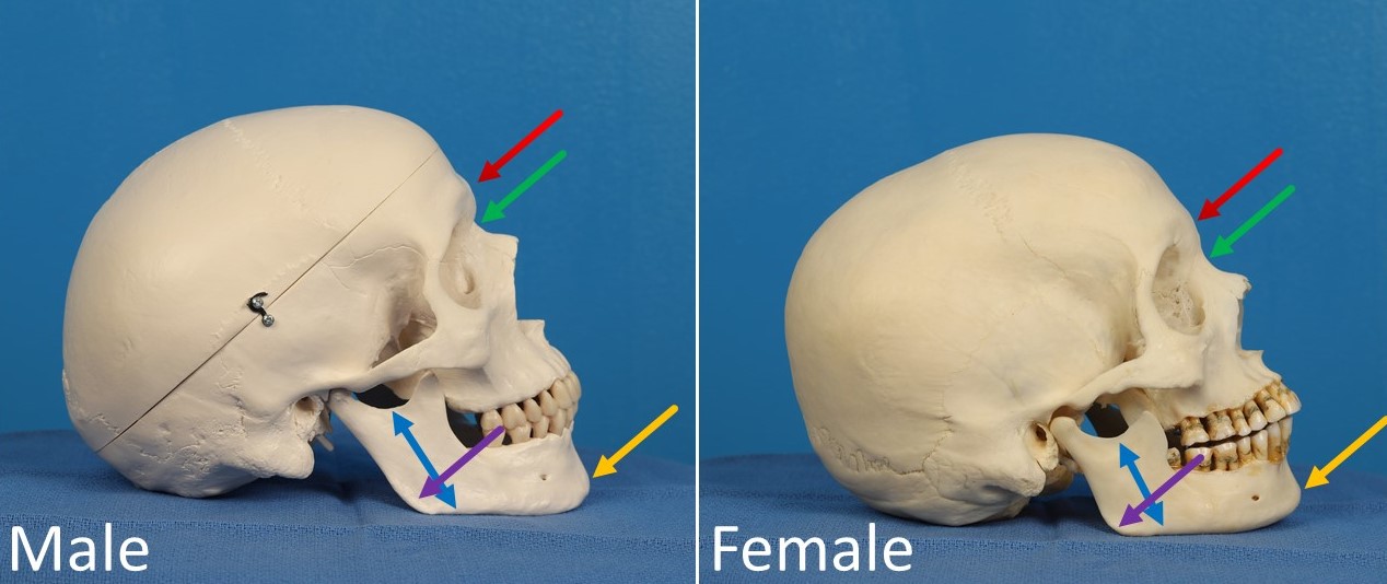
Differences Between Male and Female Skulls. The red arrow indicates frontal bossing, which is consistently more prominent in male skulls. Consequently, the nasal radix (green arrow) is deeper in the male skull. The yellow arrow demonstrates greater chin prominence in the male skull, and the blue arrows indicate greater mandibular height. In this example, the male skull has a more obtuse mandibular angle (purple arrow) than the female skull, an atypical finding that illustrates that not all sexually dimorphic traits occur in every patient.
Contributed by MH Hohman, MD, FACS
(Click Image to Enlarge)
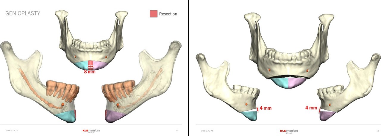
T-Osteotomies for Genioplasty. Virtual surgical planning demonstrates removal of an 8-mm central segment of the mandible to narrow the chin and a 4-mm increase in chin height. The preoperative view is shown on the left, and the projected postoperative result is shown on the right.
Contributed by MH Hohman, MD, FACS, and KLS Martin Group
(Click Image to Enlarge)
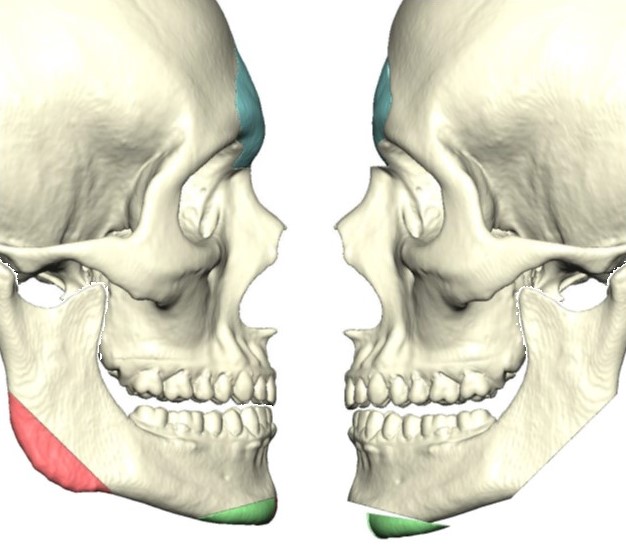
Surgical Simulation. Surgical simulation based on a 3D rendering of a preoperative CT scan demonstrating planned reductions in frontal bossing, mandibular angle, and chin projection.
Contributed by MH Hohman, MD, FACS
(Click Image to Enlarge)
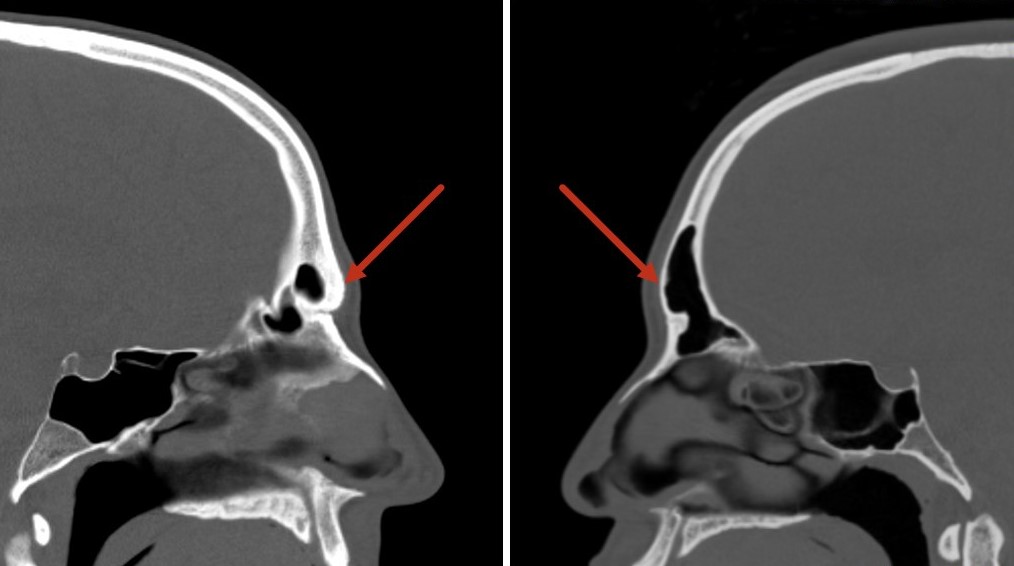
Anterior Table Thickness of the Frontal Sinuses. CT scans demonstrate varying thicknesses of the anterior tables of the frontal sinuses; bone thickness may significantly impact the surgical plan.
Contributed by MH Hohman, MD, FACS
(Click Image to Enlarge)
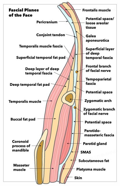
Fascial Planes of the Face. This illustration depicts the facial fascial planes, highlighting the continuity of the frontalis muscle, galea aponeurotica, temporoparietal fascia, superficial musculoaponeurotic system, platysma, and the location of the facial nerve.
Contributed by K Humphreys and MH Hohman, MD, FACS
(Click Image to Enlarge)
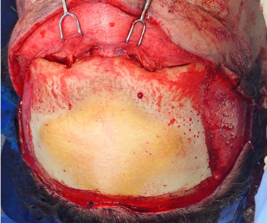
Supraorbital Rim and Frontal Bone Exposure. Supraorbital rim and frontal bone exposed via trichophytic approach, as viewed from the surgeon's perspective. Note the frontal bossing, which is more prominent in the glabella region.
Contributed by MH Hohman, MD, FACS
(Click Image to Enlarge)
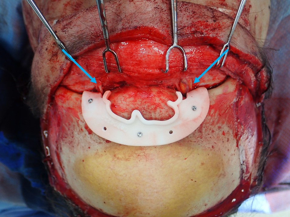
Frontal Sinus Borders Identified Using a 3D-Printed Cutting Guide. The borders of the frontal sinus were identified using a 3D-printed cutting guide based on the patient's preoperative CT scan. Blue arrows indicate supraorbital neurovascular bundles.
Contributed by MH Hohman, MD, FACS
(Click Image to Enlarge)
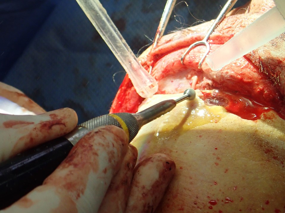
Supraorbital Ridge Reduction. The supraorbital ridge is reduced using a 5-mm diamond bur on a high-speed drill. The anterior table of the frontal sinus has already been removed.
Contributed by MH Hohman, MD, FACS
(Click Image to Enlarge)
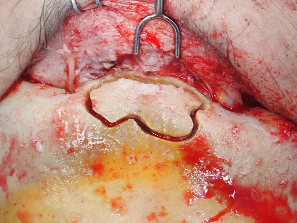
Anterior Table of the Frontal Sinus Outlined for Removal. The anterior table of the frontal sinus has been outlined using a 2-mm cutting bur on a high-speed drill and is ready for removal and thinning.
Contributed by MH Hohman, MD, FACS
(Click Image to Enlarge)
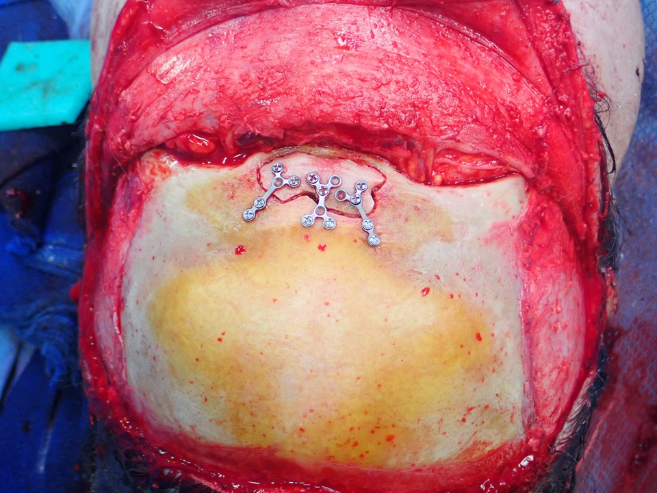
Replacement and Fixation of the Anterior Table. After thinning of the anterior table and removal of septa within the frontal sinus, the bone flap is replaced and fixated with low-profile titanium plates and screws. Note the reduction in prominence of the supraorbital ridge as well.
Contributed by MH Hohman, MD, FACS
(Click Image to Enlarge)
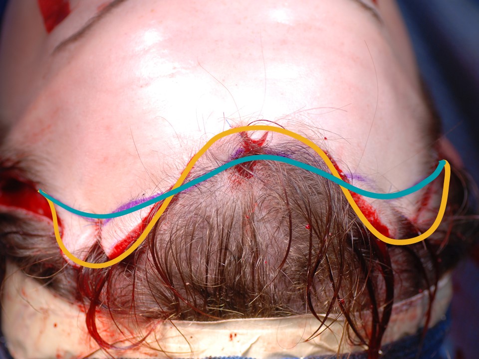
Hairline Adjustment After Frontal Cranioplasty and Brow Lift. The yellow curve indicates the preoperative hairline, and the blue curve indicates the anticipated postoperative hairline after removal of excess skin from the hairless temporal scalp and widow’s peak.
Contributed by MH Hohman, MD, FACS
(Click Image to Enlarge)
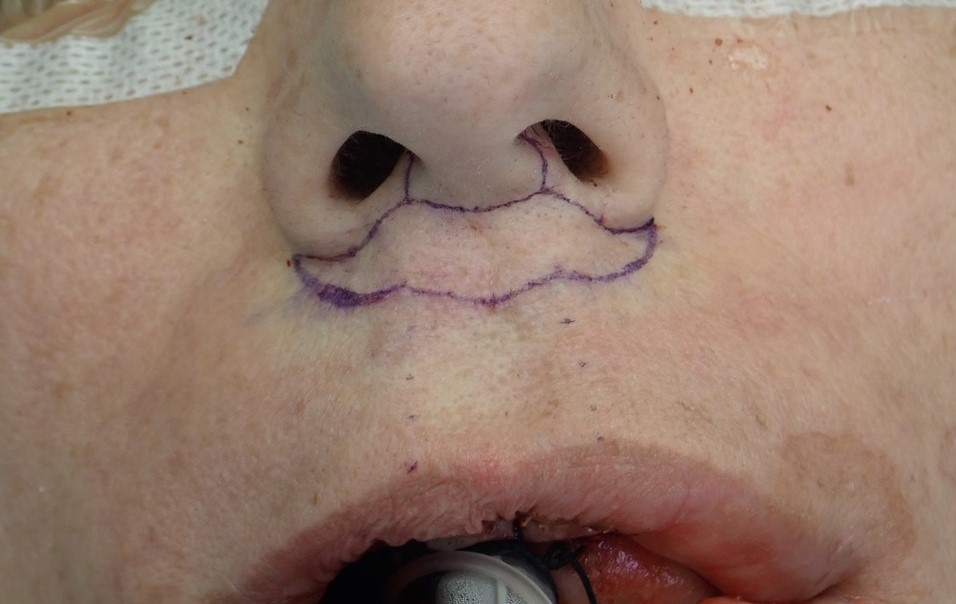
Bullhorn Lip Lift Incision. The bullhorn lip lift incision is marked to remove approximately one-third of the height of the cutaneous upper lip.
Contributed by MH Hohman, MD, FACS
(Click Image to Enlarge)
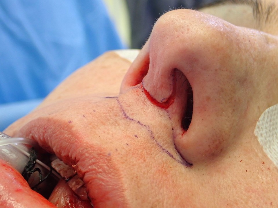
Central Bullhorn Incision for Open Rhinoplasty. The central portion of the bullhorn incision can serve as the transcolumellar incision and provide access for open rhinoplasty.
Contributed by MH Hohman, MD, FACS
(Click Image to Enlarge)
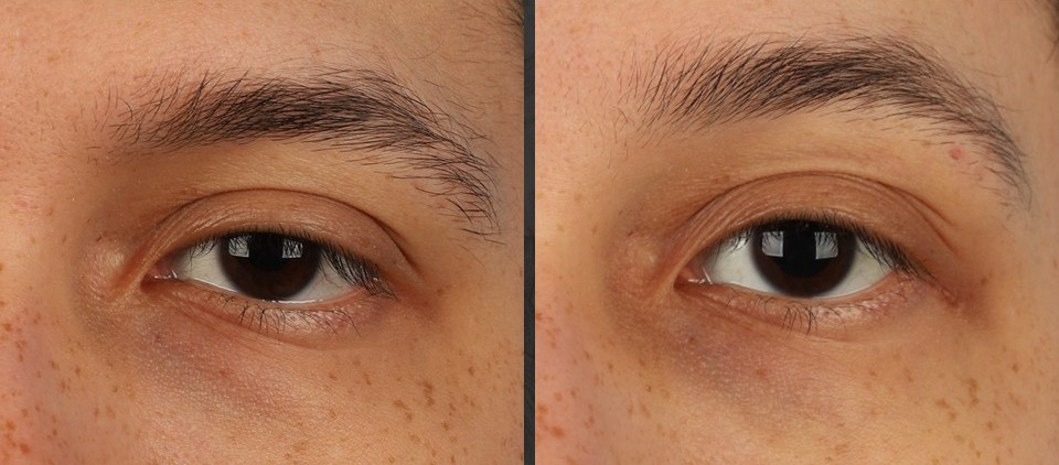
Lateral Tarsal Strip Canthoplasty. A 22-year-old transgender woman is shown before the surgical procedures (left) and 3 months after brow lift, frontal cranioplasty, and bilateral lateral tarsal strip canthoplasties (right).
Contributed by MH Hohman, MD, FACS
(Click Image to Enlarge)
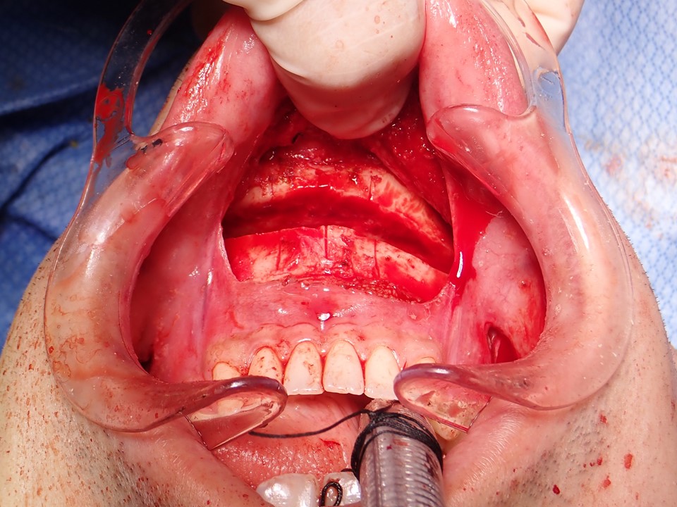
Transverse Mandibular Osteotomy for Genioplasty. A transverse mandibular osteotomy is created through a gingivolabial sulcus incision for genioplasty.
Contributed by MH Hohman, MD, and KD Smith, DMD
(Click Image to Enlarge)
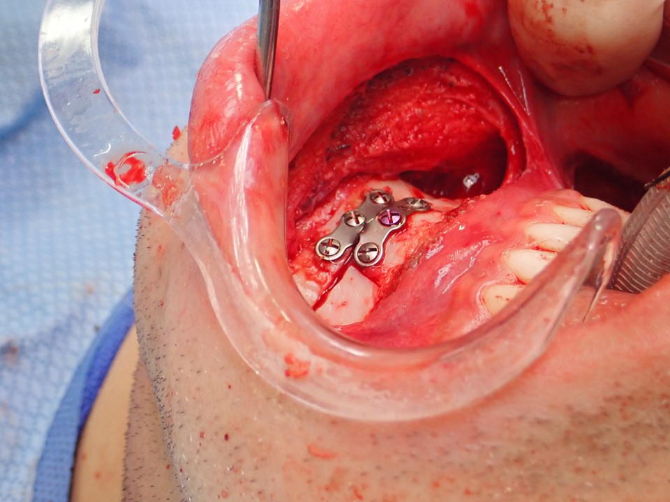
Completed Genioplasty With Chin Retrusion. Completed genioplasty demonstrates chin retrusion, with the bone segment fixed with a titanium plate and screws.
Contributed by MH Hohman, MD, FACS, and KD Smith, DMD
(Click Image to Enlarge)
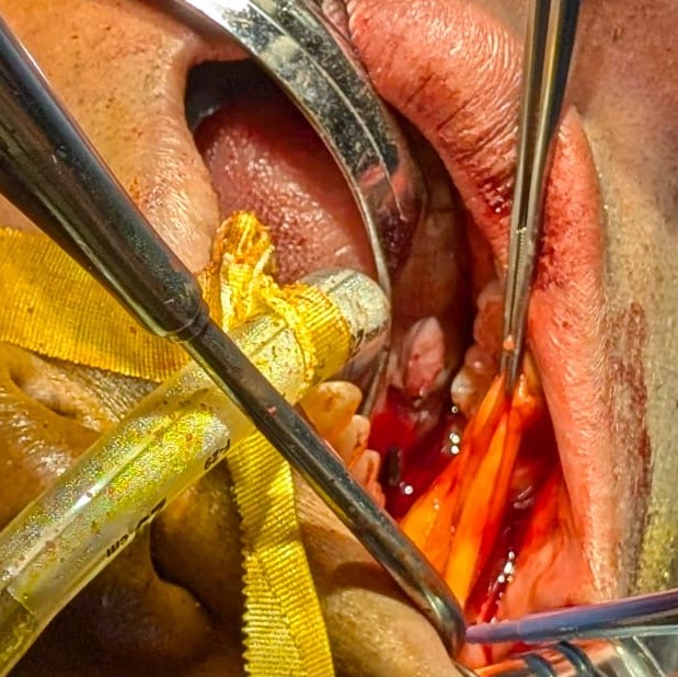
The Buccal Fat Pad. The buccal fat pad can be accessed for transfer or resection through a vertical incision in the buccal mucosa, located superior to the papilla of the parotid duct.
Contributed by MM Serra, DDS, FACS
(Click Image to Enlarge)
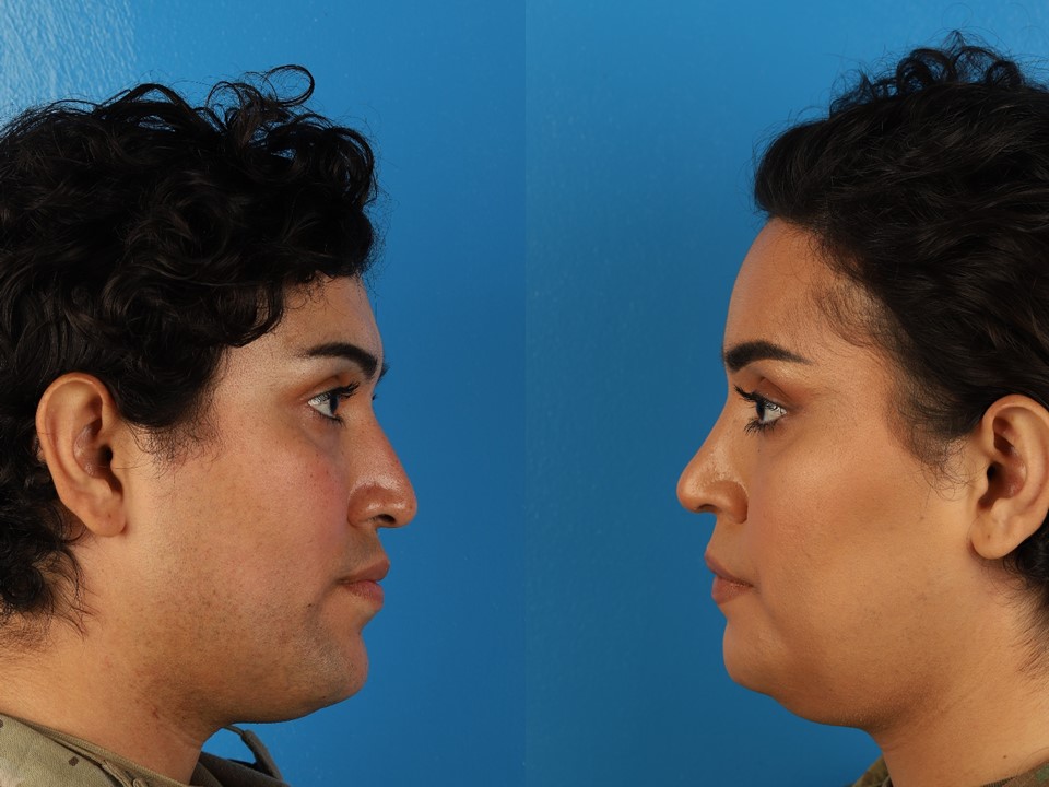
Facial Feminization Surgical Procedures in a 27-Year-Old Transgender Woman. Preoperative photograph (left) and postoperative photograph (right) obtained 1 month after a pretrichial brow lift with frontal cranioplasty and hairline advancement, rhinoplasty, lip lift, mandibular angle reduction, and reduction genioplasty.
Contributed by MH Hohman, MD, FACS
(Click Image to Enlarge)
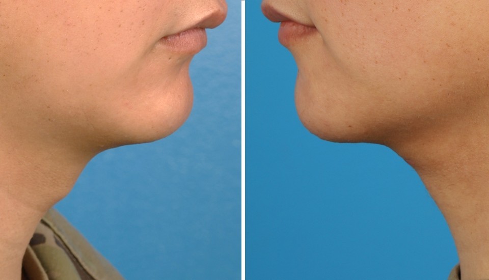
Laryngeal Chondroplasty. A 22-year-old transgender woman is shown before the surgical procedure (left) and 3 months after transcutaneous laryngeal chondroplasty (right).
Contributed by MH Hohman, MD, FACS
(Click Image to Enlarge)
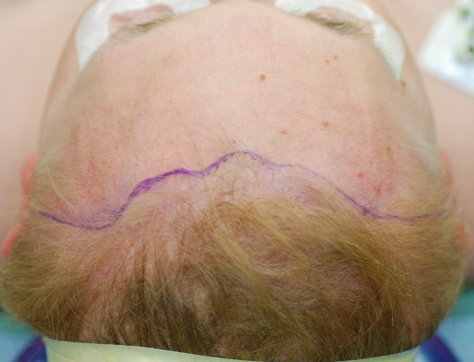
Preoperative Marking of the Pretrichial Brow Lift Incision. Note how the incision follows the frontal hairline centrally and dives into the temporal hair tufts laterally.
Contributed by MH Hohman, MD, FACS
(Click Image to Enlarge)
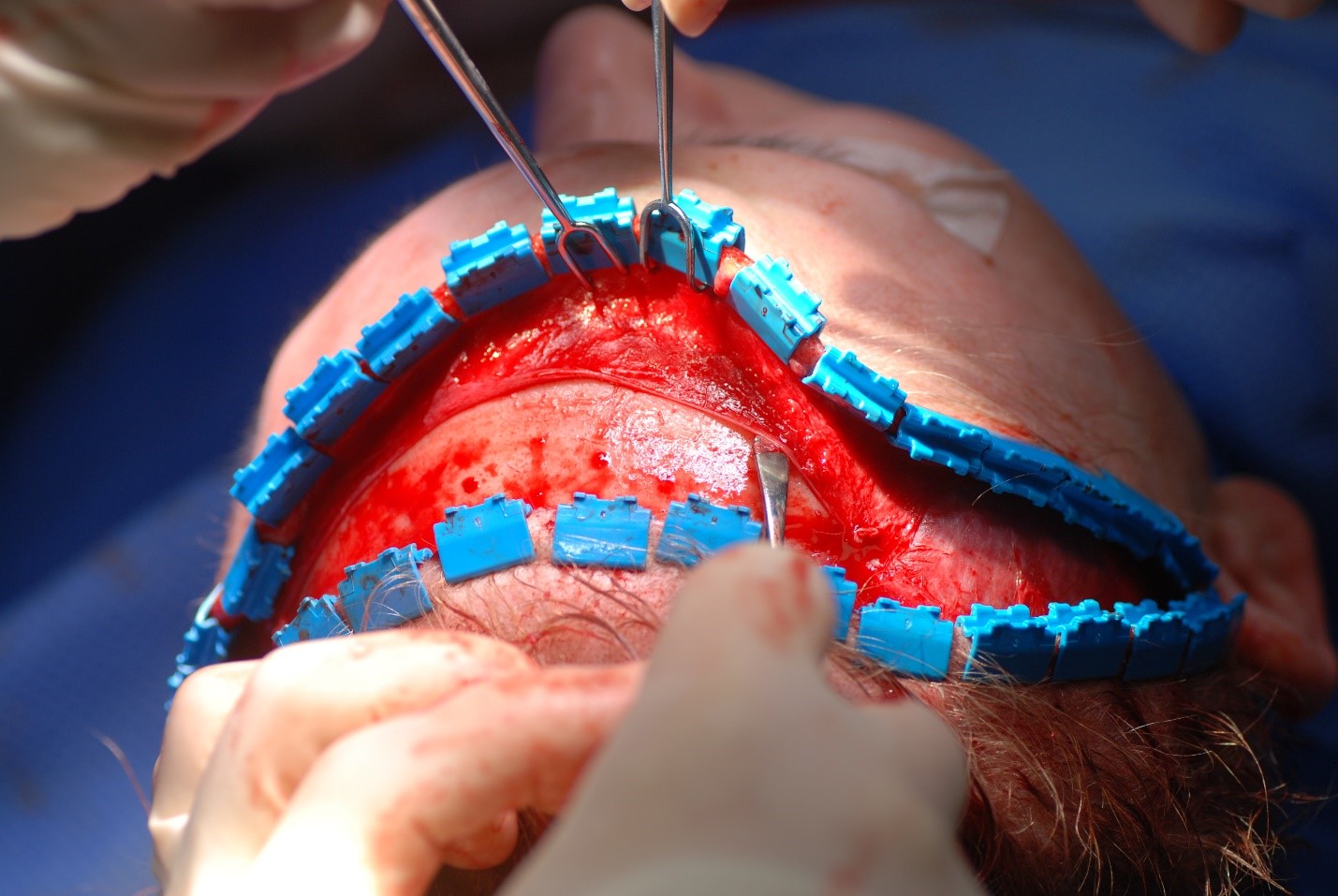
Periosteum Elevation Using a Freer Elevator. After establishing the plane, a larger periosteal elevator may be used to facilitate elevation.
Contributed by MH Hohman, MD, FACS
(Click Image to Enlarge)
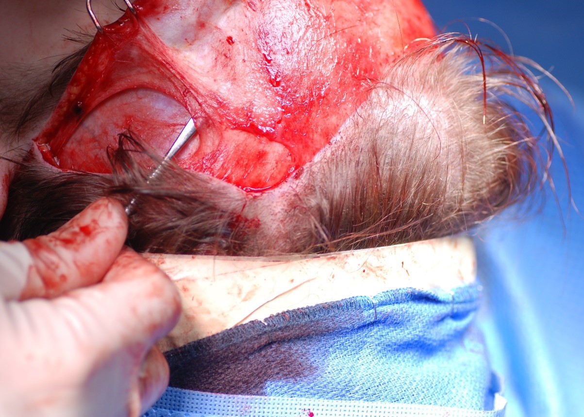
Temporoparietal Fascia Elevation. The temporoparietal fascia is elevated from the underlying temporalis muscle fascia, also known as the deep temporal fascia, while avoiding injury to the frontal branch of the facial nerve.
Contributed by MH Hohman, MD, FACS
(Click Image to Enlarge)
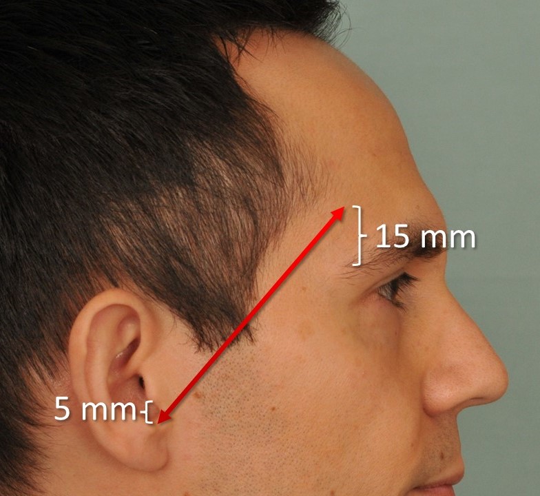
Pitanguy Line. This image illustrates the Pitanguy line, which runs from a point 5 mm inferior to the tragus to a point 15 mm superior to the lateral brow. This line approximates the course of the frontal branch of the facial nerve.
Contributed by MH Hohman, MD, FACS
(Click Image to Enlarge)
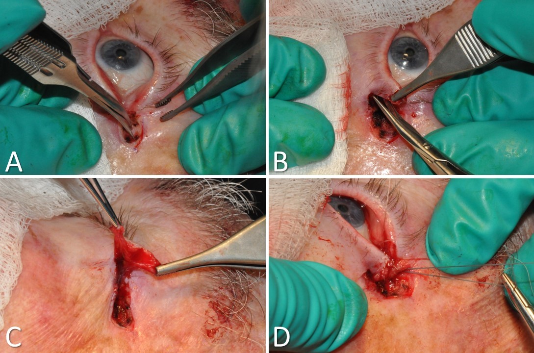
Tarsal Strip-Lateral Canthopexy. A) A lateral canthotomy and inferior cantholysis are performed. B) A full-thickness incision is made just inferior to the inferior tarsal plate. C) The skin and muscle are separated from the underlying tarsal plate and removed. The conjunctiva is scraped off the tarsal strip as well. D) The lateral aspect of the denuded tarsal strip is suspended superoposteriorly to the periorbita above the Whitnall tubercle. A sharp lateral canthal angle is then restored with a suture, and the wound is closed.
Contributed by TA Hadlock, MD, and MH Hohman, MD, FACS
(Click Image to Enlarge)
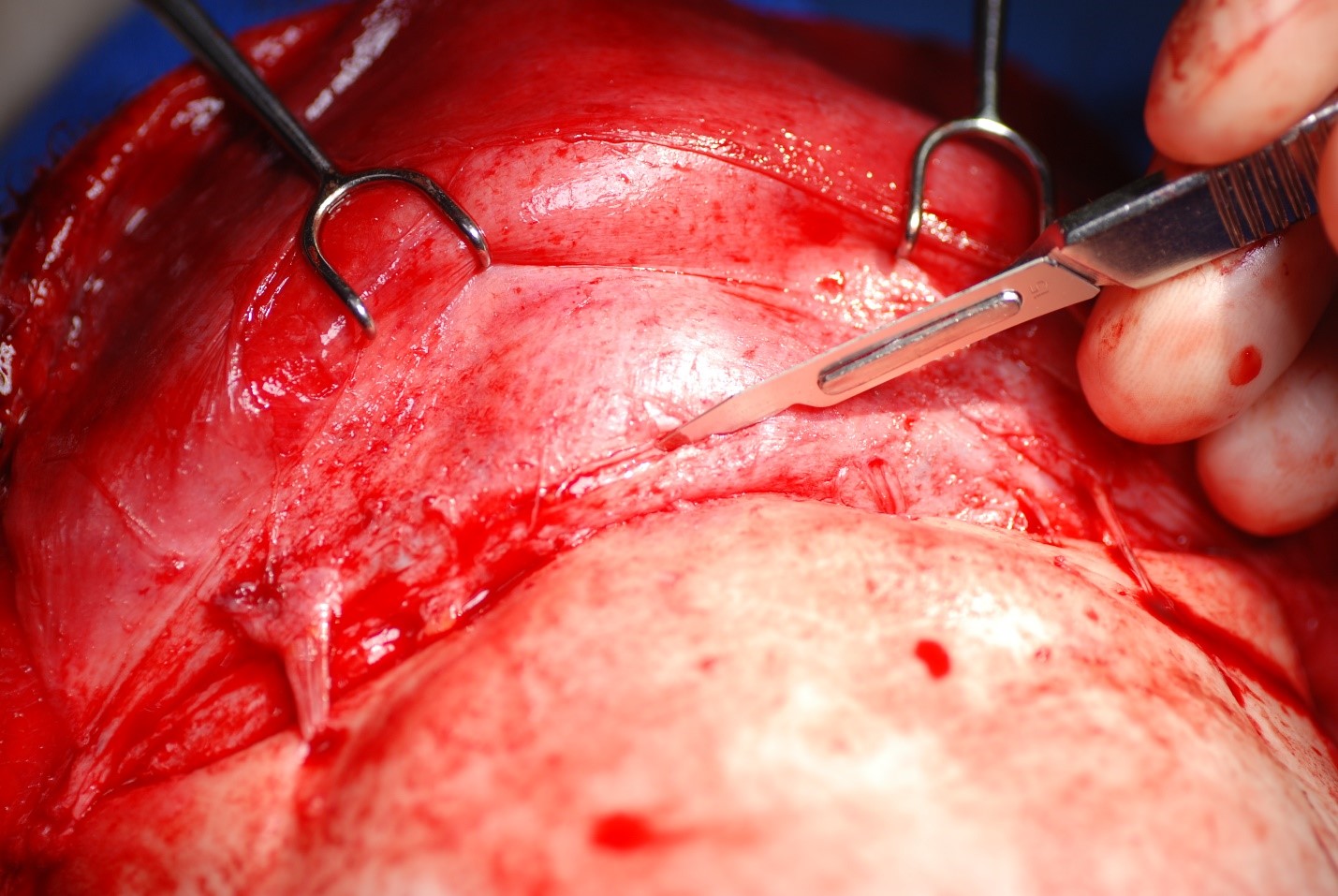
Sharp Division of the Periosteum Along the Arcus Marginalis. Sharp division of the periosteum along the arcus marginalis and across the glabella permits more effective brow elevation.
Contributed by MH Hohman, MD, FACS
References
Ewald ER, Guerino P, Dragon C, Laffan AM, Goldstein Z, Streed C Jr. Identifying Medicare Beneficiaries Accessing Transgender-Related Care in the Era of ICD-10. LGBT health. 2019 May/Jun:6(4):166-173. doi: 10.1089/lgbt.2018.0175. Epub 2019 May 14 [PubMed PMID: 31084519]
Lowry R, Johns MM, Gordon AR, Austin SB, Robin LE, Kann LK. Nonconforming Gender Expression and Associated Mental Distress and Substance Use Among High School Students. JAMA pediatrics. 2018 Nov 1:172(11):1020-1028. doi: 10.1001/jamapediatrics.2018.2140. Epub [PubMed PMID: 30264092]
Unger CA. Hormone therapy for transgender patients. Translational andrology and urology. 2016 Dec:5(6):877-884. doi: 10.21037/tau.2016.09.04. Epub [PubMed PMID: 28078219]
Ascha M, Swanson MA, Massie JP, Evans MW, Chambers C, Ginsberg BA, Gatherwright J, Satterwhite T, Morrison SD, Gougoutas AJ. Nonsurgical Management of Facial Masculinization and Feminization. Aesthetic surgery journal. 2019 Apr 8:39(5):NP123-NP137. doi: 10.1093/asj/sjy253. Epub [PubMed PMID: 30383180]
Spiegel JH. Facial determinants of female gender and feminizing forehead cranioplasty. The Laryngoscope. 2011 Feb:121(2):250-61. doi: 10.1002/lary.21187. Epub 2010 Nov 30 [PubMed PMID: 21271570]
Sadr J, Jarudi I, Sinha P. The role of eyebrows in face recognition. Perception. 2003:32(3):285-93 [PubMed PMID: 12729380]
Capitán L, Simon D, Bailón C, Bellinga RJ, Gutiérrez-Santamaría J, Tenório T, Capitán-Cañadas F. The Upper Third in Facial Gender Confirmation Surgery: Forehead and Hairline. The Journal of craniofacial surgery. 2019 Jul:30(5):1393-1398. doi: 10.1097/SCS.0000000000005640. Epub [PubMed PMID: 31299729]
Garcia-Rodriguez L, Thain LM, Spiegel JH. Scalp advancement for transgender women: Closing the gap. The Laryngoscope. 2020 Jun:130(6):1431-1435. doi: 10.1002/lary.28370. Epub 2019 Dec 4 [PubMed PMID: 31800096]
Capitán L, Simon D, Meyer T, Alcaide A, Wells A, Bailón C, Bellinga RJ, Tenório T, Capitán-Cañadas F. Facial Feminization Surgery: Simultaneous Hair Transplant during Forehead Reconstruction. Plastic and reconstructive surgery. 2017 Mar:139(3):573-584. doi: 10.1097/PRS.0000000000003149. Epub [PubMed PMID: 28234823]
Ousterhout DK. Feminization of the forehead: contour changing to improve female aesthetics. Plastic and reconstructive surgery. 1987 May:79(5):701-13 [PubMed PMID: 3575517]
Capitán L, Simon D, Kaye K, Tenorio T. Facial feminization surgery: the forehead. Surgical techniques and analysis of results. Plastic and reconstructive surgery. 2014 Oct:134(4):609-619. doi: 10.1097/PRS.0000000000000545. Epub [PubMed PMID: 24945951]
Ainsworth TA, Spiegel JH. Quality of life of individuals with and without facial feminization surgery or gender reassignment surgery. Quality of life research : an international journal of quality of life aspects of treatment, care and rehabilitation. 2010 Sep:19(7):1019-24. doi: 10.1007/s11136-010-9668-7. Epub 2010 May 12 [PubMed PMID: 20461468]
Level 2 (mid-level) evidenceJavier C, Crimston CR, Barlow FK. Surgical satisfaction and quality of life outcomes reported by transgender men and women at least one year post gender-affirming surgery: A systematic literature review. International journal of transgender health. 2022:23(3):255-273. doi: 10.1080/26895269.2022.2038334. Epub 2022 Mar 18 [PubMed PMID: 35799954]
Level 1 (high-level) evidenceCardoso da Silva D, Schwarz K, Fontanari AM, Costa AB, Massuda R, Henriques AA, Salvador J, Silveira E, Elias Rosito T, Lobato MI. WHOQOL-100 Before and After Sex Reassignment Surgery in Brazilian Male-to-Female Transsexual Individuals. The journal of sexual medicine. 2016 Jun:13(6):988-93. doi: 10.1016/j.jsxm.2016.03.370. Epub 2016 Apr 21 [PubMed PMID: 27117529]
Morrison SD, Capitán-Cañadas F, Sánchez-García A, Ludwig DC, Massie JP, Nolan IT, Swanson M, Rodríguez-Conesa M, Friedrich JB, Cederna PS, Bellinga RJ, Simon D, Capitán L, Satterwhite T. Prospective Quality-of-Life Outcomes after Facial Feminization Surgery: An International Multicenter Study. Plastic and reconstructive surgery. 2020 Jun:145(6):1499-1509. doi: 10.1097/PRS.0000000000006837. Epub [PubMed PMID: 32459779]
Level 2 (mid-level) evidenceHuynh PP, Kleinberger A, Brandstetter K, Chou DW, Pressman AR, Shih C. Facial Feminization Surgery and Mental Health Resource Utilization: A Retrospective Institutional Review. Otolaryngology--head and neck surgery : official journal of American Academy of Otolaryngology-Head and Neck Surgery. 2025 May:172(5):1585-1593. doi: 10.1002/ohn.1172. Epub 2025 Feb 25 [PubMed PMID: 39996585]
Level 2 (mid-level) evidenceBrown E, Perrett DI. What gives a face its gender? Perception. 1993:22(7):829-40 [PubMed PMID: 8115240]
Hage JJ, Becking AG, de Graaf FH, Tuinzing DB. Gender-confirming facial surgery: considerations on the masculinity and femininity of faces. Plastic and reconstructive surgery. 1997 Jun:99(7):1799-807 [PubMed PMID: 9180702]
Nusbaum BP, Fuentefria S. Naturally occurring female hairline patterns. Dermatologic surgery : official publication for American Society for Dermatologic Surgery [et al.]. 2009 Jun:35(6):907-13. doi: 10.1111/j.1524-4725.2009.01154.x. Epub 2009 Apr 6 [PubMed PMID: 19397668]
Shi LL, Lacey M, Teixeira J, Hendershot K, Hohman M, Kidwai S, Flaherty AJ, Nuara MJ. Analysis of Cephalometric Differences of the Midface and Upper Face in Males and Females: A Radiographic Study. The Journal of craniofacial surgery. 2025 Mar-Apr 01:36(2):402-406. doi: 10.1097/SCS.0000000000010416. Epub 2024 Jun 21 [PubMed PMID: 38904398]
Perez PI, Hendershot K, Teixeira JC, Hohman MH, Adidharma L, Moody M, Kidwai S, Lacey M, Johnson S, Nuara MJ. Analysis of Cephalometric Points in Male and Female Mandibles: An Application to Gender-Affirming Facial Surgery. The Journal of craniofacial surgery. 2023 Jun 1:34(4):1278-1282. doi: 10.1097/SCS.0000000000009189. Epub 2023 Jan 25 [PubMed PMID: 36727677]
Coleman E, Radix AE, Bouman WP, Brown GR, de Vries ALC, Deutsch MB, Ettner R, Fraser L, Goodman M, Green J, Hancock AB, Johnson TW, Karasic DH, Knudson GA, Leibowitz SF, Meyer-Bahlburg HFL, Monstrey SJ, Motmans J, Nahata L, Nieder TO, Reisner SL, Richards C, Schechter LS, Tangpricha V, Tishelman AC, Van Trotsenburg MAA, Winter S, Ducheny K, Adams NJ, Adrián TM, Allen LR, Azul D, Bagga H, Başar K, Bathory DS, Belinky JJ, Berg DR, Berli JU, Bluebond-Langner RO, Bouman MB, Bowers ML, Brassard PJ, Byrne J, Capitán L, Cargill CJ, Carswell JM, Chang SC, Chelvakumar G, Corneil T, Dalke KB, De Cuypere G, de Vries E, Den Heijer M, Devor AH, Dhejne C, D'Marco A, Edmiston EK, Edwards-Leeper L, Ehrbar R, Ehrensaft D, Eisfeld J, Elaut E, Erickson-Schroth L, Feldman JL, Fisher AD, Garcia MM, Gijs L, Green SE, Hall BP, Hardy TLD, Irwig MS, Jacobs LA, Janssen AC, Johnson K, Klink DT, Kreukels BPC, Kuper LE, Kvach EJ, Malouf MA, Massey R, Mazur T, McLachlan C, Morrison SD, Mosser SW, Neira PM, Nygren U, Oates JM, Obedin-Maliver J, Pagkalos G, Patton J, Phanuphak N, Rachlin K, Reed T, Rider GN, Ristori J, Robbins-Cherry S, Roberts SA, Rodriguez-Wallberg KA, Rosenthal SM, Sabir K, Safer JD, Scheim AI, Seal LJ, Sehoole TJ, Spencer K, St Amand C, Steensma TD, Strang JF, Taylor GB, Tilleman K, T'Sjoen GG, Vala LN, Van Mello NM, Veale JF, Vencill JA, Vincent B, Wesp LM, West MA, Arcelus J. Standards of Care for the Health of Transgender and Gender Diverse People, Version 8. International journal of transgender health. 2022:23(Suppl 1):S1-S259. doi: 10.1080/26895269.2022.2100644. Epub 2022 Sep 6 [PubMed PMID: 36238954]
Morrison SD, Vyas KS, Motakef S, Gast KM, Chung MT, Rashidi V, Satterwhite T, Kuzon W, Cederna PS. Facial Feminization: Systematic Review of the Literature. Plastic and reconstructive surgery. 2016 Jun:137(6):1759-1770. doi: 10.1097/PRS.0000000000002171. Epub [PubMed PMID: 27219232]
Level 1 (high-level) evidenceWright MR, Wright WK. A psychological study of patients undergoing cosmetic surgery. Archives of otolaryngology (Chicago, Ill. : 1960). 1975 Mar:101(3):145-51 [PubMed PMID: 235253]
Napoleon A. The presentation of personalities in plastic surgery. Annals of plastic surgery. 1993 Sep:31(3):193-208 [PubMed PMID: 8239409]
Knorr NJ. Feminine loss of identity in rhinoplasty. Archives of otolaryngology (Chicago, Ill. : 1960). 1972 Jul:96(1):11-5 [PubMed PMID: 5032051]
Goin MK, Burgoyne RW, Goin JM, Staples FR. A prospective psychological study of 50 female face-lift patients. Plastic and reconstructive surgery. 1980 Apr:65(4):436-42 [PubMed PMID: 7360810]
Ercolani M, Baldaro B, Rossi N, Trombini G. Five-year follow-up of cosmetic rhinoplasty. Journal of psychosomatic research. 1999 Sep:47(3):283-6 [PubMed PMID: 10576477]
Honigman RJ, Phillips KA, Castle DJ. A review of psychosocial outcomes for patients seeking cosmetic surgery. Plastic and reconstructive surgery. 2004 Apr 1:113(4):1229-37 [PubMed PMID: 15083026]
Sarwer DB, Wadden TA, Pertschuk MJ, Whitaker LA. The psychology of cosmetic surgery: a review and reconceptualization. Clinical psychology review. 1998 Jan:18(1):1-22 [PubMed PMID: 9455621]
Robin AA, Copas JB, Jack AB, Kaeser AC, Thomas PJ. Reshaping the psyche. The concurrent improvement in appearance and mental state after rhinoplasty. The British journal of psychiatry : the journal of mental science. 1988 Apr:152():539-43 [PubMed PMID: 3167406]
Beale S, Hambert G, Lisper HO, Ohlsén L, Palm B. Augmentation mammaplasty: the surgical and psychological effects of the operation and prediction of the result. Annals of plastic surgery. 1985 Jun:14(6):473-93 [PubMed PMID: 4083704]
Guyuron B, Bokhari F. Patient satisfaction following rhinoplasty. Aesthetic plastic surgery. 1996 Mar-Apr:20(2):153-7 [PubMed PMID: 8801805]
EDGERTON MT, WEBB WL Jr, SLAUGHTER R, MEYER E. SURGICAL RESULTS AND PSYCHOSOCIAL CHANGES FOLLOWING RHYTIDECTOMY; AN EVALUATION OF FACE-LIFTING. Plastic and reconstructive surgery. 1964 Jun:33():503-21 [PubMed PMID: 14171431]
King H, Kelley TP, Shatzel JJ. Gender-affirming hormone therapy in the transgender patient: influence on thrombotic risk. Hematology. American Society of Hematology. Education Program. 2024 Dec 6:2024(1):652-663. doi: 10.1182/hematology.2024000592. Epub [PubMed PMID: 39644058]
Totaro M, Palazzi S, Castellini C, Parisi A, D'Amato F, Tienforti D, Baroni MG, Francavilla S, Barbonetti A. Risk of Venous Thromboembolism in Transgender People Undergoing Hormone Feminizing Therapy: A Prevalence Meta-Analysis and Meta-Regression Study. Frontiers in endocrinology. 2021:12():741866. doi: 10.3389/fendo.2021.741866. Epub 2021 Nov 9 [PubMed PMID: 34880832]
Level 1 (high-level) evidenceLutsey PL, Steffen LM, Virnig BA, Folsom AR. Diet and incident venous thromboembolism: the Iowa Women's Health Study. American heart journal. 2009 Jun:157(6):1081-7. doi: 10.1016/j.ahj.2009.04.003. Epub [PubMed PMID: 19464420]
Blondon M, Wiggins KL, Van Hylckama Vlieg A, McKnight B, Psaty BM, Rice KM, Heckbert SR, Smith NL. Smoking, postmenopausal hormone therapy and the risk of venous thrombosis: a population-based, case-control study. British journal of haematology. 2013 Nov:163(3):418-20. doi: 10.1111/bjh.12508. Epub 2013 Aug 8 [PubMed PMID: 23927442]
Level 3 (low-level) evidencePomp ER, Rosendaal FR, Doggen CJ. Smoking increases the risk of venous thrombosis and acts synergistically with oral contraceptive use. American journal of hematology. 2008 Feb:83(2):97-102 [PubMed PMID: 17726684]
Hwang K, Son JS, Ryu WK. Smoking and Flap Survival. Plastic surgery (Oakville, Ont.). 2018 Nov:26(4):280-285. doi: 10.1177/2292550317749509. Epub 2018 Jan 9 [PubMed PMID: 30450347]
Goldminz D, Bennett RG. Cigarette smoking and flap and full-thickness graft necrosis. Archives of dermatology. 1991 Jul:127(7):1012-5 [PubMed PMID: 2064398]
Level 2 (mid-level) evidenceGetahun D, Nash R, Flanders WD, Baird TC, Becerra-Culqui TA, Cromwell L, Hunkeler E, Lash TL, Millman A, Quinn VP, Robinson B, Roblin D, Silverberg MJ, Safer J, Slovis J, Tangpricha V, Goodman M. Cross-sex Hormones and Acute Cardiovascular Events in Transgender Persons: A Cohort Study. Annals of internal medicine. 2018 Aug 21:169(4):205-213. doi: 10.7326/M17-2785. Epub 2018 Jul 10 [PubMed PMID: 29987313]
van Kesteren PJ, Asscheman H, Megens JA, Gooren LJ. Mortality and morbidity in transsexual subjects treated with cross-sex hormones. Clinical endocrinology. 1997 Sep:47(3):337-42 [PubMed PMID: 9373456]
Jones HE, Cruz C, Stewart C, Losken A. Decision Regret in Plastic Surgery: A Summary. Plastic and reconstructive surgery. Global open. 2023 Jun:11(6):e5098. doi: 10.1097/GOX.0000000000005098. Epub 2023 Jun 27 [PubMed PMID: 37383479]
Thornton SM, Edalatpour A, Gast KM. A systematic review of patient regret after surgery- A common phenomenon in many specialties but rare within gender-affirmation surgery. American journal of surgery. 2024 Aug:234():68-73. doi: 10.1016/j.amjsurg.2024.04.021. Epub 2024 Apr 24 [PubMed PMID: 38688814]
Level 1 (high-level) evidenceGoldstein Z, Khan M, Reisman T, Safer JD. Managing the risk of venous thromboembolism in transgender adults undergoing hormone therapy. Journal of blood medicine. 2019:10():209-216. doi: 10.2147/JBM.S166780. Epub 2019 Jul 10 [PubMed PMID: 31372078]
Hohman MH, Jastrzembski ME, Choe N, Nuara MJ, Teixeira JC, Vincent AG. 3D-Printed Custom Cutting Guides Facilitate Frontal Cranioplasty in Gender Affirmation Surgery. The Journal of craniofacial surgery. 2021 Oct 1:32(7):e668-e670. doi: 10.1097/SCS.0000000000007846. Epub [PubMed PMID: 34705370]
Spiegel JH. Facial Feminization for the Transgender Patient. The Journal of craniofacial surgery. 2019 Jul:30(5):1399-1402. doi: 10.1097/SCS.0000000000005645. Epub [PubMed PMID: 31299730]
Ousterhout DK. Facial Feminization Surgery: The Forehead. Surgical Techniques and Analysis of Results. Plastic and reconstructive surgery. 2015 Oct:136(4):560e-561e. doi: 10.1097/PRS.0000000000001425. Epub [PubMed PMID: 25938955]
Pitanguy I, Ramos AS. The frontal branch of the facial nerve: the importance of its variations in face lifting. Plastic and reconstructive surgery. 1966 Oct:38(4):352-6 [PubMed PMID: 5926990]
Hohman MH, Bhama PK, Hadlock TA. Epidemiology of iatrogenic facial nerve injury: a decade of experience. The Laryngoscope. 2014 Jan:124(1):260-5. doi: 10.1002/lary.24117. Epub 2013 Apr 18 [PubMed PMID: 23606475]
Level 2 (mid-level) evidenceVillepelet A, Jafari A, Baujat B. Fronto-orbital feminization technique. A surgical strategy using fronto-orbital burring with or without eggshell technique to optimize the risk/benefit ratio. European annals of otorhinolaryngology, head and neck diseases. 2018 Oct:135(5):353-356. doi: 10.1016/j.anorl.2018.04.007. Epub 2018 May 4 [PubMed PMID: 29735285]
Spiegel JH. Rhinoplasty as a Significant Component of Facial Feminization and Beautification. JAMA facial plastic surgery. 2017 May 1:19(3):181-182. doi: 10.1001/jamafacial.2016.1817. Epub [PubMed PMID: 27978546]
Davis AM, Simons RL, Rhee JS. Evaluation of the Goldman tip procedure in modern-day rhinoplasty. Archives of facial plastic surgery. 2004 Sep-Oct:6(5):301-7 [PubMed PMID: 15381575]
Level 2 (mid-level) evidenceInsalaco L, Spiegel JH. Safety of Simultaneous Lip-Lift and Open Rhinoplasty. JAMA facial plastic surgery. 2017 Mar 1:19(2):160-161. doi: 10.1001/jamafacial.2016.1396. Epub [PubMed PMID: 27832262]
Hohman MH, Vincent AG, Anderson SR, Ducic Y, Cochran S. Avoiding Complications in Functional and Aesthetic Rhinoplasty. Seminars in plastic surgery. 2020 Nov:34(4):260-264. doi: 10.1055/s-0040-1721762. Epub 2020 Dec 24 [PubMed PMID: 33380911]
Sheen JH. Spreader graft: a method of reconstructing the roof of the middle nasal vault following rhinoplasty. Plastic and reconstructive surgery. 1984 Feb:73(2):230-9 [PubMed PMID: 6695022]
Standlee AG, Hohman MH. Evaluating the Effect of Spreader Grafting on Nasal Obstruction Using the NOSE Scale. The Annals of otology, rhinology, and laryngology. 2017 Mar:126(3):219-223. doi: 10.1177/0003489416685320. Epub 2017 Jan 5 [PubMed PMID: 28056521]
Byrd HS, Meade RA, Gonyon DL Jr. Using the autospreader flap in primary rhinoplasty. Plastic and reconstructive surgery. 2007 May:119(6):1897-1902. doi: 10.1097/01.prs.0000259196.02216.a5. Epub [PubMed PMID: 17440372]
Level 3 (low-level) evidenceSpiegel JH. The Modified Bullhorn Approach for the Lip-lift. JAMA facial plastic surgery. 2019 Jan 1:21(1):69-70. doi: 10.1001/jamafacial.2018.0847. Epub [PubMed PMID: 30326516]
Raphael P, Harris R, Harris SW. The endonasal lip lift: personal technique. Aesthetic surgery journal. 2014 Mar:34(3):457-68. doi: 10.1177/1090820X14524769. Epub 2014 Mar 6 [PubMed PMID: 24604786]
Fanous N. Correction of thin lips: "lip lift". Plastic and reconstructive surgery. 1984 Jul:74(1):33-41 [PubMed PMID: 6739598]
Weston GW, Poindexter BD, Sigal RK, Austin HW. Lifting lips: 28 years of experience using the direct excision approach to rejuvenating the aging mouth. Aesthetic surgery journal. 2009 Mar-Apr:29(2):83-6. doi: 10.1016/j.asj.2009.01.013. Epub [PubMed PMID: 19371836]
Nassar A, Naba J, Demian J. Lip feminization: A review. JPRAS open. 2024 Sep:41():311-319. doi: 10.1016/j.jpra.2024.07.002. Epub 2024 Jul 11 [PubMed PMID: 39188659]
Teixeira JC, Ostrom JY, Hohman MH, Nuara MJ. Botulinum Toxin Type-A for Lip Augmentation: "Lip Flip". The Journal of craniofacial surgery. 2021 May 1:32(3):e273-e275. doi: 10.1097/SCS.0000000000007128. Epub [PubMed PMID: 33170825]
Colić MM. Lip and perioral enhancement by direct intramuscular fat autografting. Aesthetic plastic surgery. 1999 Jan-Feb:23(1):36-40 [PubMed PMID: 10022936]
Trussler AP, Kawamoto HK, Wasson KL, Dickinson BP, Jackson E, Keagle JN, Jarrahy R, Bradley JP. Upper lip augmentation: palmaris longus tendon as an autologous filler. Plastic and reconstructive surgery. 2008 Mar:121(3):1024-1032. doi: 10.1097/01.prs.0000299383.82723.ec. Epub [PubMed PMID: 18317152]
Castor SA, To WC, Papay FA. Lip augmentation with AlloDerm acellular allogenic dermal graft and fat autograft: A comparison with autologous fat injection alone. Aesthetic plastic surgery. 1999 May-Jun:23(3):218-23 [PubMed PMID: 10384022]
Hazani R, Chowdhry S, Mowlavi A, Wilhelmi BJ. Bony anatomic landmarks to avoid injury to the marginal mandibular nerve. Aesthetic surgery journal. 2011 Mar:31(3):286-9. doi: 10.1177/1090820X11398352. Epub [PubMed PMID: 21385737]
Batra AP, Mahajan A, Gupta K. Marginal mandibular branch of the facial nerve: An anatomical study. Indian journal of plastic surgery : official publication of the Association of Plastic Surgeons of India. 2010 Jan:43(1):60-4. doi: 10.4103/0970-0358.63968. Epub [PubMed PMID: 20924452]
Chowdhry S, Yoder EM, Cooperman RD, Yoder VR, Wilhelmi BJ. Locating the cervical motor branch of the facial nerve: anatomy and clinical application. Plastic and reconstructive surgery. 2010 Sep:126(3):875-879. doi: 10.1097/PRS.0b013e3181e3b374. Epub [PubMed PMID: 20463628]
Kim SK, Han JJ, Kim JT. Classification and treatment of prominent mandibular angle. Aesthetic plastic surgery. 2001 Sep-Oct:25(5):382-7 [PubMed PMID: 11692255]
Chen H, Sun J, Wang J. Reducing Prominent Mandibular Angle Osteotomy Complications: 10-Year Retrospective Review. Annals of plastic surgery. 2018 Dec:81(6S Suppl 1):S5-S9. doi: 10.1097/SAP.0000000000001372. Epub [PubMed PMID: 29481477]
Level 2 (mid-level) evidenceMorrison SD, Satterwhite T. Lower Jaw Recontouring in Facial Gender-Affirming Surgery. Facial plastic surgery clinics of North America. 2019 May:27(2):233-242. doi: 10.1016/j.fsc.2019.01.001. Epub 2019 Mar 2 [PubMed PMID: 30940389]
Deschamps-Braly J. Feminization of the Chin: Genioplasty Using Osteotomies. Facial plastic surgery clinics of North America. 2019 May:27(2):243-250. doi: 10.1016/j.fsc.2019.01.002. Epub [PubMed PMID: 30940390]
Park MC, Kang M, Lim H, Lee IJ, Shin SJ. Mandibular tubercle resection: a means of maximizing the benefits of reduction mandibuloplasty. Plastic and reconstructive surgery. 2011 May:127(5):2076-2082. doi: 10.1097/PRS.0b013e31820cf5a5. Epub [PubMed PMID: 21532435]
Kim TG, Lee JH, Cho YK. Inverted V-shape Osteotomy with Central Strip Resection: A Simultaneous Narrowing and Vertical Reduction Genioplasty. Plastic and reconstructive surgery. Global open. 2014 Oct:2(10):e227. doi: 10.1097/GOX.0000000000000169. Epub 2014 Nov 7 [PubMed PMID: 25426344]
Khafif A, Shoffel-Havakuk H, Yaish I, Tordjman K, Assadi N. Scarless Neck Feminization: Transoral Transvestibular Approach Chondrolaryngoplasty. Facial plastic surgery & aesthetic medicine. 2020 May/Jun:22(3):172-180. doi: 10.1089/fpsam.2020.0021. Epub 2020 Feb 26 [PubMed PMID: 32101468]
David AP, Knott PD, Rosen CA, Young VN, Park AM, Seth R. Clinical Feasibility and Efficacy of the Externally Scarless Transoral Chondrolaryngoplasty. Facial plastic surgery & aesthetic medicine. 2022 Nov-Dec:24(S2):S41-S43. doi: 10.1089/fpsam.2021.0295. Epub 2022 May 2 [PubMed PMID: 35506892]
Level 2 (mid-level) evidenceSturm A, Chaiet SR. Chondrolaryngoplasty-Thyroid Cartilage Reduction. Facial plastic surgery clinics of North America. 2019 May:27(2):267-272. doi: 10.1016/j.fsc.2019.01.005. Epub [PubMed PMID: 30940393]
Ihnat JM, Hu KG, Almeida MN, Parikh N, Hauc SC, Alper DP, Alperovich M. Finesse in Chondrolaryngoplasty. Plastic and reconstructive surgery. Global open. 2024 Jan:12(1):e5539. doi: 10.1097/GOX.0000000000005539. Epub 2024 Jan 22 [PubMed PMID: 38260761]
Altman K. Forehead reduction and orbital contouring in facial feminisation surgery for transgender females. The British journal of oral & maxillofacial surgery. 2018 Apr:56(3):192-197. doi: 10.1016/j.bjoms.2018.01.009. Epub 2018 Feb 7 [PubMed PMID: 29428374]
Ren T, Galenchik-Chan A, Erlichman Z, Krajewski A. Prevalence of Regret in Gender-Affirming Surgery: A Systematic Review. Annals of plastic surgery. 2024 May 1:92(5):597-602. doi: 10.1097/SAP.0000000000003895. Epub [PubMed PMID: 38685500]
Level 1 (high-level) evidenceDinno A. Homicide Rates of Transgender Individuals in the United States: 2010-2014. American journal of public health. 2017 Sep:107(9):1441-1447. doi: 10.2105/AJPH.2017.303878. Epub 2017 Jul 20 [PubMed PMID: 28727530]
Karasic DH, Fraser L. Multidisciplinary Care and the Standards of Care for Transgender and Gender Nonconforming Individuals. Clinics in plastic surgery. 2018 Jul:45(3):295-299. doi: 10.1016/j.cps.2018.03.016. Epub [PubMed PMID: 29908615]
Chen D, Hidalgo MA, Leibowitz S, Leininger J, Simons L, Finlayson C, Garofalo R. Multidisciplinary Care for Gender-Diverse Youth: A Narrative Review and Unique Model of Gender-Affirming Care. Transgender health. 2016:1(1):117-123. doi: 10.1089/trgh.2016.0009. Epub 2016 Jul 1 [PubMed PMID: 28861529]
Level 3 (low-level) evidenceLuther R, Sabino J, Llewellyn C, Hohman M, Teixeira J, Dorner E, Salter CA. Combined Multidisciplinary Gender-Affirming Surgery During the Covid-19 Pandemic: A Model to Optimize Access to Care, Resource Utilization and Medical Readiness for Military Patients. Military medicine. 2024 May 18:189(5-6):e1240-e1245. doi: 10.1093/milmed/usad372. Epub [PubMed PMID: 37756619]