 Tools
Tools
 Cameron Lesions
Introduction
Cameron lesions are linear gastric erosions or ulcers located along the mucosal folds at the diaphragmatic impression, typically seen in patients with a large hiatal hernia. Cameron and Higgins described these lesions in 1986 as a rare source of occult upper gastrointestinal tract bleeding (see Image. Cameron Lesion). The lesions were seen in patients whose chest radiographs demonstrated at least one-third of the stomach above the diaphragm. Approximately 50% of the reported cases were associated with anemia.[1][2] They can be very difficult to identify endoscopically because of their location, and they have a propensity to bleed, heal, and then recur at a later time. In many cases, multiple endoscopies are required before they can be definitively identified. When found, they can usually be treated medically or endoscopically, but in some cases, angiographic embolization or surgery is required.
Etiology
Register For Free And Read The Full Article
 Search engine and full access to all medical articles
- 10 free questions in your specialty
- Free CME/CE Activities
- Free daily question in your email
- Save favorite articles to your dashboard
- Emails offering discounts
Learn more about a Subscription to StatPearls Point-of-Care
Etiology
Cameron lesions are found in patients with usually large hiatal hernias. Gastric acid exposure and nonsteroidal anti-inflammatory drugs are clearly associated, and there is a weak association with Helicobacter pylori infection. Chronic gastritis is present in 74.4% of patients with Cameron lesions, and chronic gastritis with intestinal metaplasia in another 9.3%.[3][4]
Epidemiology
Published data on Cameron lesions are limited, and most of the available evidence comes from individual case reports and case series. Cameron lesions account for approximately 0.2% of acute upper gastrointestinal tract bleeding and are found in approximately 3% to 5% of patients with a known hiatal hernia undergoing upper endoscopy.[3][5][6] Cameron lesions may heal spontaneously within a couple of days, contributing to their underreporting.
The size of the hiatal hernia appears to correlate with the prevalence of these lesions, occurring in 10% to 20% of hernias larger than 5 cm.[6] Because the incidence of hiatal hernias increases with age, the incidence of Cameron lesions is expected to rise as well.[7] Based on available data, primarily from the hiatal hernia literature, the prevalence is higher in women and is rare in the pediatric population. See StatPearls' companion reference, "Hiatal Hernia," for further information.[8]
Pathophysiology
Although the exact etiology and pathophysiology of Cameron lesions are unknown, several mechanisms have been proposed. The most widely accepted mechanism is mechanical trauma, resulting from repetitive friction of the mucosal folds at the level of the diaphragm. This compression may cause localized ischemia. This ischemia, along with acid injury, is thought to contribute to their development.[9] During respiration, multiple forces act on the neck of the hiatal hernia sac, including upward sliding of the stomach, negative intrathoracic pressure that generates outward and upward traction, and compressive forces from the diaphragmatic crura as the stomach moves through the hiatus.[6][10]
Patients with Cameron lesions have significant variation in both anatomical and histopathological features. This variation is often related to the size of hiatal hernias in certain subgroups, suggesting that the lesions develop, at least in part, due to structural changes in and near the esophagogastric junction. Additionally, elevated levels of inflammatory and iron metabolism markers, including C-reactive protein and ferritin, across these subgroups suggest the involvement of chronic inflammatory processes and metabolic imbalances in the pathogenesis of Cameron lesions. Furthermore, the prominence of specific enzyme levels and gender-based anatomical variability highlights the multidimensional interactions among individual susceptibilities. Microbial interactions and gender-based biological differences are also involved.[4]
Histopathology
Macroscopically, the lesions appear as white, linear, superficial erosions on the crests of inflamed mucosal folds at the site of diaphragmatic indentation.[3] The diagnosis is established by direct visualization of the lesion during upper endoscopy, and biopsy is not required;[11] however, histopathologic examination reveals gastric crypt atrophy, epithelial sloughing, fibrin deposition, coagulative necrosis, and inflammatory cell infiltration and hemorrhage, consistent with ischemic gastropathy.[12]
History and Physical
Cameron lesions can present with a wide range of clinical features, but most commonly manifest as occult upper gastrointestinal tract bleeding with symptoms of progressive anemia such as fatigue, pallor, melena, palpitations, or exertional dyspnea.[1][5] Recurrent abdominal pain is a less common presentation.[13] Passage of melena or maroon stools indicates heavier or massive bleeding, respectively. Physical examination may reveal pallor and a cardiac flow murmur if the patient is anemic, while hypotension and tachycardia are indicative of hypovolemia due to blood loss.[1][14][15]
Evaluation
Evaluation of Cameron lesions is driven by the patient's clinical presentation, commonly iron-deficiency anemia or occult upper gastrointestinal bleeding in the setting of a hiatal hernia. The initial workup may include laboratory studies that typically show microcytic, hypochromic anemia with low ferritin levels and reduced iron saturation. Imaging modalities such as chest radiography, CT, or a barium swallow may identify a hiatal hernia;[13] however, Cameron lesions themselves are not visualized on these studies.
The definitive diagnosis is established with direct visualization during upper endoscopy. These lesions can be difficult to locate within the hiatal hernia, and they often bleed, heal quickly, and then recur. Cameron lesions are usually found where the diaphragm constricts the herniated portion of the stomach and can be difficult to visualize.
Because of this, identification may require one or more repeat examinations. Careful antegrade and retrograde inspection of the diaphragmatic impression is essential. Endoscopically, Cameron lesions appear as linear erosions or ulcers along the gastric folds, often with mucosal erythema and bleeding.[3][16][17][18] Advanced techniques, such as chromoendoscopy, may aid detection in subtle cases.[19]
| Pause and Reflect | A 68-year-old patient with a known large hiatal hernia presents with progressive fatigue and exertional dyspnea. Laboratory studies reveal iron deficiency anemia, and previous evaluations have not identified an obvious source of blood loss. As the clinician reviews the patient's history and considers the differential diagnosis, what is the most appropriate next step to evaluate for a potential Cameron lesion? |
Treatment / Management
Management options include medical treatment, endoscopic therapy, angiography, and surgery.[1] Medical treatment consists of acid suppression with a proton pump inhibitor to promote mucosal healing and oral or intravenous iron supplementation for iron deficiency anemia. Results from this combination therapy are usually favorable, with noticeable improvements in ulcer healing and correction of hemoglobin levels.[20]
Endoscopic management follows the algorithm for upper gastrointestinal tract bleeding,[21] unless Cameron lesions are found incidentally, in which case management is based on clinical presentation and hemodynamic stability.[22] Endoscopic band ligation, cauterization, and epinephrine injection for actively bleeding Cameron lesions yield favorable outcomes in most cases.[23] Please see StatPearls' companion reference, "Upper Gastrointestinal Bleeding," for further information. Angiographic embolization is indicated if an arterial source of bleeding is identified and cannot be adequately treated endoscopically. This therapy is considered a temporary resolution, and recurrence remains a concern.[21](B3)
Surgical options include hiatal hernia repair and fundoplication, recommended for patients with the following indications:
- Bleeding or anemia refractory to medical, endoscopic, and angiographic management
- Lesions that have been treated endoscopically or angiographically, and a more permanent solution is required
- Cameron lesions in hiatal hernias that are otherwise symptomatic with reflux, obstruction, volvulus, strangulation, incarceration, or perforation [24][25] (A1)
| Pause and Reflect | How should the healthcare team coordinate diagnostic evaluation, treatment, patient education, and follow-up if a Cameron lesion is identified as the source of the patient's anemia or gastrointestinal tract bleeding? |
Differential Diagnosis
Cameron lesions have several differential diagnoses based on the patient's age, comorbidities, and overall health. Some conditions have similar clinical features and need to be differentiated from Cameron lesions based on the presenting signs and symptoms and endoscopic findings (see Image. Cameron Lesion and Ulcer).[26]
Differential diagnoses include:
- Telangiectasias
- Erosive gastritis
- Peptic ulcers
- Esophagitis
- Mallory-Weiss tears
- Dieulafoy lesions
- Malignant neoplasms
Prognosis
The prognosis depends on the clinical presentation. Outcomes with medical treatment are excellent. Acid inhibitors and iron supplementation have been associated with good healing at 6 weeks. Results from some studies showed complications such as persistent anemia and rebleeding. Patients who have been unresponsive to medical and endoscopic therapy continue to lose blood and require multiple transfusions.[27] These individuals require angiographic therapy if there is active uncontrolled bleeding, or surgery when their condition is stable. Results from surgical treatment are generally favorable, with a low incidence of recurrent ulceration or hemorrhage.[28]
Complications
Failure or delay in the diagnosis of Cameron lesions, a common problem, or incomplete resolution with medical or endoscopic therapy may result in hemodynamic instability that may worsen preexisting comorbidities such as myocardial ischemia or symptoms of congestive heart failure.[5] Chronic occult blood loss may lead to progressive fatigue, reduced functional capacity, and repeated healthcare utilization for transfusions or iron replacement therapy. Ongoing bleeding may also necessitate repeated endoscopic evaluations, hospitalization, or surgical intervention, contributing to increased morbidity and healthcare costs.
Consultations
Gastroenterology consultation is recommended for most patients with suspected or confirmed Cameron lesions, especially those with bleeding or other recurrent symptoms requiring diagnostic or therapeutic endoscopy. An interventional radiologist should be consulted to attempt angiographic embolization when arterial bleeding is refractory to endoscopic hemostasis. A surgeon should be consulted when other measures have been ineffective in controlling bleeding or anemia, when successful interventions are thought to be temporary, or for large hiatal hernias that are otherwise symptomatic.
Deterrence and Patient Education
Patients should minimize risk factors through lifestyle modifications, including avoiding alcohol use, avoiding fried and fatty foods, and avoiding activities that increase intra-abdominal pressure. Lifestyle modifications may help reduce symptom recurrence and support treatment response. Regular follow-up with a primary care clinician for laboratory monitoring and with a gastroenterologist may help limit progression.
Enhancing Healthcare Team Outcomes
Once Cameron lesions are diagnosed and managed acutely, the patient should be prescribed appropriate medical therapy and have regular follow-up visits to ensure resolution. Patients with recurrent bleeding or persistent medically refractory anemia should undergo interdisciplinary evaluation involving a gastroenterologist and a surgeon.[27][29] Interprofessional collaboration is essential for timely diagnosis, treatment, and prevention of complications associated with Cameron lesions.
Primary care clinicians identify patients with unexplained iron-deficiency anemia or gastrointestinal tract bleeding, coordinate diagnostic testing, and determine whether a referral to gastroenterology or surgery is needed. Gastroenterologists perform diagnostic and therapeutic endoscopy, while surgeons evaluate candidates for hiatal hernia repair or fundoplication based on indications described above. Nurses monitor symptoms, assess treatment response, reinforce patient education, and facilitate follow-up care. Pharmacists prescribe acid-suppressive and iron replacement therapy, evaluate medication adherence, and identify potential drug interactions. Effective communication among team members supports shared decision-making, ensures appropriate monitoring for recurrent bleeding or anemia, reduces referral delays, and promotes patient-centered care that improves safety, quality of life, and long-term clinical outcomes.
Media
(Click Image to Enlarge)
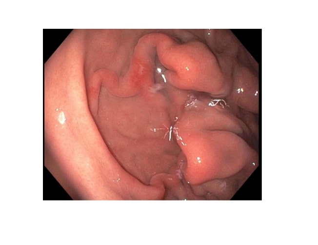
Cameron Lesion. Endoscopy showed linear erosion at the gastroesophageal junction, a finding consistent with a Cameron lesion.
Contributed by H Brar, MD
(Click Image to Enlarge)
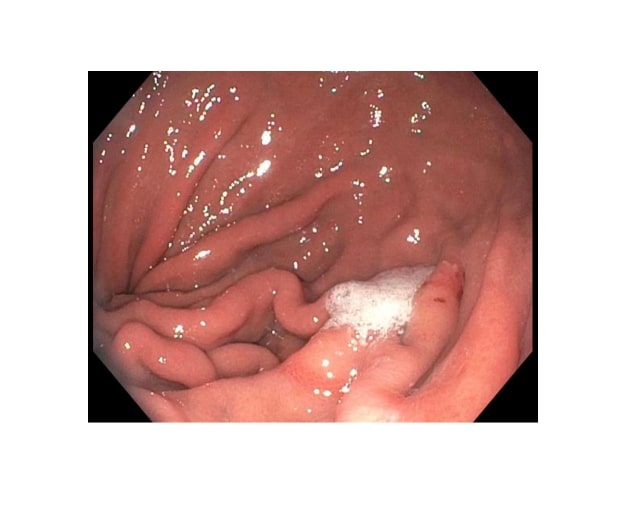
Cameron Lesion and Ulcer. Endoscopy findings of linear erosion and an ulcer at the gastroesophageal junction, consistent with a Cameron lesion.
Contributed by H Brar, MD
References
Cameron AJ, Higgins JA. Linear gastric erosion. A lesion associated with large diaphragmatic hernia and chronic blood loss anemia. Gastroenterology. 1986 Aug:91(2):338-42 [PubMed PMID: 3487479]
Moskovitz M, Fadden R, Min T, Jansma D, Gavaler J. Large hiatal hernias, anemia, and linear gastric erosion: studies of etiology and medical therapy. The American journal of gastroenterology. 1992 May:87(5):622-6 [PubMed PMID: 1595651]
Gray DM, Kushnir V, Kalra G, Rosenstock A, Alsakka MA, Patel A, Sayuk G, Gyawali CP. Cameron lesions in patients with hiatal hernias: prevalence, presentation, and treatment outcome. Diseases of the esophagus : official journal of the International Society for Diseases of the Esophagus. 2015 Jul:28(5):448-52. doi: 10.1111/dote.12223. Epub 2014 Apr 24 [PubMed PMID: 24758713]
Level 2 (mid-level) evidenceUygur FA, Aydin G, Ağirman E, Vural S, Dülger AC. A comprehensive endoscopic and clinical evaluation of Cameron ulcers: Insights from a single-center cross-sectional experience. Medicine. 2026 Apr 10:105(15):e48266. doi: 10.1097/MD.0000000000048266. Epub [PubMed PMID: 41961649]
Level 2 (mid-level) evidenceZullo A, Manta R, De Francesco V, Fiorini G, Lahner E, Vaira D, Annibale B. Cameron lesions: A still overlooked diagnosis. Case report and systematic review of literature. Clinics and research in hepatology and gastroenterology. 2018 Dec:42(6):604-609. doi: 10.1016/j.clinre.2018.05.002. Epub 2018 Jun 14 [PubMed PMID: 29910147]
Level 3 (low-level) evidenceBaumgartner T, Liu S, Li W, Giannopoulos S, Kalantar M, Selzer D, Ritter EM, Stefanidis D. Resolution and recurrence of anemia following repair of paraesophageal hernias. Surgical endoscopy. 2023 Nov:37(11):8708-8713. doi: 10.1007/s00464-023-10302-8. Epub 2023 Jul 31 [PubMed PMID: 37524917]
Nguyen N, Tam W, Kimber R, Roberts-Thomson IC. Gastrointestinal: Cameron's erosions. Journal of gastroenterology and hepatology. 2002 Mar:17(3):343 [PubMed PMID: 11982707]
Fakhre Yaseri H. Gender is a risk factor in patients with gastroesophageal reflux disease. Medical journal of the Islamic Republic of Iran. 2017:31():58. doi: 10.14196/mjiri.31.58. Epub 2017 Sep 8 [PubMed PMID: 29445687]
Verhoeff K, Dang JT, Deprato A, Kung JY, Switzer NJ, Birch DW, Wong C, Karmali S. Surgical management of hiatal hernia vs medical therapy to treat bleeding Cameron lesions: a systematic review and meta-analysis. Surgical endoscopy. 2021 Dec:35(12):7154-7162. doi: 10.1007/s00464-020-08135-w. Epub 2020 Nov 6 [PubMed PMID: 33159296]
Level 1 (high-level) evidenceRichter IA, Rabin MS. The 'riding' ulcer: A report of 3 cases. South African medical journal = Suid-Afrikaanse tydskrif vir geneeskunde. 1979 Oct 6:56(15):612-4 [PubMed PMID: 550413]
Level 3 (low-level) evidenceLe PTV, Nguyen HT, Dang C, Tran KN, Vo QC. A Case of Cameron Lesions: An Overlooked Cause of Anemia in Patients With Gastrointestinal Bleeding and Hiatal Hernia. Cureus. 2024 Jul:16(7):e65510. doi: 10.7759/cureus.65510. Epub 2024 Jul 27 [PubMed PMID: 39188491]
Level 3 (low-level) evidenceKatz J, Brar S, Sidhu JS. Histopathological characterization of a Cameron lesion. International journal of surgical pathology. 2012 Oct:20(5):528-30 [PubMed PMID: 22614163]
Li WF, Lei XP, Yu XQ, Lv L, Li XD. A case report of occult Cameron ulcer and a systematic review of the literature. Frontiers in medicine. 2025:12():1677344. doi: 10.3389/fmed.2025.1677344. Epub 2025 Sep 17 [PubMed PMID: 41041453]
Level 1 (high-level) evidenceCamus M, Jensen DM, Ohning GV, Kovacs TO, Ghassemi KA, Jutabha R, Machicado GA, Dulai GS, Hines OJ. Severe upper gastrointestinal hemorrhage from linear gastric ulcers in large hiatal hernias: a large prospective case series of Cameron ulcers. Endoscopy. 2013:45(5):397-400. doi: 10.1055/s-0032-1326294. Epub 2013 Apr 24 [PubMed PMID: 23616128]
Level 2 (mid-level) evidenceAuerbach M, DeLoughery TG, Tirnauer JS. Iron Deficiency in Adults: A Review. JAMA. 2025 May 27:333(20):1813-1823. doi: 10.1001/jama.2025.0452. Epub [PubMed PMID: 40159291]
Holt JM, Mayet FG, Warner GT, Callender ST, Gunning AJ. Iron absorption and blood loss in patients with hiatus hernia. British medical journal. 1968 Jul 6:3(5609):22-5 [PubMed PMID: 5301883]
Zaman A, Katon RM. Push enteroscopy for obscure gastrointestinal bleeding yields a high incidence of proximal lesions within reach of a standard endoscope. Gastrointestinal endoscopy. 1998 May:47(5):372-6 [PubMed PMID: 9609429]
Miller G. Linear gastric erosion associated with diaphragmatic hernia. Gastroenterology. 1987 Jan:92(1):271 [PubMed PMID: 3781200]
Level 3 (low-level) evidenceBornstein JD, Brazer SR. Cameron erosions. Gastrointestinal endoscopy. 1999 Apr:49(4 Pt 1):514 [PubMed PMID: 10202069]
Level 3 (low-level) evidenceWeston AP. Hiatal hernia with cameron ulcers and erosions. Gastrointestinal endoscopy clinics of North America. 1996 Oct:6(4):671-9 [PubMed PMID: 8899401]
Jabak S, Hashash JG. Large Cameron Ulcer Bleed. ACG case reports journal. 2020 Jan:7(1):e00308. doi: 10.14309/crj.0000000000000308. Epub 2020 Jan 17 [PubMed PMID: 32309501]
Level 3 (low-level) evidenceOrpen-Palmer J, Stanley AJ. Update on the management of upper gastrointestinal bleeding. BMJ medicine. 2022:1(1):e000202. doi: 10.1136/bmjmed-2022-000202. Epub 2022 Sep 28 [PubMed PMID: 36936565]
Lin CC, Chen TH, Ho WC, Chen TY. Endoscopic treatment of a Cameron lesion presenting as life-threatening gastrointestinal hemorrhage. Journal of clinical gastroenterology. 2001 Nov-Dec:33(5):423-4 [PubMed PMID: 11606864]
Level 3 (low-level) evidencePauwelyn KA, Verhamme M. Large hiatal hernia and iron deficiency anaemia: clinico-endoscopical findings. Acta clinica Belgica. 2005 Sep-Oct:60(4):166-72 [PubMed PMID: 16279396]
Level 2 (mid-level) evidenceRothstein R, Filipi C, Caca K, Pruitt R, Mergener K, Torquati A, Haber G, Chen Y, Chang K, Wong D, Deviere J, Pleskow D, Lightdale C, Ades A, Kozarek R, Richards W, Lembo A. Endoscopic full-thickness plication for the treatment of gastroesophageal reflux disease: A randomized, sham-controlled trial. Gastroenterology. 2006 Sep:131(3):704-12 [PubMed PMID: 16952539]
Level 1 (high-level) evidenceMalik TF, Anjum F. Dieulafoys Lesion Causing Gastrointestinal Bleeding(Archived). StatPearls. 2026 Jan:(): [PubMed PMID: 32965938]
Mehershahi S, Jog A, Ronderos DM, Shaikh D, Ihimoyan A. Cameron Ulcers: Rare Case of Overt Upper Gastrointestinal Bleed in a Patient with Alcohol Use Disorder. Cureus. 2020 Apr 12:12(4):e7644. doi: 10.7759/cureus.7644. Epub 2020 Apr 12 [PubMed PMID: 32411546]
Level 3 (low-level) evidenceCougard P, Sala JJ, Favre JP, Viard H. [Ulcers of the neck of hiatal hernias. 15 cases]. Journal de chirurgie. 1985 Jun-Jul:122(6-7):399-402 [PubMed PMID: 3876349]
Level 3 (low-level) evidenceMaganty K, Smith RL. Cameron lesions: unusual cause of gastrointestinal bleeding and anemia. Digestion. 2008:77(3-4):214-7. doi: 10.1159/000144281. Epub 2008 Jul 12 [PubMed PMID: 18622137]
Level 3 (low-level) evidence