 Tools
Tools
 Lipoid Proteinosis
Introduction
Erich Urbach and Camillo Wiethe, Austrian physicians, first described lipoid proteinosis in 1929.[1] Lipoid proteinosis is a rare genodermatosis characterized by the deposition of hyaline-like material in various tissues and organs. Lipoid proteinosis is associated with a variety of characteristic cutaneous and mucosal findings as well as several neurologic, psychiatric, and gastrointestinal manifestations.[2][3][4][5][6]
Etiology
Register For Free And Read The Full Article
 Search engine and full access to all medical articles
- 10 free questions in your specialty
- Free CME/CE Activities
- Free daily question in your email
- Save favorite articles to your dashboard
- Emails offering discounts
Learn more about a Subscription to StatPearls Point-of-Care
Etiology
Lipoid proteinosis is caused by a homozygous or compound heterozygous loss-of-function mutations in the extracellular matrix-1 (ECM1) gene located on chromosome 1q21.[7][8][9] Several mutation types (eg, missense, nonsense, deletion) and affected exons (eg, 6 and 7) have been reported, leading to variable genotypes and phenotypes.[10] Novel mutations continue to be identified.[7][9] Because lipoid proteinosis is inherited in an autosomal recessive pattern, patients often have affected family members, and some are children of consanguineous unions. The ECM1 gene encodes glycoproteins essential for the integrity of basement membranes and extracellular matrices, skin adhesion, and protein-protein interactions.[11][12]
Epidemiology
Lipoid proteinosis is extremely rare, with approximately 400 cases reported in the medical literature worldwide. Men and women are affected equally.[2] The Namaqualand region in South Africa has the largest population of individuals with lipoid proteinosis. These individuals share a common mutation, suggesting a founder effect.[8] There is also a higher incidence of lipoid proteinosis in countries with higher rates of consanguinity.[13][14][15]
Pathophysiology
ECM1 is a glycoprotein with multiple roles in skin structure and function. The ECM1 protein interacts with the basement membrane-specific heparan sulfate proteoglycan core protein perlecan, and other proteins important for extracellular matrix structure and tissue remodeling, including matrix metalloproteinase-9, fibulin-3, and laminin 332.[12][16][17] Furthermore, ultrastructural analysis of erosive lesions in a child with lipoid proteinosis revealed free-floating desmosomes and loss of connection between some desmosomes and keratinocytes, suggesting a possible role for ECM1 in the attachment of keratin filaments to desmosomes.[18] A splice variant of ECM1, ECM1b, is expressed in the stratum spinosum and granulosum, whereas the full-length ECM1a is more prevalent in the basal layer, suggesting that ECM1 contributes to keratinocyte differentiation.[19] Finally, ECM1 expression is reduced in individuals with chronic ultraviolet light exposure, signifying an additional role in the cutaneous stress response.[20]
Extracutaneous functions of ECM1 include regulation of endochondral bone and cartilage formation, endothelial cell proliferation, and angiogenesis.[21] High levels of ECM1 expression have been associated with certain cancers.[22][23] Furthermore, mutations in the ECM1 gene compromise protein-protein interactions and disrupt tissue homeostasis.[10][11][18] Hyaline-like accumulation in various tissues explains many classic clinical findings, such as hoarseness and moniliform blepharosis.
Histopathology
Histologically, lipoid proteinosis is characterized by hyaline-like periodic acid–Schiff-positive, diastase-resistant deposits within the papillary dermis and at the dermal-epidermal junction.[24] Skin microvascular changes include reduplication of blood vessel walls, enlarged vessels, and loss of the dermal capillary loop network.[25][26][27] Immunolabeling can inform diagnosis, especially early in the disease course. Reduced ECM1 protein, in conjunction with characteristic clinical findings, should heighten suspicion of lipoid proteinosis.[28] Congo red staining can help differentiate lipoid proteinosis from amyloidosis, which is often included in the differential diagnosis.[24]
History and Physical
In almost all affected individuals, the earliest manifestation is a weak or hoarse cry in early life due to hyaline-like deposits on the vocal cords.[29][30] Hoarseness and severe dysphonia can develop and persist throughout life.[31] Deposits within the larynx can occasionally obstruct the airway.[5]
Cutaneous abnormalities present early in the disease course, usually beginning with vesicles or bullae, especially on the face, that heal with prominent pock-like or acneiform scarring. (See Image. Early Cutaneous Findings of Lipoid Proteinosis.)[24][32] Over time, the skin may appear thickened and waxy, with a yellowish hue.[33] Other cutaneous manifestations include papules, nodules, and plaques on the face and lips, as well as verrucous and hyperkeratotic lesions at sites of trauma or friction.[33] Early clinical clues, including cutaneous buttock lesions, may predict a more severe clinical course with neurologic involvement.[34]
Moniliform blepharosis, characterized by beaded papules along the eyelid margin, is a pathognomonic finding identified in about 50% of patients. (See Image. Moniliform Blepharosis.)[35] Obstruction of the punctum by hyaline-like material may lead to epiphora. Similarly, infiltration of the lacrimal glands may cause dry eyes. More severe ocular manifestations, such as glaucoma and uveitis, have been rarely reported.[36][37][38]
Other notable features of lipoid proteinosis include infiltrative hyaline-like material throughout the mouth and upper aerodigestive tract. A cobblestone appearance of the oral mucosa, yellow lip nodules, lip eversion, vegetative lesions, and fissures at the lateral commissures can be seen in patients with extensive disease.[5][33] As a result of Stensen duct obstruction and secondary xerostomia, patients with lipoid proteinosis are prone to frequent upper respiratory tract infections, recurrent parotitis, and dental caries.[12][39][40] The submandibular gland can become similarly obstructed and inflamed. The tongue is often firm to palpation, with a shortened, thickened frenulum that restricts protrusion.[13]
Neurologic manifestations of lipoid proteinosis can be severe, including epilepsy, dystonia, progressive neuropsychiatric disorders (eg, memory loss and other cognitive impairment, behavioral changes, hallucinations, schizophreniform illness), and spontaneous central nervous system hemorrhage.[4][41][42][43][44] Bilateral, symmetric, comma-shaped calcifications of the amygdala are a pathognomonic radiologic finding. Calcification has also been reported in the hippocampus, basal ganglia, parahippocampal gyrus, uncinate gyrus, striatum, pineal gland, and perirhinal and parietal cortices.[3][45][46] Calcifications develop slowly and are seen more frequently in patients with longer disease duration.[3]
Results from one study of 10 patients with lipoid proteinosis showed that 6 had amygdaloid complex calcification and degeneration and 3 had decreased medial temporal lobe perfusion, suspicious for underlying calcification. However, despite these imaging findings, these 9 patients did not differ from healthy controls regarding cognitive tasks (eg, memory, attention, executive function); they differed in recognizing facial expressions, emotional processing, and odor-associative learning and recognition.[46] In findings from another analysis of 7 patients with intracranial calcifications, 4 had a seizure history, with onset in childhood or young adulthood.[41] Focal seizures were the most common seizure type, as demonstrated by video electroencephalography.
Rare reports of hyaline-like deposits throughout the gastrointestinal tract have been described, some of which regressed over time.[47] One patient presented with acute gastrointestinal tract hemorrhage due to deposits within the small bowel.[6][47] Recently, a cross-sectional study of patients with lipoid proteinosis in Turkey found simple renal cysts in 68% of patients; however, the clinical significance of these cysts was unknown because renal function and biochemical parameters were within the reference range.[48]
Evaluation
Because lipoid proteinosis follows an autosomal recessive pattern of inheritance, patients should be asked about family members with similar symptoms and about consanguinity. Synthesis of family history, physical examination, and histopathologic findings often raises suspicion of lipoid proteinosis; however, a definitive diagnosis requires identification of an ECM1 mutation. Patients with characteristic dermatologic manifestations should undergo a skin biopsy of lesions to confirm deposition of hyaline-like material.
An otolaryngologist should evaluate patients for airway involvement and potential obstruction. Similarly, patients with neurologic or neuropsychiatric symptoms should be evaluated by neurology and psychiatry. Neuroimaging may be necessary to identify intracranial calcifications and other involved brain regions. Other evaluations will depend on disease manifestations, but should include frequent dental, dermatologic, and ophthalmologic evaluations.
Treatment / Management
There have been no randomized clinical trials to establish a standard of care for lipoid proteinosis, and no Food and Drug Administration–approved treatments are available. Treatment plans should be individualized based on disease manifestations and the patient's preference for improved cosmetic outcomes.
Off-label systemic retinoids, such as acitretin (0.5 mg/kg/d), can be effective in treating cutaneous and laryngeal manifestations of lipoid proteinosis.[49][50][51] However, some patients treated with acitretin who had improved voice quality had limited, if any, cutaneous improvement.[52][53] Short-term courses of systemic corticosteroids can be effective for vesiculobullous skin lesions and oral ulceration.[2] Based on limited case reports, evidence regarding the efficacy of long-term oral dimethyl sulfoxide for the treatment of cutaneous lesions and hoarseness is conflicting.[54][55] Findings from one case report demonstrated clinical improvement with the chelating agent D-penicillamine (600 mg daily).[56](A1)
For patients with vocal cord involvement and impaired phonation, microlaryngosurgery for excision of vocal cord deposits and carbon dioxide laser procedures can be quite successful.[57][58] However, there is a risk of postoperative granulation tissue formation, fibrosis, and the need for follow-up interventions. Accordingly, some authors recommend surgical intervention only when the airway is compromised. (B3)
To minimize the appearance of scars and moniliform blepharosis, cosmetic procedures such as CO2 laser ablation, dermabrasion, cryotherapy, blepharoplasty, and erbium-doped yttrium aluminum garnet ablative laser have been employed, with varying efficacy.[59][60][61] More recently, techniques such as microwave treatments and plasma exeresis have been used successfully and safely.[62][63] (B3)
Management of neurologic and psychiatric manifestations of lipoid proteinosis should be tailored to each patient. Antiepileptic medications can minimize seizure frequency and severity. Antipsychotic medications can help manage behavioral changes and psychosis.[4][42](B2)
Differential Diagnosis
Differential diagnoses may differ between patients. Possible differential diagnoses can be stratified based on the presenting symptoms of lipoid proteinosis.
Cutaneous Lesions
- Herpes simplex infection
- Impetigo
- Epidermolysis bullosa
- Erythropoietic protoporphyria
- Lichen myxedematosus
- Systemic amyloidosis
- Nodular localized cutaneous amyloidosis
- Colloid milium
- Hyalinosis
- Xanthomas
- Scleromyxedema
- Leprosy
- Hydroa vacciniforme
- Incontinentia pigmenti
Hoarseness
- Laryngitis
- Laryngotracheitis
- Laryngotracheobronchitis
- Vocal cord nodules
- Gastroesophageal reflux disease
- Vocal cord polyps
- Laryngeal hemangiomas
- Laryngeal cysts
- Vocal cord paralysis
- Vocal cord hypertrophy
Macroglossia
- Systemic amyloidosis
- Hypothyroidism (congenital)
- Myxedema
- Acromegaly
Mesial Temporal Lobe Calcification
- Fahr disease
- Calcified glioma
- Raine syndrome
- Previous herpes encephalitis
- Dystonia
Prognosis
Lipoid proteinosis typically has a benign, slowly progressive course and is generally compatible with a normal lifespan. However, laryngeal involvement with respiratory obstruction and central nervous system complications can be life-threatening or impart substantial morbidity.
Complications
Lipoid proteinosis can present with a wide range of clinical manifestations due to hyaline deposition in the skin, mucosa, and various organs. Of the possible clinical complications, airway obstruction, seizures, and spontaneous central nervous system hemorrhage are the most severe. Hyaline deposition in the small bowel has also been linked to gastrointestinal tract bleeding.
Complications of surgical intervention on the vocal cords or larynx include bleeding, infection, and vocal cord scarring, leading to a change in voice quality and a possible need for repeated interventions. CCertain patients may be taking systemic medication to manage their disease, such as acitretin, systemic corticosteroids, antipsychotics, antiepileptics, and dopamine-modifying agents. These medications can have a variety of drug-specific adverse effects.
Consultations
An interdisciplinary approach is recommended for patients with lipoid proteinosis. Counseling will likely involve a dermatologist, an otolaryngologist, a neurologist, a psychiatrist, a dentist, and a geneticist, depending on disease manifestations. Like other autosomal recessive conditions, carrier testing and reproductive counseling should be offered to patients and their family members.
Deterrence and Patient Education
Patients with lipoid proteinosis and their families should be informed about available treatment options. Education should include the genetic basis of this condition to inform family planning. Clinicians should emphasize the importance of monitoring for potentially life-threatening complications, including airway obstruction, seizures, and central nervous system hemorrhage. Education should also include genetic counseling and a review of treatment options as well as the associated risks and adverse effects.
Enhancing Healthcare Team Outcomes
Lipoid proteinosis is a rare genetic disease caused by an autosomal recessive mutation in the ECM1 gene. Lipoid proteinosis typically presents in early childhood with a weak or hoarse cry. Children often develop crusted or vesicular lesions that heal with atrophic scarring. Other clinical features include moniliform blepharosis, inability to protrude the tongue, and seizures.
Lipoid proteinosis requires interprofessional collaboration for optimal patient care. Patients with suspected lipoid proteinosis should undergo a comprehensive evaluation by multiple specialists, including a dermatologist, neurologist, psychiatrist, otolaryngologist, dentist, and geneticist, to assess the disease burden and evaluate for potential life-threatening manifestations. Patients’ symptoms and quality of life should be assessed frequently. Additionally, genetic counseling may help patients better understand their condition and its mode of inheritance.
Media
(Click Image to Enlarge)
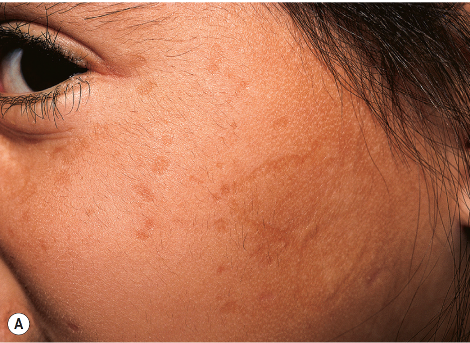
Early Cutaneous Findings of Lipoid Proteinosis. Scarring follows vesicle formation and hemorrhagic crusting early in the disease.
Paller AS, Mancini AJ. Hereditary disorders of the dermis. In: Paller AS, Mancini AJ. Hurwitz Clinical Pediatric Dermatology. 5th ed. New York, NY: Elsevier; 2016:119-135.
(Click Image to Enlarge)
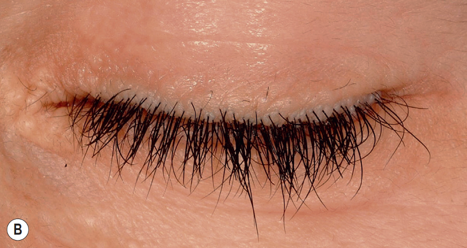
Moniliform Blepharosis. Approximately 50% of patients with lipoid proteinosis will have beaded papules along the eyelid margin.
Paller AS, Mancini AJ. Hereditary disorders of the dermis. In: Paller AS, Mancini AJ. Hurwitz Clinical Pediatric Dermatology. 5th ed. New York, NY: Elsevier; 2016:119-135.
References
KATZENELLENBOGEN I, UNGAR H. Lipoid proteinosis; reinvestigation of a case previously reported by Urbach and Wiethe in 1929. Dermatologica. 1957 Jul:115(1):23-35 [PubMed PMID: 13461538]
Level 3 (low-level) evidenceAn İ. Cutaneous findings and treatment responses of lipoid proteinosis patients. International journal of dermatology. 2025 Jan:64(1):119-129. doi: 10.1111/ijd.17331. Epub 2024 Jun 14 [PubMed PMID: 38876475]
Appenzeller S, Chaloult E, Velho P, de Souza EM, Araújo VZ, Cendes F, Li LM. Amygdalae calcifications associated with disease duration in lipoid proteinosis. Journal of neuroimaging : official journal of the American Society of Neuroimaging. 2006 Apr:16(2):154-6 [PubMed PMID: 16629738]
Level 3 (low-level) evidenceShukla S, Prajapati A, Parikh A, Oza Y, Parikh D, Gediya A. Neuropsychiatric Sequelae of Urbach- Wiethe Disease in Siblings: A Case Series. Indian journal of psychological medicine. 2024 Sep 25:():02537176241280018. doi: 10.1177/02537176241280018. Epub 2024 Sep 25 [PubMed PMID: 39564342]
Level 2 (mid-level) evidenceAlly M, Kinshuck AJ, Sandison A, Sandhu GS. The management of laryngeal lipoid proteinosis. The Journal of laryngology and otology. 2018 Oct:132(10):936-939. doi: 10.1017/S0022215118001329. Epub 2018 Aug 13 [PubMed PMID: 30099970]
Caccamo D, Jaen A, Telenta M, Varela E, Tiscornia O. Lipoid proteinosis of the small bowel. Archives of pathology & laboratory medicine. 1994 May:118(5):572-4 [PubMed PMID: 7514865]
Level 3 (low-level) evidenceLiu YL, Zhang ZY, Chen XM. A Sporadic Family of Lipoid Proteinosis with Novel ECM1 Gene Mutations. Clinical, cosmetic and investigational dermatology. 2024:17():885-889. doi: 10.2147/CCID.S452127. Epub 2024 Apr 18 [PubMed PMID: 38651074]
Van Hougenhouck-Tulleken W, Chan I, Hamada T, Thornton H, Jenkins T, McLean WH, McGrath JA, Ramsay M. Clinical and molecular characterization of lipoid proteinosis in Namaqualand, South Africa. The British journal of dermatology. 2004 Aug:151(2):413-23 [PubMed PMID: 15327549]
Level 2 (mid-level) evidenceLi M, Fischer J, Safwat S, Shoman W, Chazli YE, Alter S, Has C, Abdalla E. Lipoid proteinosis: Novel ECM1 pathogenic variants and intrafamilial variability in four unrelated Arab families. Pediatric dermatology. 2023 Jan:40(1):113-119. doi: 10.1111/pde.15105. Epub 2022 Aug 4 [PubMed PMID: 36670503]
Hamada T, Wessagowit V, South AP, Ashton GH, Chan I, Oyama N, Siriwattana A, Jewhasuchin P, Charuwichitratana S, Thappa DM, Jeevankumar B, Lenane P, Krafchik B, Kulthanan K, Shimizu H, Kaya TI, Erdal ME, Paradisi M, Paller AS, Seishima M, Hashimoto T, McGrath JA. Extracellular matrix protein 1 gene (ECM1) mutations in lipoid proteinosis and genotype-phenotype correlation. The Journal of investigative dermatology. 2003 Mar:120(3):345-50 [PubMed PMID: 12603844]
Level 3 (low-level) evidenceHamada T, McLean WH, Ramsay M, Ashton GH, Nanda A, Jenkins T, Edelstein I, South AP, Bleck O, Wessagowit V, Mallipeddi R, Orchard GE, Wan H, Dopping-Hepenstal PJ, Mellerio JE, Whittock NV, Munro CS, van Steensel MA, Steijlen PM, Ni J, Zhang L, Hashimoto T, Eady RA, McGrath JA. Lipoid proteinosis maps to 1q21 and is caused by mutations in the extracellular matrix protein 1 gene (ECM1). Human molecular genetics. 2002 Apr 1:11(7):833-40 [PubMed PMID: 11929856]
Chan I, Liu L, Hamada T, Sethuraman G, McGrath JA. The molecular basis of lipoid proteinosis: mutations in extracellular matrix protein 1. Experimental dermatology. 2007 Nov:16(11):881-90 [PubMed PMID: 17927570]
Frenkel B, Vered M, Taicher S, Yarom N. Lipoid proteinosis unveiled by oral mucosal lesions: a comprehensive analysis of 137 cases. Clinical oral investigations. 2017 Sep:21(7):2245-2251. doi: 10.1007/s00784-016-2017-7. Epub 2016 Nov 29 [PubMed PMID: 27900487]
Level 3 (low-level) evidenceKhan MAH, Reza MA, Sharaf IM, Alam MJ, Rahman MM, Chandra P, Anwar KS, Salam MA. Lipoid Proteinosis: Identification of a Novel Nonsense Mutation c.1246C}T:p.R416X in ECM1 gene from Bangladesh. Pakistan journal of medical sciences. 2023 Jul-Aug:39(4):1212-1215. doi: 10.12669/pjms.39.4.7437. Epub [PubMed PMID: 37492299]
Youssefian L, Vahidnezhad H, Daneshpazhooh M, Abdollahzadeh S, Talari H, Khoshnevisan A, Chams-Davatchi C, Mobasher R, Li Q, Uitto J, Akhondzadeh S, Tabrizi M. Lipoid proteinosis: phenotypic heterogeneity in Iranian families with c.507delT mutation in ECM1. Experimental dermatology. 2015 Mar:24(3):220-2. doi: 10.1111/exd.12620. Epub [PubMed PMID: 25529926]
Level 3 (low-level) evidenceSercu S, Lambeir AM, Steenackers E, El Ghalbzouri A, Geentjens K, Sasaki T, Oyama N, Merregaert J. ECM1 interacts with fibulin-3 and the beta 3 chain of laminin 332 through its serum albumin subdomain-like 2 domain. Matrix biology : journal of the International Society for Matrix Biology. 2009 Apr:28(3):160-9. doi: 10.1016/j.matbio.2009.02.003. Epub 2009 Mar 9 [PubMed PMID: 19275936]
Fujimoto N, Terlizzi J, Aho S, Brittingham R, Fertala A, Oyama N, McGrath JA, Uitto J. Extracellular matrix protein 1 inhibits the activity of matrix metalloproteinase 9 through high-affinity protein/protein interactions. Experimental dermatology. 2006 Apr:15(4):300-7 [PubMed PMID: 16512877]
Dyer JA, Yu QC, Paller AS. "Free-floating" desmosomes in lipoid proteinosis: an inherent defect in keratinocyte adhesion? Pediatric dermatology. 2006 Jan-Feb:23(1):1-6 [PubMed PMID: 16445401]
Level 3 (low-level) evidenceSmits P, Poumay Y, Karperien M, Tylzanowski P, Wauters J, Huylebroeck D, Ponec M, Merregaert J. Differentiation-dependent alternative splicing and expression of the extracellular matrix protein 1 gene in human keratinocytes. The Journal of investigative dermatology. 2000 Apr:114(4):718-24 [PubMed PMID: 10733679]
Sander CS, Sercu S, Ziemer M, Hipler UC, Elsner P, Thiele JJ, Merregaert J. Expression of extracellular matrix protein 1 (ECM1) in human skin is decreased by age and increased upon ultraviolet exposure. The British journal of dermatology. 2006 Feb:154(2):218-24 [PubMed PMID: 16433788]
Mongiat M, Fu J, Oldershaw R, Greenhalgh R, Gown AM, Iozzo RV. Perlecan protein core interacts with extracellular matrix protein 1 (ECM1), a glycoprotein involved in bone formation and angiogenesis. The Journal of biological chemistry. 2003 May 9:278(19):17491-9 [PubMed PMID: 12604605]
Han Z, Ni J, Smits P, Underhill CB, Xie B, Chen Y, Liu N, Tylzanowski P, Parmelee D, Feng P, Ding I, Gao F, Gentz R, Huylebroeck D, Merregaert J, Zhang L. Extracellular matrix protein 1 (ECM1) has angiogenic properties and is expressed by breast tumor cells. FASEB journal : official publication of the Federation of American Societies for Experimental Biology. 2001 Apr:15(6):988-94 [PubMed PMID: 11292659]
Rickman DS, Bobek MP, Misek DE, Kuick R, Blaivas M, Kurnit DM, Taylor J, Hanash SM. Distinctive molecular profiles of high-grade and low-grade gliomas based on oligonucleotide microarray analysis. Cancer research. 2001 Sep 15:61(18):6885-91 [PubMed PMID: 11559565]
Nanda A, Alsaleh QA, Al-Sabah H, Ali AM, Anim JT. Lipoid proteinosis: report of four siblings and brief review of the literature. Pediatric dermatology. 2001 Jan-Feb:18(1):21-6 [PubMed PMID: 11207965]
Level 3 (low-level) evidenceKowalewski C, Kozłowska A, Chan I, Górska M, Woźniak K, Jabłońska S, McGrath JA. Three-dimensional imaging reveals major changes in skin microvasculature in lipoid proteinosis and lichen sclerosus. Journal of dermatological science. 2005 Jun:38(3):215-24 [PubMed PMID: 15927815]
Level 3 (low-level) evidenceFabrizi G, Porfiri B, Borgioli M, Serri F. Urbach-Wiethe disease. Light and electron microscopic study. Journal of cutaneous pathology. 1980 Feb:7(1):8-20 [PubMed PMID: 7358884]
Level 3 (low-level) evidenceMoy LS, Moy RL, Matsuoka LY, Ohta A, Uitto J. Lipoid proteinosis: ultrastructural and biochemical studies. Journal of the American Academy of Dermatology. 1987 Jun:16(6):1193-201 [PubMed PMID: 2439554]
Level 3 (low-level) evidenceChan I, South AP, McGrath JA, Oyama N, Bhogal BS, Black MM, Hamada T. Rapid diagnosis of lipoid proteinosis using an anti-extracellular matrix protein 1 (ECM1) antibody. Journal of dermatological science. 2004 Aug:35(2):151-3 [PubMed PMID: 15265527]
Level 3 (low-level) evidenceSriwastava S, Desai A, Yuliati A, Watson CR, Sivaswamy L. A Child With a Hoarse Cry and Intracranial Calcification. Pediatric neurology. 2018 Oct:87():78-79. doi: 10.1016/j.pediatrneurol.2018.08.013. Epub 2018 Aug 22 [PubMed PMID: 30501889]
Regester RM, Antonson M, Harter NN. Clinicopathological Challenge: Five Males Presenting With Progressive Skin Thickening and Hoarse Voices. International journal of dermatology. 2025 Jul:64(7):1161-1163. doi: 10.1111/ijd.17766. Epub 2025 Apr 18 [PubMed PMID: 40248982]
Cocchi C, Milanese A, Abdul-Messie L, Vestri AR, Longo L. Laryngeal features in Lipoid proteinosis: a systematic review and meta-analysis of individual participant data. European archives of oto-rhino-laryngology : official journal of the European Federation of Oto-Rhino-Laryngological Societies (EUFOS) : affiliated with the German Society for Oto-Rhino-Laryngology - Head and Neck Surgery. 2024 Sep:281(9):4555-4564. doi: 10.1007/s00405-024-08713-x. Epub 2024 May 7 [PubMed PMID: 38713291]
Level 1 (high-level) evidenceNayak S, Acharjya B. Lipoid proteinosis in a six-year-old child. Indian dermatology online journal. 2012 Jan:3(1):25-7. doi: 10.4103/2229-5178.93490. Epub [PubMed PMID: 23130256]
Level 3 (low-level) evidenceHamada T. Lipoid proteinosis. Clinical and experimental dermatology. 2002 Nov:27(8):624-9 [PubMed PMID: 12472532]
Hamie L, Knio Z, Abbas O, Akel R, Bardawil T, Kibbi AG, Kurban M. Clinical clues early in the lives of individuals with lipoid proteinosis can determine the course of the disease. Clinical and experimental dermatology. 2017 Jun:42(4):428-430. doi: 10.1111/ced.13075. Epub 2017 Feb 28 [PubMed PMID: 28244239]
Belliveau MJ, Alkhotani A, Ali A. Moniliform Blepharosis of Lipoid Proteinosis. JAMA ophthalmology. 2015 Jul:133(7):e150688. doi: 10.1001/jamaophthalmol.2015.0688. Epub 2015 Jul 9 [PubMed PMID: 26158437]
Ng XG, Mpt J, Ang EL. A Rare Ocular Manifestation of Lipoid Proteinosis. Cureus. 2025 Apr:17(4):e82423. doi: 10.7759/cureus.82423. Epub 2025 Apr 17 [PubMed PMID: 40385837]
Ali MJ, Mishra DK, Naik MN. Punctal and peri-punctal involvement in Urbach-Wiethe syndrome: case report and review of literature. Orbit (Amsterdam, Netherlands). 2019 Dec:38(6):474-476. doi: 10.1080/01676830.2018.1550791. Epub 2018 Dec 20 [PubMed PMID: 30569803]
Level 3 (low-level) evidenceAbtahi SM, Kianersi F, Abtahi MA, Abtahi SH, Zahed A, Fesharaki HR, Abtahi ZA, Baradaran S, Mazloumi M, Naghiabadi S. Urbach-wiethe syndrome and the ophthalmologist: review of the literature and introduction of the first instance of bilateral uveitis. Case reports in medicine. 2012:2012():281516. doi: 10.1155/2012/281516. Epub 2012 Jul 31 [PubMed PMID: 22899939]
Level 3 (low-level) evidenceDi Giandomenico S, Masi R, Cassandrini D, El-Hachem M, De Vito R, Bruno C, Santorelli FM. Lipoid proteinosis: case report and review of the literature. Acta otorhinolaryngologica Italica : organo ufficiale della Societa italiana di otorinolaringologia e chirurgia cervico-facciale. 2006 Jun:26(3):162-7 [PubMed PMID: 17063986]
Level 3 (low-level) evidenceGunes B, Karadag R, Uzun L, Sımsek BC, Karadag AS. Is it always blepharitis? Urbach-Wiethe syndrome (lipoid proteinosis). Arquivos brasileiros de oftalmologia. 2019 May-Jun:82(3):242-244. doi: 10.5935/0004-2749.20190051. Epub 2019 Mar 25 [PubMed PMID: 30916217]
Oguz Akarsu E, Dinçsoy Bir F, Baykal C, Taşdemir V, Kara B, Bebek N, Gürses C, Uyguner O, Baykan B. The Characteristics and Long-Term Course of Epilepsy in Lipoid Proteinosis: A Spectrum From Mild to Severe Seizures in Relation to ECM1 Mutations. Clinical EEG and neuroscience. 2018 May:49(3):192-196. doi: 10.1177/1550059417705280. Epub 2017 Apr 23 [PubMed PMID: 28434238]
Thornton HB, Nel D, Thornton D, van Honk J, Baker GA, Stein DJ. The neuropsychiatry and neuropsychology of lipoid proteinosis. The Journal of neuropsychiatry and clinical neurosciences. 2008 Winter:20(1):86-92. doi: 10.1176/jnp.2008.20.1.86. Epub [PubMed PMID: 18305289]
Teive HA, Ruschel E, Munhoz RP. Spontaneous intracerebral hemorrhage in Urbach-Wiethe disease. Neurology. 2013 Apr 30:80(18):1720-1. doi: 10.1212/01.wnl.0000430259.37814.f5. Epub [PubMed PMID: 23628933]
Level 3 (low-level) evidenceAgredano PM, Del Barrio CM, Martinez MC, Cabrera CA. Intracranial calcifications associated with epilepsy: A case report of lipoid proteinosis. Seizure. 2020 Dec:83():172-174. doi: 10.1016/j.seizure.2020.10.027. Epub 2020 Nov 4 [PubMed PMID: 33161246]
Level 3 (low-level) evidenceChandrasekaran S, Nanjundan M, Natarajan S, Ramadhas K. Radiologic presentation of lipoid proteinosis with symmetrical medial temporal lobe calcifications. Radiology case reports. 2015:10(2):1121. doi: 10.2484/rcr.v10i2.1121. Epub 2016 Feb 17 [PubMed PMID: 27398129]
Level 3 (low-level) evidenceSiebert M, Markowitsch HJ, Bartel P. Amygdala, affect and cognition: evidence from 10 patients with Urbach-Wiethe disease. Brain : a journal of neurology. 2003 Dec:126(Pt 12):2627-37 [PubMed PMID: 12937075]
Custódio Lima J, Nagasako CK, Montes CG, Barcelos IH, de Carvalho RB, Mesquita MA. Gastrointestinal involvement in lipoid proteinosis: a ten-year follow-up of a brazilian female patient. Case reports in medicine. 2014:2014():952038. doi: 10.1155/2014/952038. Epub 2014 Jun 19 [PubMed PMID: 25045357]
Level 3 (low-level) evidenceYilmaz K, An I, Sahin S, Sahutoglu T. Are the kidneys involved in lipoid proteinosis? Study of 22 patients. International journal of clinical practice. 2021 Aug:75(8):e14329. doi: 10.1111/ijcp.14329. Epub 2021 May 17 [PubMed PMID: 33999483]
Bueno-Molina RC, Hernández-Rodríguez JC, Cabrera-Fuentes R, Cabrera-Pérez R, Conejo-Mir Sánchez J, Pereyra-Rodríguez JJ. Advances in treatment for lipoid proteinosis (Urbach-Wiethe disease): a case report and systematic review. Clinical and experimental dermatology. 2024 May 21:49(6):547-555. doi: 10.1093/ced/llae039. Epub [PubMed PMID: 38308656]
Level 1 (high-level) evidenceBakry OA, Samaka RM, Houla NS, Basha MA. Two Egyptian cases of lipoid proteinosis successfully treated with acitretin. Journal of dermatological case reports. 2014 Mar 31:8(1):29-34. doi: 10.3315/jdcr.2014.1168. Epub 2014 Mar 31 [PubMed PMID: 24748909]
Level 3 (low-level) evidenceAkoglu G, Karaduman A, Ergin S, Erkin G, Gokoz O, Unal OF, Hamada T. Clinical and histopathological response to acitretin therapy in lipoid proteinosis. The Journal of dermatological treatment. 2011 Jun:22(3):178-83. doi: 10.3109/09546631003639407. Epub 2010 Jul 28 [PubMed PMID: 20666665]
Level 3 (low-level) evidenceToosi S, Ehsani AH. Treatment of lipoid proteinosis with acitretin: a case report. Journal of the European Academy of Dermatology and Venereology : JEADV. 2009 Apr:23(4):482-3. doi: 10.1111/j.1468-3083.2008.02928.x. Epub [PubMed PMID: 18808438]
Level 3 (low-level) evidenceGündüz O, Sahiner N, Atasoy P, Senyücel C. Acitretin treatment for lipoid proteinosis. Case reports in dermatological medicine. 2012:2012():324506. doi: 10.1155/2012/324506. Epub 2012 Aug 9 [PubMed PMID: 23259080]
Level 3 (low-level) evidenceWong CK, Lin CS. Remarkable response of lipoid proteinosis to oral dimethyl sulphoxide. The British journal of dermatology. 1988 Oct:119(4):541-4 [PubMed PMID: 3191019]
Level 3 (low-level) evidenceOzkaya-Bayazit E, Ozarmağan G, Baykal C, Uluğ T. [Oral DMSO therapy in 3 patients with lipoidproteinosis. Results of long-term therapy]. Der Hautarzt; Zeitschrift fur Dermatologie, Venerologie, und verwandte Gebiete. 1997 Jul:48(7):477-81 [PubMed PMID: 9333627]
Level 3 (low-level) evidenceKaya TI, Kokturk A, Tursen U, Ikizoglu G, Polat A. D-penicillamine treatment for lipoid proteinosis. Pediatric dermatology. 2002 Jul-Aug:19(4):359-62 [PubMed PMID: 12220287]
Level 3 (low-level) evidenceXu W, Wang L, Zhang L, Liu HG, Zan F, Hu R, Liu WB, Han DM. [Manifestation and treatment of lipoid proteinosis in larynx]. Zhonghua er bi yan hou tou jing wai ke za zhi = Chinese journal of otorhinolaryngology head and neck surgery. 2010 Apr:45(4):301-4 [PubMed PMID: 20627049]
Harper JI, Duguid KP, Staughton RC, Moffat DA. Oropharyngeal and laryngeal lesions in lipoid proteinosis. The Journal of laryngology and otology. 1983 Sep:97(9):877-80 [PubMed PMID: 6886551]
Level 3 (low-level) evidenceRosenthal G, Lifshitz T, Monos T, Kachco L, Argov S. Carbon dioxide laser treatment for lipoid proteinosis (Urbach-Wiethe syndrome) involving the eyelids. The British journal of ophthalmology. 1997 Mar:81(3):253 [PubMed PMID: 9135394]
Level 3 (low-level) evidenceBuchan NG, Kemble JV. Successful surgical treatment of lipoid proteinosis. The British journal of dermatology. 1974 May:90(5):561-6 [PubMed PMID: 4833793]
Çalıskan E, Açıkgöz G, Tunca M, Koç E, Arca E, Akar A. Treatment of lipoid proteinosis with ablative Er:YAG laser resurfacing. Dermatologic therapy. 2015 Sep-Oct:28(5):291-5. doi: 10.1111/dth.12247. Epub 2015 Jun 2 [PubMed PMID: 26031844]
Zhang B, Zeng X, Jiang Y, Chen L, Xu X. Microwave treatment ameliorates beaded eyelid papules caused by lipoid proteinosis. Pediatric dermatology. 2024 Nov-Dec:41(6):1248-1250. doi: 10.1111/pde.15685. Epub 2024 Jun 23 [PubMed PMID: 38923547]
Araghi F, Moravvej Farshi H. A New Method to Remove Eyelid Lesions in Lipoid Proteinosis. Dermatologic surgery : official publication for American Society for Dermatologic Surgery [et al.]. 2024 Jun 1:50(6):594-595. doi: 10.1097/DSS.0000000000004143. Epub 2024 Mar 26 [PubMed PMID: 38530982]