 Tools
Tools
 Diagnostic Ultrasound Imaging: Physics Principles and Clinical Applications
Definition/Introduction
The development of ultrasound, or medical ultrasound, was built on the understanding and research of sound, which can be traced as far back as the sixth century. Specifically, sonography can be traced back to the 1700s, when an Italian physicist, Lazaro Spallanzani, studied bats' navigation.[1] In the early 20th century, French physicist Paul Langevin studied more applicable uses of ultrasound. He eventually laid the groundwork for SONAR (sound navigation and ranging) as he was commissioned to investigate the sunken Titanic. One of the first medical applications of ultrasound was by Ian Donald in the 1950s. As Ian Donald extrapolated ultrasound's ability to detect flaws and cracks in steel welds, he began researching and applying it to obstetrics and gynecology. He published a fetus at 14 weeks' gestational age in the prestigious academic journal The Lancet.[2]
Basic Ultrasound Physics
As the name implies, ultrasound uses sound waves at frequencies higher than those audible to the human ear. For example, the average human ear hears frequencies from 20 hertz to 20 kHz (kilohertz), whereas the typical clinical ultrasound uses frequencies from 1 to 20 MHz (megahertz). A basic understanding of sinusoidal waves is useful for better understanding ultrasound. Wavelength (λ) is inversely proportional to frequency (f) and proportionate to the velocity (v), as depicted in the following equation.
- λ = v/f
Modern-day ultrasounds use piezoelectric technology to convert electrical energy into sound waves and back again, using lead zirconate titanate (PZT), a human-made crystal (also called a ceramic). The vibration occurs when these crystals are electrically stimulated, producing longitudinal sound waves at the previously described frequency, thereby generating ultrasound.[3] PZT crystals are arranged in unique configurations within an ultrasound transducer (also known as a probe) for clinical use in various situations. Further details on the variation of these transducers will be discussed later.
As a rock dropped into a still pond, with waves bouncing off the shore, the sound waves emitted by the transducer resonate and refract back to the transducer. When the ultrasound reverberates off tissue and returns, it induces vibration into the PZT crystals, which are converted back into an electric signal. The processing computer within the ultrasound machine then generates the signals into 2D (2-dimensional) and, in more recent times, 3D (3-dimensional) images.
The frequency (and, inversely, wavelength) of the sound waves can be adjusted to achieve greater penetration through soft tissue by varying the power or gain. The lower the frequency, the deeper or greater the sound wave penetrance. Unfortunately, the higher the penetrance, the poorer the image quality. Lastly, some common terminology unique to the ultrasound field will be discussed. Hyperechoic is defined by an object that is brighter than the surrounding tissue. Hypoechoic or anechoic is the opposite, in which an object is darker. Both of which are determined by the object's density.
Instrumentation
The following section focuses on a variety of transducers. Each transducer is uniquely shaped and has a specific, intentional arrangement of PZT crystals to produce a distinct image. Each ceramic arrangement has a specific use for various clinical scenarios.
- Phased array transducer: The phased array transducer is one of the physically smaller probes with a usefully small footprint. Specifically, the phased array probe’s footprint can be angled between ribs to better visualize the heart or lungs. Due to the probe's size, the peripheral and superficial images have poorer focus and detail than those from other transducers. In addition to the footprint, the phased-array transducers use every crystal in the probe to produce the image.[4] Phased-array transducers are key to endoscopic ultrasound image quality, enabling electronic beam steering and focusing without mechanical movement, making them ideal for confined spaces. See Image. Ultrasound Phased Array Probe.
- Linear transducer: The linear transducer does not directly utilize every crystal, but each element is electrically stimulated or vibrated and subsequently excites adjacent crystals. Each crystal in the linear transducer is not directly electrically stimulated. The advantage of the linear probe is a larger field of view and a higher superficial focus. Common clinical applications of this probe are within the soft tissue and more superficial structures. See Image. Ultrasound Linear Probe.
- Endocavity probes: Endocavity probes are among the most distinctive in appearance. The wand-like shape and long neck allow sonographers to extend into orifices such as the oropharynx, rectum, or vagina (also called a transvaginal probe). Common clinical applications include surveillance of early pregnancies. Transabdominal imaging may not provide optimal imaging in smaller fetuses, but it can be used to diagnose and treat parapharyngeal abscesses. See Image. Endocavity Ultrasound Probe.
- Curvilinear (convex) transducer: Lastly, the curvilinear or curved-array transducer has a convex shape, whereas the 2 previously described transducers have a more linear shape. The curvilinear probe typically has the largest footprint of the 3. This probe is utilized in the endocervical, intrauterine, or abdominal cavity. The image produced by the probe is “C”-shaped, reflecting the probe’s shape. Superficial focus is poor, but the advantage is a wider overall field of view.[5] See Image. Ultrasound Curvilinear Probe. With advances in ultrasonography, new research has brought products to market that utilize a silicon chip in place of piezo crystals, enhancing ultrasound machines' ability to capture multiple images and guiding care beyond PZT imaging.
- Emerging transducer technologiesWith advances in ultrasonography, new research has brought products to market that utilize silicon-based capacitive micromachined ultrasonic transducers (CMUTs) in place of PZT crystals, enhancing ultrasound machines’ ability to acquire multiple images and guiding care beyond PZT imaging. A study proposed a silicon nanocolumn capacitive micromachined ultrasonic transducer (snCMUT) array for real-time wearable ultrasound imaging in disposable patches. Using a lead-free design, snCMUT achieves high transmission efficiency, flexibility, and low power consumption. Phantom imaging demonstrated superior performance with high axial and lateral resolutions (0.52 and 0.55 mm) and depth penetration (~70 mm) at low voltage (8.9 VPP). The device was successfully applied to monitor both sides of the human carotid arteries, offering clear ultrasound images and continuous blood pressure waveform monitoring. This innovation presents significant potential for continuous medical imaging and cardiovascular health assessment while addressing environmental concerns and reducing manufacturing costs (<$20).[6]
Issues of Concern
Register For Free And Read The Full Article
 Search engine and full access to all medical articles
- 10 free questions in your specialty
- Free CME/CE Activities
- Free daily question in your email
- Save favorite articles to your dashboard
- Emails offering discounts
Learn more about a Subscription to StatPearls Point-of-Care
Issues of Concern
Ultrasound is acoustic energy that interacts with human tissues, producing bioeffects that may be hazardous, especially in sensitive organs (brain, eye, heart, lung, and digestive tract) and embryos/fetuses. Two basic mechanisms of ultrasound interaction with biological systems have been identified: thermal and nonthermal. As a result, thermal and mechanical indices have been developed to assess the potential for biological effects from exposure to diagnostic US.[7] For diagnostic and research purposes, the ultrasound has been officially declared safe, and no harmful biological effects in humans have yet been demonstrated with new imaging modalities; however, physicians should be adequately informed about the potential risks of biological effects. US exposure, according to the ALARA (as low as reasonably achievable) principle, should be as low as reasonably possible.[7]
The mechanical index and thermal index are displayed on modern ultrasound machines as safety indicators. The mechanical index estimates the risk of mechanical bioeffects (primarily cavitation), while the thermal index estimates the risk of tissue heating.[8] These indices account for the physical mechanism of interaction between ultrasound and biological tissue, which depends on the temporal and spatial parameters of the acoustic field generated by ultrasound transducers.[8] Clinicians should monitor these indices during examinations, particularly in sensitive applications such as obstetric, pediatric, and ophthalmic ultrasound, and adhere to the ALARA principle
Image Optimization and Artifacts
To obtain ideal images when scanning human tissue, a sonographer must understand how to optimize the resolution. The following section focuses on the properties of ultrasound used to produce such images and the common artifacts arising from imperfections in both the tissue and the ultrasound that may cause major issues in the final images.
A sound wave penetrates deeper into a particular cavity; more interference is met. As such, the reverberant sound waves weaken as they reflect on the transducer. To optimize image quality, one must alter the gain. Altering the gain does not affect the sound-wave impulse emitted by the transducer; instead, it amplifies the echo returned to and received by the PZT crystals.
If the resulting image remains suboptimal despite optimizing the gain, the intensity or power of the emitted sound wave can be adjusted. If the image produced is too dim, the sound-wave intensity may be adjusted by varying the power. The greater the power of the sound waves, the deeper the waves typically penetrate. Power is measured in decibels (dB). As described above, increasing the frequency of PZT crystals produces a louder sound wave with greater power that penetrates deeper, but at the cost of more artifacts in the resulting image.
Despite proper optimization, several errors or artifacts can occur during image production. In a perfect world, there are some assumptions used to make these images. These assumptions are that sound waves travel in a straight line and reflect in that same line, sound travels at 1540 m/s, and that the only source of sound wave production is the transducer; any deviation from these assumptions produces artifacts.
When a sound wave encounters a medium such as air, through which sound waves poorly pass compared to water-based tissue, the artifact known as a shadow is produced. This appears as a hypoechoic or anechoic area seen distal to the area of gas or air, which obscures any views within the shadow. Besides air, a common cause of shadowing is gallstones, for which the shadow can be diagnostic in identifying gallstones within the gallbladder. See Image. Gallstone on Point-of-Care Ultrasound.
Comet tails typically have the appearance opposite of a shadow. A comet tail has a hyperechoic, or white, presentation. Common causes of this artifact include 2 closely approximated objects, such as a mechanical heart valve. With 2 objects close together, reverberation produces a hyperechoic streak. The last artifact to be discussed is a mirror image.
The artificial mirror image is always produced in parallel with the sound beam emitted from an ultrasound transducer. Gas or air is a common culprit behind various artifacts, and when a smooth, flat pocket of gas interacts with sound waves, it reflects sound in the same way a common household mirror does. Sonographers can utilize the mirror artifact and reflective surfaces to obtain views to their advantage.[9][10][11]
Clinical Significance
The clinical significance of sonography spans across multiple medical specialties and fields. Equally important is the training and education surrounding ultrasound, as it is a heavily user-dependent skill and tool. A poor sonographer, even with the highest-quality ultrasound, can produce inadequate images. Competent training and experience are crucial.
There are 3 regulatory organizations for sonographic certification: Cardiovascular Credentialing International, the American Registry of Radiologic Technologists, and the American Registry for Diagnostic Medical Sonography. Formal training takes 12 to 18 months and combines written and practical knowledge.[12] Outside of ultrasound technician training, medical residents and fellows study ultrasound. Specifically, cardiology fellows focus on cardiac applications, nephrology on renal imaging, and formal fellowships for ultrasound use by emergency medicine residents and attendings. Lastly, a radiology residency provides formal training in obtaining and interpreting sonographic images.[13]
Simulation-based training allows healthcare professionals to learn, practice, and improve their ultrasound imaging skills in a safe learning environment. Simulation-Based Medical Education enhances residents’ performance in both simulated environments and real clinical settings and is more effective than traditional clinical training methods. Results from a recent randomized controlled trial demonstrated that simulator-based training was more effective than traditional training, with a large effect size.[14]
Point-of-care ultrasound (POCUS) is a portable, affordable, and versatile diagnostic and procedural tool that enhances bedside decision-making. The simplicity and safety of this modality make it especially valuable in low- and middle-income countries, where access to advanced imaging is limited. POCUS helps bridge diagnostic gaps by enabling real-time, noninvasive assessment and procedural guidance across anesthesiology, perioperative care, critical care, and emergency medicine.[15]
Nursing, Allied Health, and Interprofessional Team Interventions
The integration of POCUS into nursing and allied health practice enhances diagnostic accuracy, patient safety, and care coordination, particularly in resource-limited settings. Bedside ultrasonography provides nurses with a valuable health assessment tool, yet barriers such as a lack of formal training, quality assurance mechanisms, and documentation pathways remain significant. Nurse practitioners can achieve proficiency with structured mentorship and simulation-based training. Task-sharing models in low- and middle-income countries have successfully trained midwives and nurses to perform obstetric POCUS, yielding safe, scalable workforce capacity building with high image-interpretation accuracy and positive impacts on patient management. Interprofessional education using blended learning, simulation, and team-based approaches has demonstrated significant improvements in ultrasound competence across medical and nursing staff.
Effective interprofessional ultrasound practice requires standardized credentialing, quality assurance, and ethical frameworks. Collaborative models that leverage radiology department expertise to train and credential physicians and advanced practitioners have shown significant growth in POCUS utilization and low false-positive rates when routine quality audits are performed. Ethical best practices include obtaining informed consent for educational POCUS, maintaining patient privacy, and coordinating disclosure of incidental findings with the primary clinical team. Close collaboration among radiologists, nurses, sonographers, and safety experts ensures proper equipment calibration, contrast administration protocols, and emergency preparedness, ultimately improving team performance and patient-centered outcomes.
Just as Ian Donald saw the use of ultrasound in obstetrics, many have since recognized its utility in other medical fields: cardiology, vascular surgery, anesthesiology, pain medicine, radiology, general surgery, nephrology, ophthalmology, urology, emergency medicine, obstetrics, and gynecology.[4][16] Ultrasound is a quick, efficient, and safe diagnostic and therapeutic tool in medicine. Team efforts can be taken to influence medical care and decision-making. As described above, the training to properly utilize medical ultrasound has many different pathways. Collectively, technicians, nursing staff, and clinicians benefit from the information obtained from sonography. The coordination subsequently increases patient care and eventual outcomes.
From the foundation of Lazaro Spallanzani’s research on sonography in bats to Ian Donald extrapolating its use from iron welds to the field of obstetrics, ultrasound is now an immediately available and safe diagnostic and therapeutic tool. The utility and applicability of ultrasound range from outpatient obstetrical offices to the acquisition of critical information in intensive care units. As technology develops, ultrasound machines are becoming smaller and more portable.[17] The future of ultrasound utility continues to expand and likely spans almost all medical specialties.
Media
(Click Image to Enlarge)
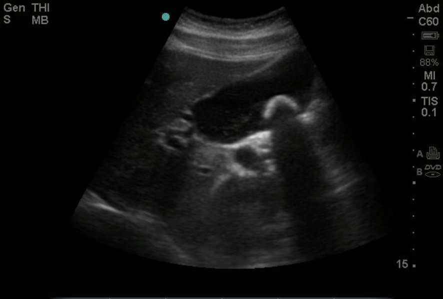
Gallstone, Ultrasound Image. This image shows a gallstone on point-of-care ultrasound.
Emory Emergency Medicine Ultrasound Section
(Click Image to Enlarge)
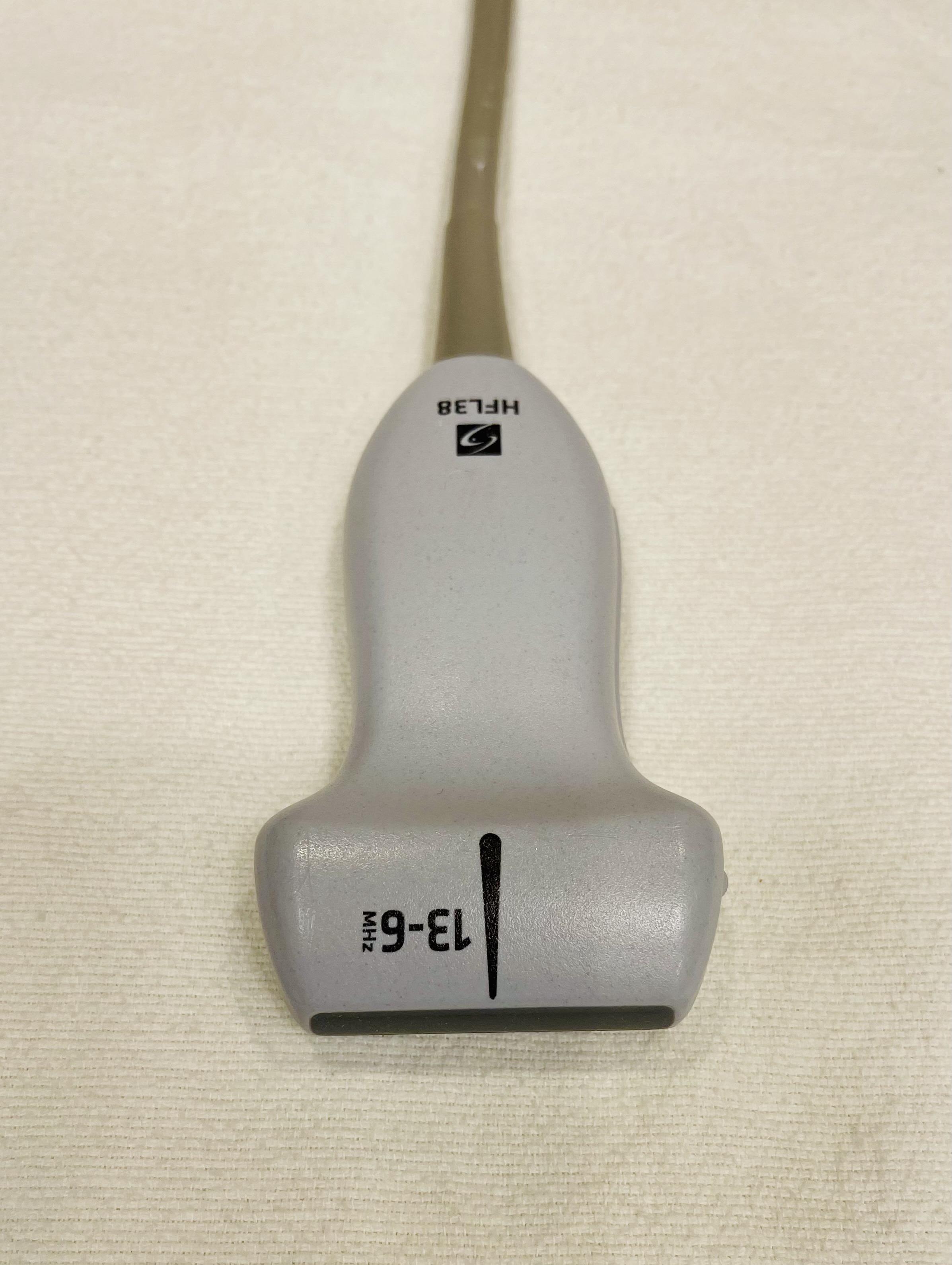
Ultrasound Linear Probe.
Contributed by C Borowy, DO
(Click Image to Enlarge)
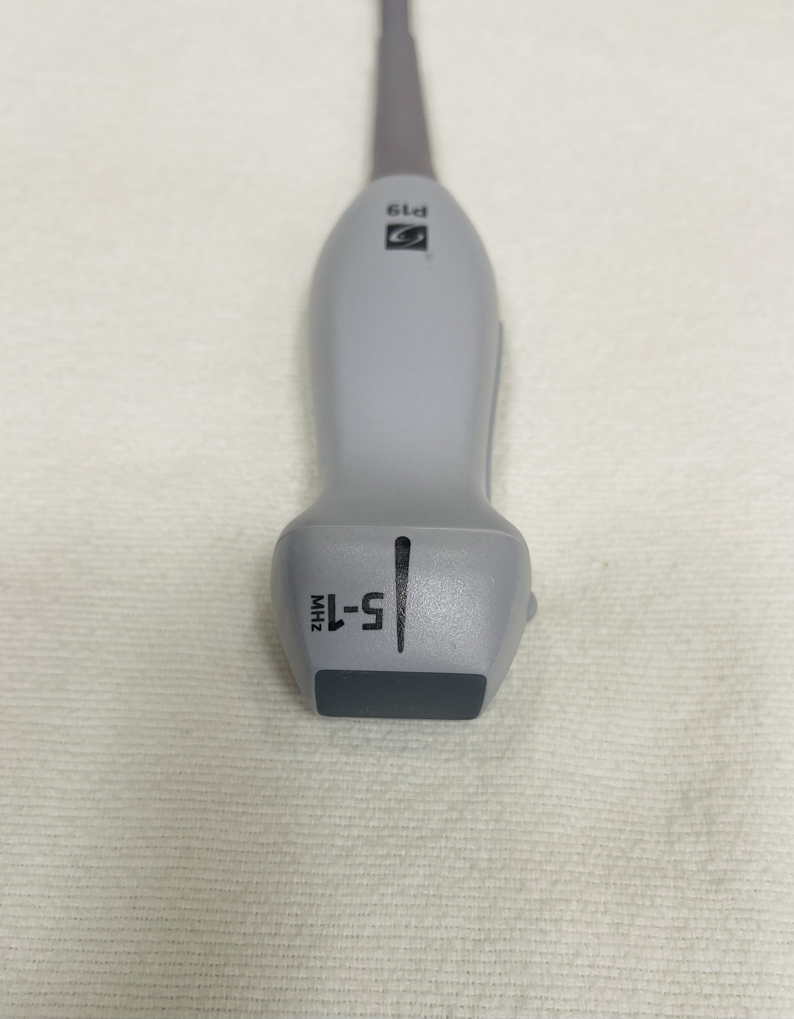
Ultrasound Phased Array Probe.
Contributed by C Borowy, DO
(Click Image to Enlarge)
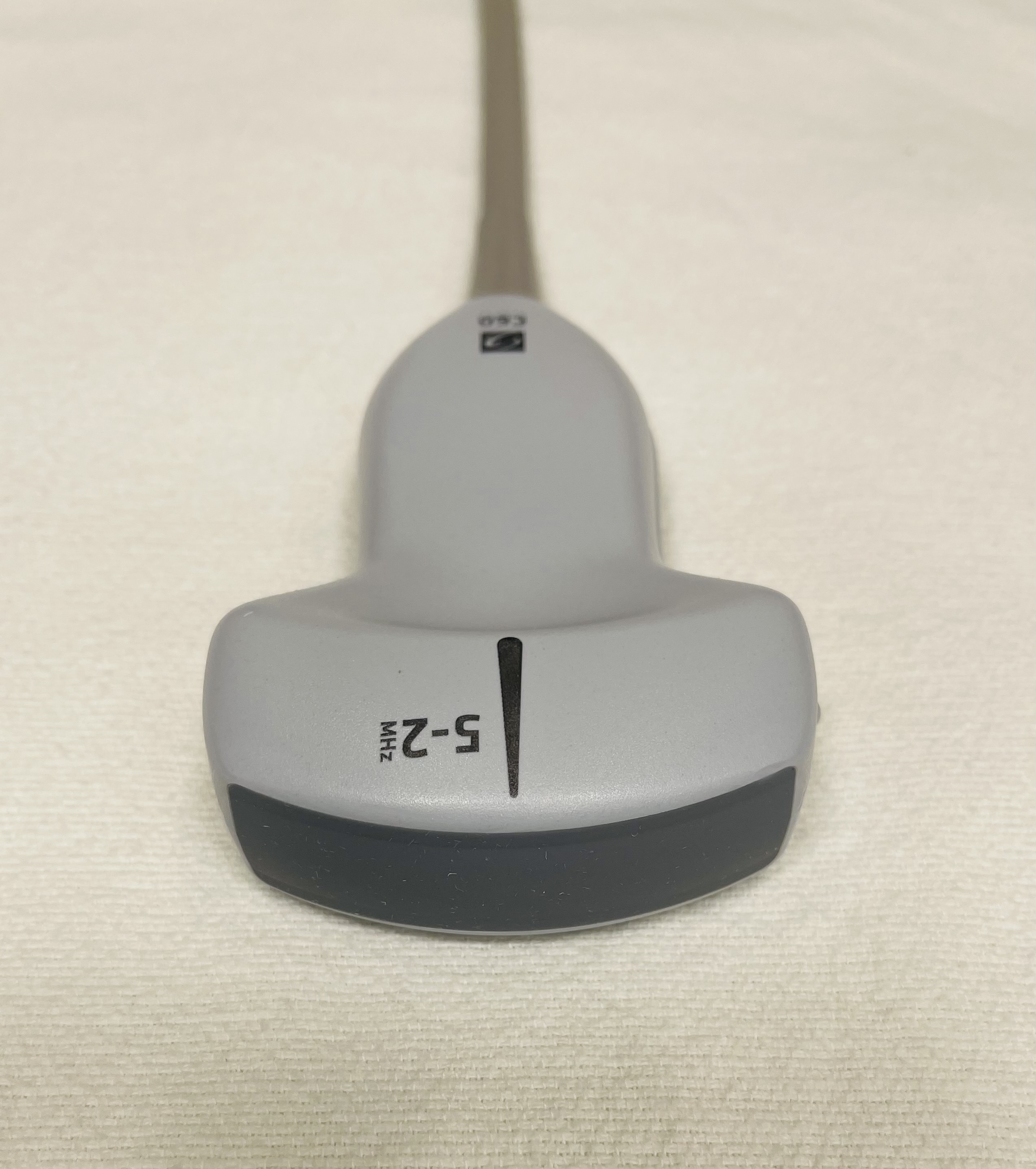
Ultrasound Curvilinear Probe.
Contributed by C Borowy, DO
(Click Image to Enlarge)
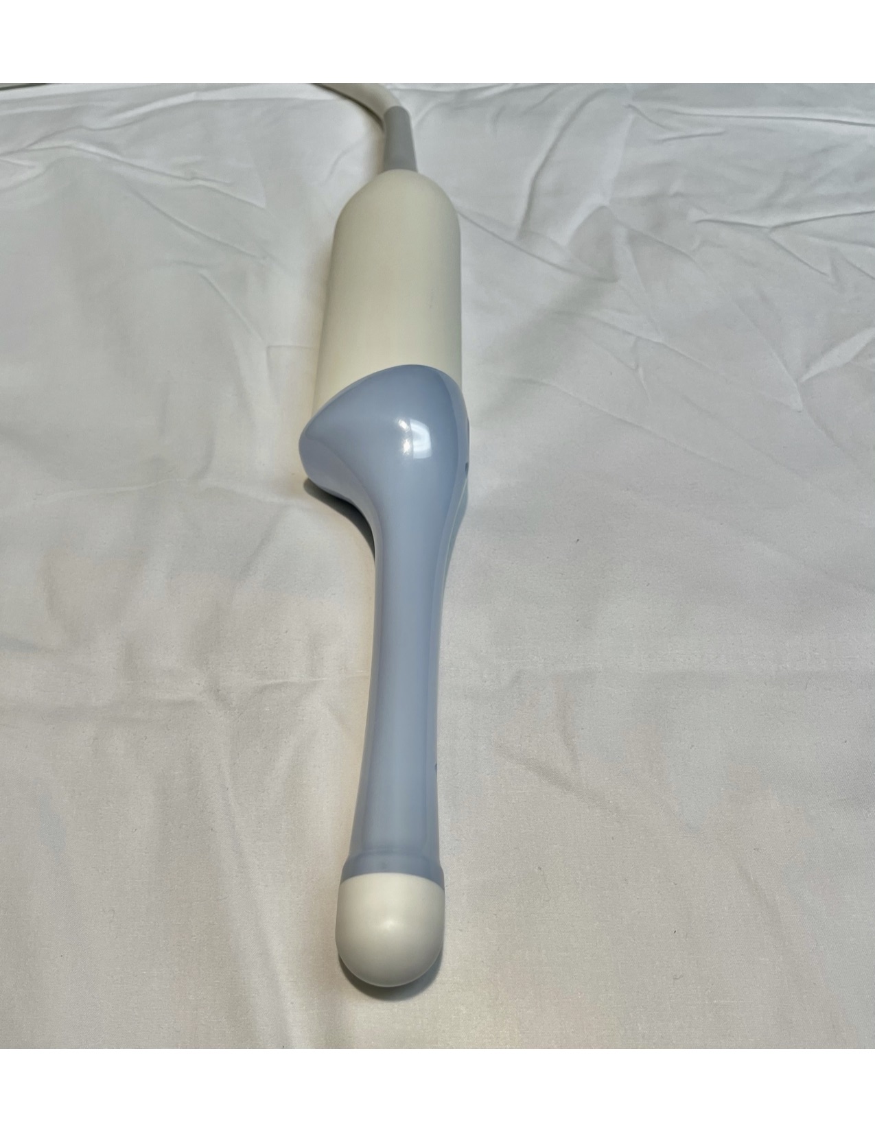
Endocavity Ultrasound Probe.
Contributed by C Borowy, DO
References
Ledermann D W. [Reading Spallanzani today]. Revista chilena de infectologia : organo oficial de la Sociedad Chilena de Infectologia. 2020 Feb:37(1):64-68. doi: 10.4067/S0716-10182020000100064. Epub [PubMed PMID: 32730402]
Chenhao Z, Ruqi Z, Xiaozhuo S, Jiabo W, Jiao L, Yajuan M. Ultrasound in medicine from 2014 to 2024: A bibliometric review. Medicine. 2026 Jan 9:105(2):e46890. doi: 10.1097/MD.0000000000046890. Epub [PubMed PMID: 41517659]
Level 2 (mid-level) evidenceMantsevich SN, Yushkov KB. Optimization of piezotransducer dimensions for quasicollinear paratellurite AOTF. Ultrasonics. 2021 Apr:112():106335. doi: 10.1016/j.ultras.2020.106335. Epub 2020 Dec 30 [PubMed PMID: 33395592]
Prabhu M, Raju D, Pauli H. Transesophageal echocardiography: instrumentation and system controls. Annals of cardiac anaesthesia. 2012 Apr-Jun:15(2):144-55. doi: 10.4103/0971-9784.95080. Epub [PubMed PMID: 22508208]
Matthias I, Panebianco NL, Maltenfort MG, Dean AJ, Baston C. Effect of Machine Settings on Ultrasound Assessment of B-lines. Journal of ultrasound in medicine : official journal of the American Institute of Ultrasound in Medicine. 2020 Dec 2:40(10):2039-46. doi: 10.1002/jum.15581. Epub 2020 Dec 2 [PubMed PMID: 33289208]
Kang DH, Cho S, Kim HY, Shim S, Kim DH, Jeong B, Lee YS, Park EA, Lee W, Kim H, Khuri-Yakub BT, Im M, Jeong JW, Lee BC. Silicon nanocolumn-based disposable and flexible ultrasound patches. Nature communications. 2025 Jul 18:16(1):6609. doi: 10.1038/s41467-025-61903-x. Epub 2025 Jul 18 [PubMed PMID: 40676025]
Quarato CMI, Lacedonia D, Salvemini M, Tuccari G, Mastrodonato G, Villani R, Fiore LA, Scioscia G, Mirijello A, Saponara A, Sperandeo M. A Review on Biological Effects of Ultrasounds: Key Messages for Clinicians. Diagnostics (Basel, Switzerland). 2023 Feb 23:13(5):. doi: 10.3390/diagnostics13050855. Epub 2023 Feb 23 [PubMed PMID: 36899998]
Nowicki A. Safety of ultrasonic examinations; thermal and mechanical indices. Medical ultrasonography. 2020 May 11:22(2):203-210. doi: 10.11152/mu-2372. Epub [PubMed PMID: 32399527]
Goffi A, Kruisselbrink R, Volpicelli G. The sound of air: point-of-care lung ultrasound in perioperative medicine. Canadian journal of anaesthesia = Journal canadien d'anesthesie. 2018 Apr:65(4):399-416. doi: 10.1007/s12630-018-1062-x. Epub 2018 Feb 6 [PubMed PMID: 29411300]
Sites BD, Brull R, Chan VW, Spence BC, Gallagher J, Beach ML, Sites VR, Hartman GS. Artifacts and pitfall errors associated with ultrasound-guided regional anesthesia. Part I: understanding the basic principles of ultrasound physics and machine operations. Regional anesthesia and pain medicine. 2007 Sep-Oct:32(5):412-8 [PubMed PMID: 17961841]
Level 3 (low-level) evidenceSchafer ME. Fundamentals of High-Resolution Ultrasound in Breast Implant Screening for Plastic Surgeons. Clinics in plastic surgery. 2021 Jan:48(1):59-69. doi: 10.1016/j.cps.2020.08.001. Epub 2020 Sep 19 [PubMed PMID: 33220905]
Haynes KW. The Importance of Professional Values From Radiologic Technologists' Perspective. Radiologic technology. 2020 Jul:91(6):525-532 [PubMed PMID: 32606230]
Level 3 (low-level) evidenceShokoohi H, Duggan NM, Adhikari S, Selame LA, Amini R, Blaivas M. Point-of-care ultrasound stewardship. Journal of the American College of Emergency Physicians open. 2020 Dec:1(6):1326-1331. doi: 10.1002/emp2.12279. Epub 2020 Oct 11 [PubMed PMID: 33392540]
Zhou J, Zhao Y, Zhou P, Tang X, Liu W, Guo S, Wang J, Li L. Evaluating the effectiveness of high-fidelity simulation-based training on clinical skill transfer in obstetric ultrasound residents: a prospective randomized controlled trial. BMC medical education. 2026 Feb 17:26(1):. doi: 10.1186/s12909-026-08835-2. Epub 2026 Feb 17 [PubMed PMID: 41699591]
Level 1 (high-level) evidenceDoyal AS, Sholes P, Drum E, Tesfay B, Sileshi B. Point-of-Care Ultrasound: A High-Tech Solution for Low- and Middle-Income Countries. Cureus. 2025 May:17(5):e83520. doi: 10.7759/cureus.83520. Epub 2025 May 5 [PubMed PMID: 40470437]
Guner NG, Yurumez Y, Yucel M, Alacam M, Guner ST, Ercan B. Effects of Point-of-care Ultrasonography on the Diagnostic Process of Patients Admitted to the Emergency Department with Chest Pain: A Randomised Controlled Trial. Journal of the College of Physicians and Surgeons--Pakistan : JCPSP. 2020 Dec:30(12):1262-1268. doi: 10.29271/jcpsp.2020.12.1262. Epub [PubMed PMID: 33397050]
Level 1 (high-level) evidenceAl-Naser Y, Alshadeedi F. Bringing imaging to the people: Enhancing access and equity in healthcare through mobile imaging. Journal of medical imaging and radiation sciences. 2024 Dec:55(4):101715. doi: 10.1016/j.jmir.2024.101715. Epub 2024 Jul 23 [PubMed PMID: 39047372]