 Tools
Tools
 Diphtheria
Introduction
Diphtheria is a vaccine-preventable infectious disease caused primarily by Corynebacterium diphtheriae, a gram-positive bacillus that can produce a potent exotoxin. Clinical presentations range from asymptomatic carriage to cutaneous disease and respiratory tract infection, with respiratory tract diphtheria generally representing the more severe form. A hallmark feature of respiratory tract diphtheria is the formation of an adherent pharyngeal pseudomembrane, a classic diagnostic finding. The term diphtheria is derived from the Greek word diphthera, meaning leather or hide, reflecting the leathery appearance of this membrane.[1]
Humans are the only known reservoir for Corynebacterium diphtheriae, which primarily colonizes the upper respiratory tract. Transmission occurs mainly through respiratory droplets from infected individuals or asymptomatic carriers, although direct contact with cutaneous lesions may also contribute to spread. Diphtheria occurs more frequently during the winter and spring months. In untreated individuals, transmission may persist for approximately 2 to 6 weeks. Individuals at greatest risk include those who are inadequately immunized, have waning antitoxin immunity, or have close contact with infected persons or asymptomatic carriers.[1]
Etiology
Register For Free And Read The Full Article
 Search engine and full access to all medical articles
- 10 free questions in your specialty
- Free CME/CE Activities
- Free daily question in your email
- Save favorite articles to your dashboard
- Emails offering discounts
Learn more about a Subscription to StatPearls Point-of-Care
Etiology
Corynebacterium diphtheriae organisms are non–spore-forming, gram-positive bacilli that lack motility and a capsule. Microscopically, the organism characteristically appears in palisade formations or angular arrangements resembling V- or L-shapes.[2] Respiratory tract diphtheria is most commonly associated with toxigenic strains of C diphtheriae. The 4 recognized biotypes are gravis, intermedius, mitis, and belfanti, all of which have been implicated in both endemic disease and outbreak settings.[2]
Other Corynebacterium spp may also produce diphtheria-like illness. Corynebacterium ulcerans is primarily associated with cutaneous disease but has occasionally been reported as a cause of respiratory tract diphtheria. In some cases, infection can progress to severe toxin-mediated illness requiring prompt treatment with diphtheria antitoxin and appropriate antimicrobial therapy, including macrolides. Unlike C diphtheriae, C ulcerans is considered a zoonotic pathogen, and evidence supporting sustained person-to-person transmission remains limited.[3][CDC. Toxigenic Corynebacterium ulcerans in Humans and Household Pets]
Epidemiology
Transmission and Reservoir
Corynebacterium diphtheriae organisms spread primarily through respiratory secretions from infected individuals or asymptomatic carriers.[1] Transmission occurs mainly via respiratory droplets, while direct contact with cutaneous lesions may also contribute. Humans are the primary reservoir, with colonization occurring predominantly in the upper respiratory tract. No clear racial or sex predilection has been consistently demonstrated. Disease outcomes are influenced primarily by immunization status, access to antitoxin, early diagnosis, and supportive care. Diphtheria occurs year-round but is more common during colder months in temperate climates.[4]
Global Burden of Disease
Before widespread vaccination, diphtheria was a major cause of childhood morbidity and mortality worldwide. Routine immunization with diphtheria toxoid led to a marked decline in incidence and transformed the disease into a rare infection in many resource-rich countries.[5] Despite these advances, diphtheria remains endemic in parts of Africa, Asia, the Middle East, and Latin America, particularly in regions with low vaccine coverage, limited healthcare access, humanitarian crises, or population displacement. Recent outbreaks in Europe, Africa, and Latin America highlight the continued risk of reemergence when herd immunity declines.[6][7]
Historically, diphtheria primarily affected children younger than 12 years. Following widespread childhood vaccination, disease epidemiology shifted toward adolescents and adults, particularly individuals with incomplete immunization or waning immunity. Adults older than 40 years now account for an increasing proportion of cases in many settings.[8]
Risk factors for infection include:
- Lack of vaccination or incomplete immunization
- Failure to receive booster doses
- Waning antitoxin immunity
- Residence in endemic areas
- International travel to regions with active transmission
- Crowded living conditions, homelessness, and refugee settings
- Exposure to infected individuals or asymptomatic carriers
- Limited healthcare access and disrupted public health infrastructure [4][9]
Pathophysiology
The clinical manifestations of diphtheria result from both localized infection and the effects of diphtheria exotoxin. Diphtheria typically involves the respiratory tract mucosa or skin, producing local inflammation and tissue injury, while systemic complications arise from hematogenous dissemination of the toxin.[4] Virulence depends primarily on the production of diphtheria toxin by toxigenic strains of Corynebacterium diphtheriae. Toxin synthesis occurs only when the organism is infected by a lysogenic bacteriophage carrying the tox gene. Nontoxigenic strains generally cause milder disease and are less likely to produce severe systemic complications.[10]
Diphtheria toxin is composed of subunits A and B. The B subunit mediates attachment to host cells by binding to the heparin-binding epidermal growth factor–like precursor, which serves as the cellular receptor. Following receptor-mediated endocytosis, the toxin enters the cytoplasm, where the A subunit exerts enzymatic activity. The catalytic fragment transfers adenosine diphosphate-ribose from nicotinamide adenine dinucleotide to elongation factor 2, thereby inhibiting protein synthesis and leading to cell death.[2][10]
At the site of infection, the host's inflammatory response, combined with toxin-mediated tissue necrosis, results in the formation of the characteristic gray-white pseudomembrane involving the tonsils, pharynx, or larynx. This membrane consists of necrotic epithelial cells, fibrin, inflammatory cells, and bacterial colonies and may contribute to airway obstruction in severe cases.[7] Local tissue injury facilitates lymphatic and vascular spread of the toxin to distant organs. The myocardium and peripheral nervous system are particularly susceptible to toxin-mediated damage, leading to complications such as myocarditis, conduction abnormalities, cranial neuropathies, and peripheral neuropathy. Renal injury and other systemic manifestations may also occur in severe disease. The extent of toxin production and host immunity largely determines disease severity and clinical outcome.[2][10]
History and Physical
History
Infection caused by Corynebacterium diphtheriae most commonly presents as respiratory or cutaneous diphtheria. Although disease is frequently localized, toxin-mediated systemic complications may involve the cardiovascular, neurologic, and renal systems, resulting in myocarditis, neuropathies, and kidney injury. The incubation period is generally 2 to 5 days, with a range of 1 to 10 days. Clinicians should obtain travel history to endemic regions and vaccination status, particularly in areas where diphtheria is uncommon.
Respiratory diphtheria classically presents with the formation of a thick, gray, adherent pseudomembrane involving the tonsils, pharynx, or larynx. Early findings often include mild pharyngeal erythema that progresses to membrane formation. The pseudomembrane consists of fibrin, necrotic epithelial cells, inflammatory cells, erythrocytes, leukocytes, and bacterial debris. Removal of the membrane frequently causes bleeding because of its firm attachment to the underlying tissue.[11] Common symptoms include low-grade fever, sore throat, malaise, cervical lymphadenopathy, and headache.[4]
Marked cervical lymphadenopathy and soft-tissue edema may produce the characteristic bull-neck appearance. Airway obstruction due to extensive pseudomembrane formation is a major cause of mortality. Cutaneous diphtheria typically appears as chronic, nonhealing ulcerative lesions covered by a gray membrane. These lesions usually remain localized without deep tissue invasion and often develop in areas of prior skin injury, trauma, or underlying dermatologic disease.[12]
Physical Examination
Patients typically exhibit a low-grade fever but appear toxic, often accompanied by a swollen neck. General symptoms include fever, tachycardia, halitosis, and anxiety. A thick, gray, leathery membrane covers the tonsils, oropharynx, soft palate, nasopharynx, and uvula (see Image. Diphtheria in the Oral Cavity). Scraping the pseudomembrane causes bleeding of the underlying mucosa. A patient may hold the head extended due to a bull neck. Dysphonia may occur occasionally. Respiratory distress manifests as stridor, wheeze, cyanosis, and retractions.[9] Cardiac involvement usually occurs 1 to 2 weeks into the illness, following improvement in the pharyngeal phase.[13] Cardiac involvement may manifest as follows:
- Myocarditis can present acutely with congestive heart failure and circulatory collapse. Patients can have more subtle features, such as progressive dyspnea, cardiac chamber dilatation, diminished heart sounds, and weakness.[14]
- Electrocardiographic changes may include ST segment and T wave changes, atrioventricular blocks, and various dysrhythmias.
- Endocarditis may occur, particularly in the presence of an artificial valve.[15]
Neurological involvement depends on the extent of the pharyngeal infection. Deficits include the following:
- Cranial nerve deficits
- Stocking and glove peripheral sensory neuropathy
- Peripheral neuritis [16]
Evaluation
Diphtheria is primarily a clinical diagnosis supported by microbiologic confirmation. Suspected cases require immediate isolation, notification of public health authorities, and prompt treatment; therapy should not be delayed while awaiting confirmation.[CDC. Guidance for Diphtheria]
Microbiologic Evaluation
When diphtheria is suspected, culture of the site of infection, such as the throat, nasopharynx, pseudomembrane, or skin wound, should be performed. Specimens should be collected before antibiotics are started, when feasible. Clinicians should notify the laboratory when diphtheria is suspected so appropriate selective or enrichment media can be used.
Colonies of gram-positive bacilli isolated on culture can then be further identified by additional tests, including biochemical assays, matrix-assisted laser desorption/ionization-time-of-flight mass spectrometry, or polymerase chain reaction (PCR).[17]. Culture may be performed on media such as Löffler medium, cystine-tellurite blood agar, Tinsdale medium, or tellurite-containing agar, which support the isolation of Corynebacterium diphtheriae.[CDC. Laboratory Testing for Diphtheria]
Toxigenicity testing: If C diphtheriae is isolated, further testing is required to determine whether the strain is toxigenic. Testing usually involves detection of the diphtheria toxin gene by polymerase chain reaction and confirmation of actual toxin production, most commonly by the Elek immunodiffusion test or another validated toxin-production assay. Identification of C diphtheriae alone, or detection of the tox gene alone, is insufficient to confirm toxigenic diphtheria because some strains carry the gene but do not produce toxin. Testing is typically coordinated through local or state public health laboratories, with CDC support in the US [CDC. Clinical Guidance for Diphtheria].
Additional Evaluation for Complications
- Complete blood count:
- May show leukocytosis
- Nonspecific finding
- Cardiac evaluation (if myocarditis suspected):
- Electrocardiography
- Troponin levels
- Echocardiography for heart failure, conduction abnormalities, or hemodynamic instability
- Imaging (when clinically indicated):
- Chest radiography for pneumonia or aspiration
- Neck imaging for airway compromise
Serology
- Diphtheria antitoxin serology may assess immune status
- Not recommended for the diagnosis of acute infection
Treatment / Management
The primary treatment approaches for diphtheria involve prompt administration of diphtheria antitoxin and antibiotics. Clinicians should also perform a thorough assessment for potential respiratory and cardiovascular instability. In suspected cases of diphtheria, antitoxin should be administered urgently based on clinical judgment without waiting for laboratory confirmation. Suspected cases must be isolated, and stringent droplet precautions should be implemented. Additionally, clinicians should evaluate the patient for signs of respiratory distress, and airway protection is imperative when indicated. Early management also involves continuous cardiac monitoring to address potential cardiovascular complications.
Diphtheria Antitoxin
Diphtheria antitoxin, derived from horse antiserum, is crucial for treatment.[18] The antitoxin neutralizes unbound diphtheria toxin circulating in the blood. However, the antitoxin becomes ineffective once the toxin has bound to the cell membrane. The antitoxin dose is tailored to the patient's clinical state and the severity of the condition, and it can be administered intramuscularly or intravenously. Before antitoxin administration, the patient should undergo hypersensitivity testing, and emergency medications for anaphylaxis must be readily available at the bedside.[4](A1)
Antibiotic Treatment
The antibiotics of choice for treating diphtheria are erythromycin or penicillin G.[19] Erythromycin is prescribed at a dose of 500 mg 4 times daily for 2 weeks. Penicillin G is administered intramuscularly at a dose of 300,000 units every 12 hours for patients weighing 10 kg or less and 600,000 units every 12 hours for those weighing more than 10 kg. Once the patient is able to take oral medication, oral penicillin V can be initiated at 250 mg 4 times daily for 2 weeks. Early initiation of antibiotics is crucial to promptly eliminate the organism, limit toxin release, expedite patient recovery, and prevent the infection from spreading to close contacts. In cases of antibiotic resistance, linezolid or vancomycin may be considered.(A1)
Close contacts, including household members and individuals in direct contact with the infected patient, should receive postexposure prophylaxis. Healthcare professionals exposed to a patient's respiratory secretions should also receive postexposure prophylaxis. Postexposure prophylaxis involves a single dose of penicillin G administered as follows:
- 600,000 units intramuscularly for individuals younger than 6 years
- 1.2 million units intramuscularly for individuals aged 6 years and older [20] (B3)
Alternatively, oral erythromycin can be given at a dose of 500 mg 4 times daily for 7 to 10 days. Timely, comprehensive antibiotic treatment is essential for treating infected individuals, preventing potential outbreaks, and minimizing the risk of complications within the broader community.
Differential Diagnosis
Distinguishing diphtheria from other upper respiratory tract infections with similar presentations is crucial. Relevant differential diagnoses to consider during the diagnostic process include the following:
- Epiglottitis: Characterized by acute inflammation of the epiglottis and surrounding structures.[21][22]
- Retropharyngeal abscess: Manifests with high-grade fever and requires urgent drainage.[23]
- Angioedema: Presents as generalized swelling involving the lower dermis and subcutaneous or submucosal tissues.[24]
- Infectious mononucleosis: Features include fatigue, malaise, sore throat, fever, nausea, anorexia, and cough. The classic presentation in children includes fever, pharyngitis, and lymphadenopathy.[25][26]
- Pharyngitis: Exhibits sudden onset of sore throat, odynophagia, fever, and cough.[27]
- Oral candidiasis: The grayish pseudomembrane in diphtheria must be differentiated from the whitish appearance of oral candidiasis.[28][29]
- Vincent angina: Involves infection of the gums, presenting with painful, bleeding gums and ulcerative gingiva necrosis.
Prognosis
Although overall mortality has declined substantially since vaccine introduction, diphtheria remains associated with significant morbidity and mortality when diagnosis or treatment is delayed. Reported case-fatality rates are approximately 5% to 10%, although mortality may exceed 20% among children younger than 5 years and adults older than 40 years.[20][30][31] Most deaths result from airway obstruction caused by pseudomembrane formation or toxin-mediated complications, particularly myocarditis. Invasive disease with bacteremia carries substantially higher mortality rates.[31][32]
Complications
The primary complications of diphtheria often involve myocarditis and neuritis.[30][31] Fatality occurs in 5% to 10% of cases. Another critical complication is pseudomembrane formation in the upper respiratory tract, which can cause respiratory obstruction and necessitate urgent mechanical ventilation and intubation.[32]
Cardiac Complications
Diphtheria can manifest with myocarditis, characterized by cardiac arrhythmias that may present as first-, second-, or third-degree heart block and often lead to circulatory collapse.[33] Electrocardiogram changes in affected patients include a prolonged PR interval, ST segment and T wave changes.[13][34]
Neurologic Complications
Neurological complications in diphtheria include nerve weakness or paralysis, particularly involving the cranial nerves and peripheral nerves, leading to extremity muscle weakness. Pharyngeal muscle and soft palate involvement can result in regurgitation of foods and fluids through the nose. Although uncommon, encephalitis stemming from diphtheria complications can manifest in children.[35]
Consultations
For the effective management of diphtheria, the following consultations may be essential:
- Centers for Disease Control and Prevention: Given the unavailability of commercially sourced antitoxins, immediate contact with the Centers for Disease Control and Prevention is imperative to notify public health authorities and discuss the case before obtaining the necessary antitoxin.
- Infectious disease center: Suspected cases must be promptly reported to the contagious disease centers for early intervention.
- Cardiology: In instances of cardiac complications, cardiology consultation is crucial. This consultation ensures a comprehensive assessment of the disease's extent and facilitates the implementation of measures required to treat cardiac dysrhythmias and heart block.
- Critical care service: Patients with severe forms of the disease and those experiencing septicemia should be admitted to the intensive care unit, requiring treatment by critical care services.
- Otolaryngology and anesthesia: Consultations with an otolaryngologist and anesthesia clinicians are essential to determine the extent of the disease spread. Their expertise becomes crucial in cases requiring interventions for respiratory distress, such as intubation.
- Pulmonology: Pulmonology consultations help evaluate the extent of disease in the respiratory tract, contributing to a comprehensive understanding of the condition.
Deterrence and Patient Education
Toxigenic C diphtheriae strains are generally considered noncontagious after completion of at least 48 hours of appropriate antibiotic therapy. However, eradication should be confirmed in cases caused by toxigenic strains by obtaining 2 consecutive negative cultures collected at least 24 hours apart after completion of antibiotic treatment. This recommendation applies to both respiratory and cutaneous diphtheria.[CDC. Clinical Guidance for Diphtheria]
Infection Control Precautions
Respiratory diphtheria: Maintain droplet precautions until completion of antibiotic therapy and documentation of culture negativity.[20]
Cutaneous diphtheria: Maintain contact precautions until culture results are negative.[20]
Vaccination
Vaccination is the cornerstone of preventing diphtheria.[36] Vaccines against diphtheria are formulated as toxoids, which are denatured proteins (bacterial toxins) with an intact receptor-binding site capable of eliciting antibody production. Generally, diphtheria vaccination is administered in combination with vaccines targeting tetanus and pertussis. Various forms of the combined diphtheria vaccine include the following:
- DTaP: Protection against diphtheria, tetanus, and pertussis.
- Tdap: Protection against tetanus, diphtheria, and pertussis
- DT: Protection against diphtheria and tetanus
- Td: Protection against tetanus and diphtheria
In the US, the DTaP vaccination schedule is implemented for infants, with doses administered at ages 2, 4, and 6 months. The fourth dose is administered between ages 15 and 18 months, and the fifth dose is administered between ages 4 and 6 years. The booster vaccine is administered at age 11 to 12 years, using the Tdap vaccine. Subsequently, booster doses of Tdap or Td are given every 10 years throughout life. Previously fully immunized pregnant individuals should receive a Tdap vaccine between 27 and 36 weeks of gestation.[CDC. ACIP Vaccine Recommendations]
Pearls and Other Issues
Key facts to know about diphtheria include the following:
- Diphtheria, caused by C diphtheriae, primarily affects the respiratory and integumentary systems.
- Clinical presentation includes a distinctive pseudomembrane on the tonsils and throat.
- Timely diagnosis is crucial and involves isolation, antitoxin therapy, and antibiotic treatment.
- Patients with suspected or confirmed diphtheria require immediate isolation and appropriate medical treatment.
- Incomplete immunization and low antitoxin levels increase susceptibility.
- Vaccination is the primary preventive measure, with DTaP and Tdap vaccines recommended at specific ages.
- Timely booster doses help sustain immunity over time.
Enhancing Healthcare Team Outcomes
Diphtheria often presents with nonspecific symptoms, which may delay recognition, particularly in regions with low disease prevalence. Early identification requires clinicians to obtain a thorough history, including immunization status, recent travel, and exposure risk factors. Recognition of characteristic findings, such as pseudomembrane formation and cervical lymphadenopathy, is essential for prompt diagnosis and treatment.
Optimal management of diphtheria requires an interprofessional approach involving primary care clinicians, pediatricians, infectious disease specialists, otorhinolaryngologists, dermatologists, nurses, pharmacists, laboratory personnel, and public health professionals. Nurses play a key role in patient monitoring and early detection of complications, while pharmacists assist with antimicrobial therapy, antitoxin administration, and immunization recommendations. Collaboration with cardiologists and neurologists is important for identifying and managing toxin-mediated complications such as myocarditis and neuropathy. Public health authorities should be notified promptly to facilitate contact tracing, outbreak control, and implementation of vaccination strategies. Effective interprofessional communication and coordinated care improve early diagnosis, reduce disease transmission, and enhance patient outcomes while minimizing diphtheria-associated morbidity and mortality.
Media
(Click Image to Enlarge)
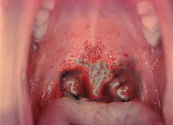
Diphtheria in the Oral Cavity. The distinctive thick, gray pseudomembrane forms on the tonsils and throat, potentially causing respiratory obstruction.
Contributed by S Bhimji, MD
References
Sharma NC, Efstratiou A, Mokrousov I, Mutreja A, Das B, Ramamurthy T. Diphtheria. Nature reviews. Disease primers. 2019 Dec 5:5(1):81. doi: 10.1038/s41572-019-0131-y. Epub 2019 Dec 5 [PubMed PMID: 31804499]
Hadfield TL, McEvoy P, Polotsky Y, Tzinserling VA, Yakovlev AA. The pathology of diphtheria. The Journal of infectious diseases. 2000 Feb:181 Suppl 1():S116-20 [PubMed PMID: 10657202]
Helleren R, Fløystad HK, Tellefsen OA, Boskovic M, Skraastad T, Mengshoel AT, Alfsnes K, Skogen V. Severe Respiratory Diphtheria-Like Illness Caused by Toxigenic Corynebacterium ulcerans. Emerging infectious diseases. 2026 Feb:32(2):285-287. doi: 10.3201/eid3202.250908. Epub [PubMed PMID: 41714899]
Truelove SA, Keegan LT, Moss WJ, Chaisson LH, Macher E, Azman AS, Lessler J. Clinical and Epidemiological Aspects of Diphtheria: A Systematic Review and Pooled Analysis. Clinical infectious diseases : an official publication of the Infectious Diseases Society of America. 2020 Jun 24:71(1):89-97. doi: 10.1093/cid/ciz808. Epub [PubMed PMID: 31425581]
Level 1 (high-level) evidenceHarnisch JP, Tronca E, Nolan CM, Turck M, Holmes KK. Diphtheria among alcoholic urban adults. A decade of experience in Seattle. Annals of internal medicine. 1989 Jul 1:111(1):71-82 [PubMed PMID: 2472081]
Edrees WH, Al-Ghoury AA, Humaid AA, Abdullah QY, Al-Hadheq AA, Al-Shehari WA, Al-Halani AA, Alsebaeai M. Epidemiological trend and clinical features of diphtheria in Hajjah Governorate, Yemen: a seven-years retrospective analysis (2018-2024). BMC infectious diseases. 2026 Feb 20:26(1):. doi: 10.1186/s12879-026-12917-3. Epub 2026 Feb 20 [PubMed PMID: 41715022]
Level 2 (mid-level) evidenceClarke KEN, MacNeil A, Hadler S, Scott C, Tiwari TSP, Cherian T. Global Epidemiology of Diphtheria, 2000-2017(1). Emerging infectious diseases. 2019 Oct:25(10):1834-1842. doi: 10.3201/eid2510.190271. Epub [PubMed PMID: 31538559]
Hoefer A, Seth-Smith H, Palma F, Schindler S, Freschi L, Dangel A, Berger A, D'Aeth J, Cordery R, Delgado-Rodriguez E, Gruner E, Flury D, Hinic V, Kofler J, Lienhard R, Mariman R, Nolte O, Schibli A, Toubiana J, Traugott M, Jacquinet S, Indra A, Fry NK, Palm D, Sing A, Brisse S, Egli A, 2022 European Diphtheria Consortium. Corynebacterium diphtheriae Outbreak in Migrant Populations in Europe. The New England journal of medicine. 2025 Jun 19:392(23):2334-2345. doi: 10.1056/NEJMoa2311981. Epub 2025 Jun 4 [PubMed PMID: 40466062]
Jayashree M, Shruthi N, Singhi S. Predictors of outcome in patients with diphtheria receiving intensive care. Indian pediatrics. 2006 Feb:43(2):155-60 [PubMed PMID: 16528112]
Parveen S, Bishai WR, Murphy JR. Corynebacterium diphtheriae: Diphtheria Toxin, the tox Operon, and Its Regulation by Fe2(+) Activation of apo-DtxR. Microbiology spectrum. 2019 Jul:7(4):. doi: 10.1128/microbiolspec.GPP3-0063-2019doi: 10.1128/microbiolspec.gpp3-0063-2019. Epub [PubMed PMID: 31267892]
NAIDITCH MJ, BOWER AG. Diphtheria; a study of 1,433 cases observed during a ten-year period at the Los Angeles County Hospital. The American journal of medicine. 1954 Aug:17(2):229-45 [PubMed PMID: 13180531]
Level 3 (low-level) evidenceOkechukwu C, Rebellato S, Pitfield H, Magnusson K, Sereshk R, Slavic D, McClinchey H, Wilson S, Kus J, Lee C. Isolation of toxigenic Corynebacterium diphtheriae from cutaneous lesions in a donkey in Ontario, Canada, 2024: Implications for zoonotic disease transmission and One Health approach. Canada communicable disease report = Releve des maladies transmissibles au Canada. 2026 Feb:52(1-2):8-14. doi: 10.14745/ccdr.v52i0102a02. Epub 2026 Feb 19 [PubMed PMID: 41822011]
Kneen R, Nguyen MD, Solomon T, Pham NG, Parry CM, Nguyen TT, Ha TL, Taylor A, Vo TT, Nguyen TT, Day NP, White NJ. Clinical features and predictors of diphtheritic cardiomyopathy in Vietnamese children. Clinical infectious diseases : an official publication of the Infectious Diseases Society of America. 2004 Dec 1:39(11):1591-8 [PubMed PMID: 15578357]
Singh S, Gupta N, Saple P. Diphtheritic myocarditis: A case series and review of literature. Journal of family medicine and primary care. 2020 Nov:9(11):5769-5771. doi: 10.4103/jfmpc.jfmpc_1396_20. Epub 2020 Nov 30 [PubMed PMID: 33532430]
Level 2 (mid-level) evidencePatris V, Argiriou O, Konstantinou C, Lama N, Georgiou H, Katsanevakis E, Argiriou M, Charitos C. Corynebacterium diphtheriae endocarditis with multifocal septic emboli: can prompt diagnosis help avoid surgery? The American journal of case reports. 2014 Aug 25:15():352-4. doi: 10.12659/AJCR.890855. Epub 2014 Aug 25 [PubMed PMID: 25153519]
Level 3 (low-level) evidenceMuttaiyah S, Best EJ, Freeman JT, Taylor SL, Morris AJ, Roberts SA. Corynebacterium diphtheriae endocarditis: a case series and review of the treatment approach. International journal of infectious diseases : IJID : official publication of the International Society for Infectious Diseases. 2011 Sep:15(9):e584-8. doi: 10.1016/j.ijid.2011.04.003. Epub 2011 Jun 8 [PubMed PMID: 21641260]
Level 3 (low-level) evidenceRau J, Berger A, Dangel A, Dyk M, Eisenberg T, Hiller E, Hoffmann C, Kutzer P, Martel A, Sing A, Sting R. Up-to-date MALDI-TOF MS-based identification of the Corynebacterium diphtheriae species complex for improved diagnostics. Journal of clinical microbiology. 2026 Jun 10:64(6):e0132425. doi: 10.1128/jcm.01324-25. Epub 2026 May 22 [PubMed PMID: 42171534]
Ciok AE. Horses and the diphtheria antitoxin. Academic medicine : journal of the Association of American Medical Colleges. 2000 Apr:75(4):396 [PubMed PMID: 10893126]
Level 3 (low-level) evidenceKneen R, Pham NG, Solomon T, Tran TM, Nguyen TT, Tran BL, Wain J, Day NP, Tran TH, Parry CM, White NJ. Penicillin vs. erythromycin in the treatment of diphtheria. Clinical infectious diseases : an official publication of the Infectious Diseases Society of America. 1998 Oct:27(4):845-50 [PubMed PMID: 9798043]
Level 1 (high-level) evidenceFarizo KM, Strebel PM, Chen RT, Kimbler A, Cleary TJ, Cochi SL. Fatal respiratory disease due to Corynebacterium diphtheriae: case report and review of guidelines for management, investigation, and control. Clinical infectious diseases : an official publication of the Infectious Diseases Society of America. 1993 Jan:16(1):59-68 [PubMed PMID: 8448320]
Level 3 (low-level) evidenceGietzen L, Kury D. Epiglottitis. JAAPA : official journal of the American Academy of Physician Assistants. 2018 Aug:31(8):53. doi: 10.1097/01.JAA.0000534985.92038.83. Epub [PubMed PMID: 30048357]
Baiu I, Melendez E. Epiglottitis. JAMA. 2019 May 21:321(19):1946. doi: 10.1001/jama.2019.3468. Epub [PubMed PMID: 31112260]
Vinckenbosch P, Guilcher P, Lambercy K, Richard C. [Retropharyngeal abscess in children]. Revue medicale suisse. 2017 Oct 4:13(577):1698-1702 [PubMed PMID: 28980783]
Pattanaik D, Lieberman JA. Pediatric Angioedema. Current allergy and asthma reports. 2017 Aug 8:17(9):60. doi: 10.1007/s11882-017-0729-7. Epub 2017 Aug 8 [PubMed PMID: 28791569]
Schwartzkopf J. Infectious mononucleosis. JAAPA : official journal of the American Academy of Physician Assistants. 2018 Nov:31(11):52-53. doi: 10.1097/01.JAA.0000546488.73851.dd. Epub [PubMed PMID: 30358681]
Thompson AE. JAMA Patient Page. Infectious mononucleosis. JAMA. 2015 Mar 17:313(11):1180. doi: 10.1001/jama.2015.159. Epub [PubMed PMID: 25781456]
Oliveira Pereira C, Ramos D, Mação P, Januário G, Januário L. [Diagnosis and treatment of acute pharyngitis -Is there any benefit on ten-day course of antibiotics?]. Anales de pediatria. 2018 Jun:88(6):335-339. doi: 10.1016/j.anpedi.2017.06.010. Epub 2017 Aug 7 [PubMed PMID: 28781106]
Millsop JW, Fazel N. Oral candidiasis. Clinics in dermatology. 2016 Jul-Aug:34(4):487-94. doi: 10.1016/j.clindermatol.2016.02.022. Epub 2016 Mar 2 [PubMed PMID: 27343964]
Hellstein JW, Marek CL. Candidiasis: Red and White Manifestations in the Oral Cavity. Head and neck pathology. 2019 Mar:13(1):25-32. doi: 10.1007/s12105-019-01004-6. Epub 2019 Jan 29 [PubMed PMID: 30693459]
Cooper AR. Diphtheria. Paediatrics & child health. 2002 Mar:7(3):205 [PubMed PMID: 20046292]
Skogmar S, Tham J. Severe diphtheria with neurologic and myocardial involvement in a Swedish patient: a case report. BMC infectious diseases. 2018 Jul 31:18(1):359. doi: 10.1186/s12879-018-3264-9. Epub 2018 Jul 31 [PubMed PMID: 30064365]
Level 3 (low-level) evidenceJain A, Samdani S, Meena V, Sharma MP. Diphtheria: It is still prevalent!!! International journal of pediatric otorhinolaryngology. 2016 Jul:86():68-71. doi: 10.1016/j.ijporl.2016.04.024. Epub 2016 Apr 21 [PubMed PMID: 27260583]
Samdani S, Jain A, Meena V, Meena CB. Cardiac complications in diphtheria and predictors of outcomes. International journal of pediatric otorhinolaryngology. 2018 Jan:104():76-78. doi: 10.1016/j.ijporl.2017.10.032. Epub 2017 Oct 31 [PubMed PMID: 29287886]
Lumio JT, Groundstroem KW, Melnick OB, Huhtala H, Rakhmanova AG. Electrocardiographic abnormalities in patients with diphtheria: a prospective study. The American journal of medicine. 2004 Jan 15:116(2):78-83 [PubMed PMID: 14715320]
Foo JC, Rahmat K, Mumin NA, Koh MT, Gan CS, Ramli N, Fong CY. Diphtheric encephalitis and brain neuroimaging features. Journal of clinical neuroscience : official journal of the Neurosurgical Society of Australasia. 2017 Nov:45():155-157. doi: 10.1016/j.jocn.2017.08.024. Epub 2017 Sep 1 [PubMed PMID: 28867361]
Weinberger B. Adult vaccination against tetanus and diphtheria: the European perspective. Clinical and experimental immunology. 2017 Jan:187(1):93-99. doi: 10.1111/cei.12822. Epub 2016 Jul 12 [PubMed PMID: 27279025]
Level 3 (low-level) evidence