 Tools
Tools
 Histology, Osteoprogenitor Cells
Introduction
Osteoprogenitor cells, also known as osteogenic cells, are stem cells in the bone that play a prodigal role in bone repair and growth (see Image. Cells in the Bone).[1] These cells are the precursors to the more specialized bone cells (osteocytes and osteoblasts) and reside in the bone marrow. Osteoprogenitor cells originate from infant mesenchymal cells and turn into spindle cells at the surface of matured bones. In developing bones, they appear more frequently and activate multifunctional stages to remodel the bones. The body loses the ability to synthesize or utilize more osteoprogenitor cells with age. Dysfunction of osteoprogenitor cells may delay ossification and lead to a spectrum of diseases such as dwarfism and Kashin-Beck disease.[2]
Structure
Register For Free And Read The Full Article
 Search engine and full access to all medical articles
- 10 free questions in your specialty
- Free CME/CE Activities
- Free daily question in your email
- Save favorite articles to your dashboard
- Emails offering discounts
Learn more about a Subscription to StatPearls Point-of-Care
Structure
Osteoprogenitor cells are often referred to as preosteoblasts. They can be present within the endosteum, the cellular layer of the periosteum, and the lining of the osteogenic cells. Osteoprogenitor cells exist as flattened spindle-shaped structures in matured bones that no longer display active bone remodeling or formation. They attach to the bone surface and are called inactive osteoblasts during this period. In maturing bones, however, these cells appear in their largest form. During fetal development or high turnover periods in adult osteogenesis, numerous osteoprogenitor cells function to give rise to osteoblasts. These structures display plump oval nuclei and emboldened abundant spindle-shaped cytoplasm at this stage, later converting to characteristic cuboidal active osteoblasts.[3][4]
Function
Osteoprogenitors can self-proliferate and self-renew. They participate in osteogenic differentiation and play a role in regulating angiogenesis.[5] Osteoprogenitor cells can differentiate into osteoblasts through mitotic division or by dividing into 2 stem cells via a highly regulated mechanism, which remains static during the proliferation process. After DNA synthesis and cell expansion, the cell retains its original genetic information. The endocrine system and local factors (growth factors and cytokines) mainly regulate osteogenesis.
Neuropeptide Y is a potent regulator of osteogenesis, and recent studies have confirmed that osteoprogenitor cells originating from hypothalamic Y2-/- mice have enhanced osteogenic activities.[6] Proline-rich tyrosine kinase 2 supposedly regulates the differentiation of early osteoprogenitors, and proline-rich tyrosine kinase 2 inhibitors promote osteogenesis and could act as a treatment for osteoporosis.[7] Osteoprogenitor cells also reside in the perichondrium. These osteoprogenitors hyper-regulate bone morphogenetic proteins during differentiation into mature osteoblasts and are responsible for the production of bone matrix.[8]
Tissue Preparation
Bone marrow stromal cells (BMSCs) yield testable osteoprogenitors. The cell samples are diluted with phosphate-buffered saline (PBS) at a ratio of 1:3, and then nucleated cells are isolated with a density gradient solution. Preparatory phase complete medium (CM) consists of 0.1 mmol/L nonessential amino acids, alpha-MEM with 10% fetal bovine serum, 4.5 mg/mL d-glucose, 1 mmol/L sodium pyruvate, 100 U/mL penicillin, 100 mmol/L HEPES buffer, 100 μg/mL streptomycin, and 0.29 mg/mL l-glutamine.[9]
The collected nucleated cells are plated at 100,000 cells/cm^2 density in supplemented CM. The CM supplement requires an additional 5 ng/mL fibroblast growth factor-2 (GF2) and 10 nmol/L dexamethasones; both elements proliferate osteogenic commitment of bone marrow stromal cells. The supplemental process undergoes humidification at 37 degrees C in a 5% CO2 incubator.[10]
Histochemistry and Cytochemistry
Several research groups have studied the development of osteoprogenitor cells. A study suggests that the perichondrial Thy-1–positive cells demonstrate potential osteogenic activity and participate in osteoblast formation during endochondral ossification. Another study shows intense alkaline phosphatase activity 2 weeks after osteogenic induction and the presence of mineralized nodules. The expression of bone sialoprotein, dentin matrix protein-1, and osteocalcin increases due to ALP proliferation. As confirmed by flow cytometry, 55 to 65% of cells display the cell-surface markers Sca1+ and Thy1+ in vitro expansion for an alpha-SMA-GFP positive population. The alpha-SMA–GFP-positive population also exhibits higher proliferative and osteogenic probabilities compared to an alpha-SMA–GFP-negative population.[8][11]
Microscopy, Light
Final visualization of osteoprogenitor cells under the microscope involves a delicate preparation phase: the explants are fixed in 4% buffered formalin for 1 whole day, and samples are decalcified with 0.5 mol/L ethylenediaminetetraacetic acid (pH 8) for 7 to 10 days. The samples are deposited in paraffin, and the cross-sections of the samples are prepared with 5 um thickness at 3 different strata.[12]
The samples are stained with hematoxylin/eosin and Masson/Trichrome, and both a qualitative exam for the presence of bone tissue and a quantitative exam by computerized bone histomorphometry is conducted. For each hematoxylin/eosin-stained cross-section, 3 or 4 images (adequate to fill the construct cross-sections) are required to calculate the bone tissue area and available area for tissue ingrowth (net implant area - undegraded scaffold area) by digital imaging analysis. Under the scanning electron microscopy (SEM) visuals, the red color in the slides indicates that the Masson/Trichrome stains indicate lamellar and remodeled bone, while the blue color shows freshly deposited and immature bone.[9]
Pathophysiology
Bone tumors are formed within osteoprogenitor or stromal cell lineage during Paget disease (PD), which likely derive from genetic alterations related to familial Paget’s disease. During Paget’s disease, the endosteal surface undergoes active remodeling, and abnormal osteoclasts bearing nuclear inclusions occur. The fibrotic tissues in bone biopsies from PD patients are considered to be made of surplus elongated, sophisticatedly branched stromal cells that display high alkaline phosphatase levels; these cells are similar to the pre-osteogenic stromal cells located within the normal bone marrow.[13]
Early and full-blown pagetic lesions show dynamic transformations in the arrangement, number, and function of stromal cells within endosteal/medullary tissue. An excessive amount of bone marrow osteoprogenitor cells in pagetic lesion areas verify earlier data on static and dynamic histomorphometry in patients with Paget’s disease. Previous data revealed that the rate of osteogenesis increases during Paget disease, but research has confirmed that there is also a surge in the birthrate of osteoblasts.[14]
The quality of the new bones depends on nature and hypermineralization. Most stromal/osteoblastic abnormalities result from bone mass increment, characterized by denser and thicker trabecular structures. Another characteristic is compromised mechanical integrity caused by poor architectural organization and transformations within the mineralized matrix material.[15]
Clinical Significance
Mesenchymal stem cells (MSCs) collected from adipose and bone marrow tissue hold therapeutic value for various bone disease treatments. Current studies demonstrate the benefit of bone grafts based on combinations of MSC, biomimetic scaffolds, and growth factor delivery, which showed an increased osteogenic regeneration rate with minimal side effects. The specific mechanisms of cellular signaling in bone remodeling are essential in understanding the incorporation of newer effective treatment methods for numerous bone diseases.[16]
Various transplant therapies involving osteogenic autologous bone grafts are less frequently used; patient-specific cell therapies involving autologous BM-MNCs (bone marrow mononuclear cells) composed of stem cells, monocytes, lymphocytes, and dendritic cells are rising in popularity for bone pathology treatments. All patients suffering from long-bone pseudoarthrosis attained full bone consolidation when treated with autologous BM-MNCs and allogeneic cancellous bone grafts. Bone marrow aspirate utilized during osteonecrosis treatment via minimally invasive decompression of the femoral head decreased disease progress and yielded overall pain and symptom relief. Bone marrow concentrated injections lead to better osteogenic unions, resulting in complete recovery in post-operative achondroplastic dwarf patients within 2 to 10 months after femoral lightning surgeries. Intravenous BMC injections, mixed with iloprost, proliferate fracture healing in patients with avascular necrosis. Patients receiving Intra-articular BMNc displayed better chewing and maximum interincisal opening with integral pain relief.[17][16]
Media
(Click Image to Enlarge)
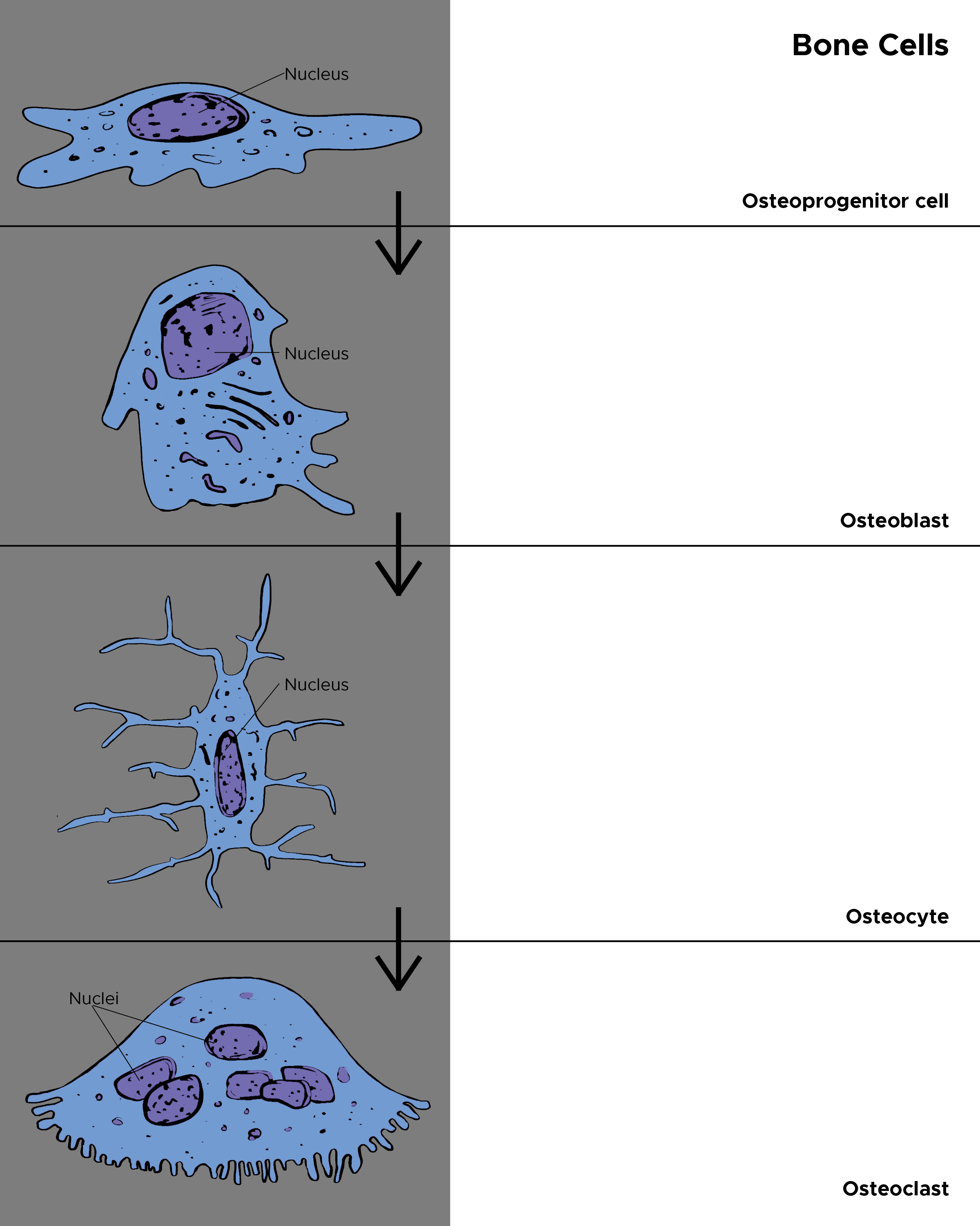
Cells in the Bone. The figure depicts the types of cells in the bone: osteoprogenitor cells, osteoblasts, osteocytes, and osteoclasts.
Contributed by C Rowe
References
Friedenstein AJ, Chailakhyan RK, Gerasimov UV. Bone marrow osteogenic stem cells: in vitro cultivation and transplantation in diffusion chambers. Cell and tissue kinetics. 1987 May:20(3):263-72 [PubMed PMID: 3690622]
Level 3 (low-level) evidenceRen X, Zhou Q, Foulad D, Tiffany AS, Dewey MJ, Bischoff D, Miller TA, Reid RR, He TC, Yamaguchi DT, Harley BAC, Lee JC. Osteoprotegerin reduces osteoclast resorption activity without affecting osteogenesis on nanoparticulate mineralized collagen scaffolds. Science advances. 2019 Jun:5(6):eaaw4991. doi: 10.1126/sciadv.aaw4991. Epub 2019 Jun 12 [PubMed PMID: 31206025]
Level 3 (low-level) evidenceXu J, Wang Y, Hsu CY, Gao Y, Meyers CA, Chang L, Zhang L, Broderick K, Ding C, Peault B, Witwer K, James AW. Human perivascular stem cell-derived extracellular vesicles mediate bone repair. eLife. 2019 Sep 4:8():. doi: 10.7554/eLife.48191. Epub 2019 Sep 4 [PubMed PMID: 31482845]
Clines GA. Prospects for osteoprogenitor stem cells in fracture repair and osteoporosis. Current opinion in organ transplantation. 2010 Feb:15(1):73-8. doi: 10.1097/MOT.0b013e328333d52c. Epub [PubMed PMID: 19935065]
Level 3 (low-level) evidenceIbrahim A, Bulstrode NW, Whitaker IS, Eastwood DM, Dunaway D, Ferretti P. Nanotechnology for Stimulating Osteoprogenitor Differentiation. The open orthopaedics journal. 2016:10():849-861. doi: 10.2174/1874325001610010849. Epub 2016 Dec 30 [PubMed PMID: 28217210]
Lundberg P, Allison SJ, Lee NJ, Baldock PA, Brouard N, Rost S, Enriquez RF, Sainsbury A, Lamghari M, Simmons P, Eisman JA, Gardiner EM, Herzog H. Greater bone formation of Y2 knockout mice is associated with increased osteoprogenitor numbers and altered Y1 receptor expression. The Journal of biological chemistry. 2007 Jun 29:282(26):19082-91 [PubMed PMID: 17491022]
Level 3 (low-level) evidenceHan S, Mistry A, Chang JS, Cunningham D, Griffor M, Bonnette PC, Wang H, Chrunyk BA, Aspnes GE, Walker DP, Brosius AD, Buckbinder L. Structural characterization of proline-rich tyrosine kinase 2 (PYK2) reveals a unique (DFG-out) conformation and enables inhibitor design. The Journal of biological chemistry. 2009 May 8:284(19):13193-201. doi: 10.1074/jbc.M809038200. Epub 2009 Feb 25 [PubMed PMID: 19244237]
Nakamura H, Yukita A, Ninomiya T, Hosoya A, Hiraga T, Ozawa H. Localization of Thy-1-positive cells in the perichondrium during endochondral ossification. The journal of histochemistry and cytochemistry : official journal of the Histochemistry Society. 2010 May:58(5):455-62. doi: 10.1369/jhc.2010.955393. Epub 2010 Feb 1 [PubMed PMID: 20124093]
Level 3 (low-level) evidenceJaquiéry C, Schaeren S, Farhadi J, Mainil-Varlet P, Kunz C, Zeilhofer HF, Heberer M, Martin I. In vitro osteogenic differentiation and in vivo bone-forming capacity of human isogenic jaw periosteal cells and bone marrow stromal cells. Annals of surgery. 2005 Dec:242(6):859-67, discussion 867-8 [PubMed PMID: 16327496]
Level 3 (low-level) evidenceDupree MA, Pollack SR, Levine EM, Laurencin CT. Fibroblast growth factor 2 induced proliferation in osteoblasts and bone marrow stromal cells: a whole cell model. Biophysical journal. 2006 Oct 15:91(8):3097-112 [PubMed PMID: 16861274]
Level 3 (low-level) evidenceSan Miguel SM, Fatahi MR, Li H, Igwe JC, Aguila HL, Kalajzic I. Defining a visual marker of osteoprogenitor cells within the periodontium. Journal of periodontal research. 2010 Feb:45(1):60-70. doi: 10.1111/j.1600-0765.2009.01201.x. Epub 2009 Apr 30 [PubMed PMID: 19453851]
Level 3 (low-level) evidenceDhouskar S, Tamgadge S, Tamgadge A, Periera T, Mudaliar U, Pillai A. Comparison of Hematoxylin and Eosin Stain with Modified Gallego's Stain for Differentiating Mineralized Components in Ossifying Fibroma, Cemento-ossifying Fibroma, and Cementifying Fibroma. Journal of microscopy and ultrastructure. 2019 Jul-Sep:7(3):124-129. doi: 10.4103/JMAU.JMAU_2_19. Epub [PubMed PMID: 31548923]
Roodman GD, Windle JJ. Paget disease of bone. The Journal of clinical investigation. 2005 Feb:115(2):200-8 [PubMed PMID: 15690073]
Level 3 (low-level) evidenceSabharwal R, Gupta S, Sepolia S, Panigrahi R, Mohanty S, Subudhi SK, Kumar M. An Insight in to Paget's Disease of Bone. Nigerian journal of surgery : official publication of the Nigerian Surgical Research Society. 2014 Jan:20(1):9-15. doi: 10.4103/1117-6806.127098. Epub [PubMed PMID: 24665195]
Cortini M, Baldini N, Avnet S. New Advances in the Study of Bone Tumors: A Lesson From the 3D Environment. Frontiers in physiology. 2019:10():814. doi: 10.3389/fphys.2019.00814. Epub 2019 Jun 26 [PubMed PMID: 31316395]
Level 3 (low-level) evidencePoon Z, Lee WC, Guan G, Nyan LM, Lim CT, Han J, Van Vliet KJ. Bone marrow regeneration promoted by biophysically sorted osteoprogenitors from mesenchymal stromal cells. Stem cells translational medicine. 2015 Jan:4(1):56-65. doi: 10.5966/sctm.2014-0154. Epub 2014 Nov 19 [PubMed PMID: 25411477]
Level 3 (low-level) evidenceHutchings G, Moncrieff L, Dompe C, Janowicz K, Sibiak R, Bryja A, Jankowski M, Mozdziak P, Bukowska D, Antosik P, Shibli JA, Dyszkiewicz-Konwińska M, Bruska M, Kempisty B, Piotrowska-Kempisty H. Bone Regeneration, Reconstruction and Use of Osteogenic Cells; from Basic Knowledge, Animal Models to Clinical Trials. Journal of clinical medicine. 2020 Jan 4:9(1):. doi: 10.3390/jcm9010139. Epub 2020 Jan 4 [PubMed PMID: 31947922]
Level 3 (low-level) evidenceTrompet D, Melis S, Chagin AS, Maes C. Skeletal stem and progenitor cells in bone development and repair. Journal of bone and mineral research : the official journal of the American Society for Bone and Mineral Research. 2024 Jul 23:39(6):633-654. doi: 10.1093/jbmr/zjae069. Epub [PubMed PMID: 38696703]
Matsushita Y, Liu J, Chu AKY, Ono W, Welch JD, Ono N. Endosteal stem cells at the bone-blood interface: A double-edged sword for rapid bone formation: Bone marrow endosteal stem cells provide a robust source of bone-making osteoblasts both in normal and abnormal bone formation. BioEssays : news and reviews in molecular, cellular and developmental biology. 2024 Mar:46(3):e2300173. doi: 10.1002/bies.202300173. Epub 2023 Dec 31 [PubMed PMID: 38161246]
Julien A, Perrin S, Martínez-Sarrà E, Kanagalingam A, Carvalho C, Luka M, Ménager M, Colnot C. Skeletal Stem/Progenitor Cells in Periosteum and Skeletal Muscle Share a Common Molecular Response to Bone Injury. Journal of bone and mineral research : the official journal of the American Society for Bone and Mineral Research. 2022 Aug:37(8):1545-1561. doi: 10.1002/jbmr.4616. Epub 2022 Jun 29 [PubMed PMID: 35652423]
Jeffery EC, Mann TLA, Pool JA, Zhao Z, Morrison SJ. Bone marrow and periosteal skeletal stem/progenitor cells make distinct contributions to bone maintenance and repair. Cell stem cell. 2022 Nov 3:29(11):1547-1561.e6. doi: 10.1016/j.stem.2022.10.002. Epub 2022 Oct 21 [PubMed PMID: 36272401]
Komori T. Regulation of Skeletal Development and Maintenance by Runx2 and Sp7. International journal of molecular sciences. 2024 Sep 20:25(18):. doi: 10.3390/ijms251810102. Epub 2024 Sep 20 [PubMed PMID: 39337587]
Wu S, Ohba S, Matsushita Y. Single-Cell RNA-Sequencing Reveals the Skeletal Cellular Dynamics in Bone Repair and Osteoporosis. International journal of molecular sciences. 2023 Jun 6:24(12):. doi: 10.3390/ijms24129814. Epub 2023 Jun 6 [PubMed PMID: 37372962]
Zhang W, Wang N, Yang M, Sun T, Zhang J, Zhao Y, Huo N, Li Z. Periosteum and development of the tissue-engineered periosteum for guided bone regeneration. Journal of orthopaedic translation. 2022 Mar:33():41-54. doi: 10.1016/j.jot.2022.01.002. Epub 2022 Feb 16 [PubMed PMID: 35228996]
Dillard LJ, Rosenow WT, Calabrese GM, Mesner LD, Al-Barghouthi BM, Abood A, Farber EA, Onengut-Gumuscu S, Tommasini SM, Horowitz MA, Rosen CJ, Yao L, Qin L, Farber CR. Single-Cell Transcriptomics of Bone Marrow Stromal Cells in Diversity Outbred Mice: A Model for Population-Level scRNA-Seq Studies. Journal of bone and mineral research : the official journal of the American Society for Bone and Mineral Research. 2023 Sep:38(9):1350-1363. doi: 10.1002/jbmr.4882. Epub 2023 Jul 27 [PubMed PMID: 37436066]
Level 2 (mid-level) evidenceLiao X, Koehnken Sawall J, Seeley R, Song F, Ji X, Liu X, Song C, Long F. An adipo-osteoprogenitor population in the endosteal niche contributes to bone and fat formation in adult mouse bone marrow. Proceedings of the National Academy of Sciences of the United States of America. 2025 Nov 25:122(47):e2502436122. doi: 10.1073/pnas.2502436122. Epub 2025 Nov 17 [PubMed PMID: 41248279]
Shi F, Yuan G, Wu Z, Luo Z, Chen Z, Liu Q, Li N, Xu R. Identification and function of periosteal skeletal stem cells in skeletal development, homeostasis, and disease. Journal of orthopaedic translation. 2025 Mar:51():177-186. doi: 10.1016/j.jot.2025.01.010. Epub 2025 Mar 14 [PubMed PMID: 40160808]
Jin A, Xu H, Gao X, Sun S, Yang Y, Huang X, Wang X, Liu Y, Zhu Y, Dai Q, Bian Q, Jiang L. ScRNA-Seq Reveals a Distinct Osteogenic Progenitor of Alveolar Bone. Journal of dental research. 2023 Jun:102(6):645-655. doi: 10.1177/00220345231159821. Epub 2023 May 6 [PubMed PMID: 37148259]
Wang L, Ruan M, Bu Q, Zhao C. Signaling Pathways Driving MSC Osteogenesis: Mechanisms, Regulation, and Translational Applications. International journal of molecular sciences. 2025 Feb 4:26(3):. doi: 10.3390/ijms26031311. Epub 2025 Feb 4 [PubMed PMID: 39941080]
Li Y, Fu G, Gong Y, Li B, Li W, Liu D, Yang X. BMP-2 promotes osteogenic differentiation of mesenchymal stem cells by enhancing mitochondrial activity. Journal of musculoskeletal & neuronal interactions. 2022 Mar 1:22(1):123-131 [PubMed PMID: 35234167]
Collins JM, Lang A, Parisi C, Moharrer Y, Nijsure MP, Thomas Kim JH, Ahmed S, Szeto GL, Qin L, Gottardi R, Dyment NA, Nowlan NC, Boerckel JD. YAP and TAZ couple osteoblast precursor mobilization to angiogenesis and mechanoregulation in murine bone development. Developmental cell. 2024 Jan 22:59(2):211-227.e5. doi: 10.1016/j.devcel.2023.11.029. Epub 2023 Dec 22 [PubMed PMID: 38141609]
Level 2 (mid-level) evidenceLiu J, Yang L, Liu K, Gao F. Hydrogel scaffolds in bone regeneration: Their promising roles in angiogenesis. Frontiers in pharmacology. 2023:14():1050954. doi: 10.3389/fphar.2023.1050954. Epub 2023 Feb 13 [PubMed PMID: 36860296]
Zhao X, Tang L, Le TP, Nguyen BH, Chen W, Zheng M, Yamaguchi H, Dawson B, You S, Martinez-Traverso IM, Erhardt S, Wang J, Li M, Martin JF, Lee BH, Komatsu Y, Wang J. Yap and Taz promote osteogenesis and prevent chondrogenesis in neural crest cells in vitro and in vivo. Science signaling. 2022 Oct 25:15(757):eabn9009. doi: 10.1126/scisignal.abn9009. Epub 2022 Oct 25 [PubMed PMID: 36282910]
Xiong Z, Song Y, Wu J, Lu W, Xiong J, Zhu M, Wang L, Chen Y, Xu L, Li X, Wang T. Bidirectional regulation factor of bone marrow mesenchymal stromal cells differentiation: a focus on bone-fat balance in osteoporosis. Stem cell research & therapy. 2025 Dec 2:17(1):14. doi: 10.1186/s13287-025-04838-w. Epub 2025 Dec 2 [PubMed PMID: 41331786]
Rong X, Kou Y, Zhang Y, Yang P, Tang R, Liu H, Li M. ED-71 Prevents Glucocorticoid-Induced Osteoporosis by Regulating Osteoblast Differentiation via Notch and Wnt/β-Catenin Pathways. Drug design, development and therapy. 2022:16():3929-3946. doi: 10.2147/DDDT.S377001. Epub 2022 Nov 15 [PubMed PMID: 36411860]
Lang E, Semon JA. Mesenchymal stem cells in the treatment of osteogenesis imperfecta. Cell regeneration (London, England). 2023 Feb 2:12(1):7. doi: 10.1186/s13619-022-00146-3. Epub 2023 Feb 2 [PubMed PMID: 36725748]
Mejias Rivera L, Shore EM, Mourkioti F. Cellular and Molecular Mechanisms of Heterotopic Ossification in Fibrodysplasia Ossificans Progressiva. Biomedicines. 2024 Apr 2:12(4):. doi: 10.3390/biomedicines12040779. Epub 2024 Apr 2 [PubMed PMID: 38672135]
Zhang T, Li J, Sun Z, Tu B, Wang W, Luo G, He Y, Jiang S, Fan C. Human osteoprogenitor cells obtained from traumatic heterotopic ossification samples showed enhanced osteogenic differentiation potential and ERK/hedgehog signaling than that from normal bone. IUBMB life. 2022 Nov:74(11):1081-1093. doi: 10.1002/iub.2670. Epub 2022 Aug 27 [PubMed PMID: 35964153]
Kaplan FS, Shore EM, Pignolo RJ. Fibrodysplasia ossificans progressiva emerges from obscurity. Trends in molecular medicine. 2025 Feb:31(2):106-116. doi: 10.1016/j.molmed.2024.08.010. Epub 2024 Sep 18 [PubMed PMID: 39299836]
Timberlake AT, Kiziltug E, Jin SC, Nelson-Williams C, Loring E, Yale Center for Genome Analysis, Allocco A, Marlier A, Banka S, Stuart H, Passos-Buenos MR, Rosa R, Rogatto SR, Tonne E, Stiegler AL, Boggon TJ, Alperovich M, Steinbacher D, Staffenberg DA, Flores RL, Persing JA, Kahle KT, Lifton RP. De novo mutations in the BMP signaling pathway in lambdoid craniosynostosis. Human genetics. 2023 Jan:142(1):21-32. doi: 10.1007/s00439-022-02477-2. Epub 2022 Aug 23 [PubMed PMID: 35997807]
Murthy A, Lee B, Zavala A, Tirado CA. ETV6::RUNX1-like Acute Lymphoblastic Leukemia. Journal of the Association of Genetic Technologists. 2024:50(2):61-63 [PubMed PMID: 38824653]
Perrin S, Colnot C. Periosteal Skeletal Stem and Progenitor Cells in Bone Regeneration. Current osteoporosis reports. 2022 Oct:20(5):334-343. doi: 10.1007/s11914-022-00737-8. Epub 2022 Jul 13 [PubMed PMID: 35829950]
Hall AF, Wang DV. A cortical-hippocampal communication undergoes rebalancing after new learning. bioRxiv : the preprint server for biology. 2025 Mar 29:():. pii: 2025.03.26.645547. doi: 10.1101/2025.03.26.645547. Epub 2025 Mar 29 [PubMed PMID: 40196557]
Wang Y, Wang B, Liu D, Xu L, Hu X, Liu X, Zheng H, Wang Z, Cheng M, Yan W. Osteoimmunology uncovered: How macrophages and biomaterials revolutionize bone healing. Materials today. Bio. 2026 Feb:36():102647. doi: 10.1016/j.mtbio.2025.102647. Epub 2025 Dec 13 [PubMed PMID: 41560822]